The objective of this study was to enhance the transdermal delivery of citalopram HBr (CTP) by incorporating it into novasomes (Citalosomes) prepared via probe sonication. Previous studies have shown limited transdermal absorption and short-term therapeutic effects of conventional citalopram formulations, representing a research gap that this study aims to address. The influence of cholesterol-to-surfactant ratio and oleic acid content on formulation characteristics was investigated. Particle size, zeta potential, and entrapment efficiency (EE
Naproxen is one of the non-steroidal anti-inflammatory drugs which show some side effects via oral administration. Percutaneous delivery of this drug can reduce these adverse effects. The bioavailability of naproxen could be promoted by several methods including administration of naproxen-loaded nanoparticles. In this research solid lipid nanoparticles (SLN) based on glyceryl behenate were developed as drug carriers and after characterization, the percutaneous absorption of optimum nanoparticles was compared with plain topical naproxen preparation. Naproxen-loaded SLNs were prepared by emulsification followed by ultrasonication method. Several nanoparticles were formulated with different amounts of glyceryl behenate as lipid phase and some ratios of Tween 60 and Span 80 as emulsifiers. Selected formulation showed a mean particle size of 259.6 +/- 10.2 nm, a polydispersity index (PDI) of 0.39 +/- 0.04, a zeta potential of -10.7 +/- 0.6 mV and a drug loading of 63.9% +/- 5.6%. These data were confirmed by SEM (Scanning Electron Microscope) analysis. DSC (Differential Scanning Calorimetry) and FTIR (Fourier Transform Infrared) spectroscopy proved the drug loading and showed no chemical interaction between formulation ingredients. The percutaneous absorption study via rat skin showed significant drug absorption of naproxen SLN after 24 h via rat skin which was 261.22 +/- 86.25 mu g/cm(2) of rat skin, in comparison with simple naproxen solution, 41.66 +/- 10.88 mu g/cm(2) of rat skin (p < 0.0001). This observation was made regarding the drug retained in the skin after 24 h (p < 0.0001). These results indicated SLN prepared based on glyceryl behenate can improve naproxen permeation via skin and can be used as a novel system for dermal drug delivery.
Background and purpose: Ascorbic acid is a crystalline, water-soluble powder that is used in topical preparations as an antioxidant, a skin lightener, a wound-healing agent, and an anti-wrinkle agent. However, skin penetration following topical administration is limited due to its hydrophilic characteristics and high-water solubility, and this challenge is further exacerbated by the vitamin’s low chemical stability in aqueous solutions. In this study, a niosomal delivery system was employed to enhance the skin permeability of ascorbic acid. Materials and methods: In this experimental study, ascorbic acid-loaded niosomes were prepared by the thin-layer hydration method, followed by sonication. The effects of the surfactant system and the sonication process on nanoparticle properties were investigated. An optimal formulation, based on particle size, polydispersity index (PDI), zeta potential (ZP), and entrapment efficiency (EE), was selected for evaluation of transdermal absorption and drug release across a dialysis membrane. Results: The results showed that the optimal formulation had a mean particle size of 168.15 ± 7.3 nm, a PDI of 0.31 ± 0.06, and an entrapment efficiency (EE) of 34.7 ± 5.2%. Transmission electron microscopy (TEM) images demonstrated the homogeneity and morphology of the niosomes. Attenuated total reflectance–Fourier transform infrared (ATR-FTIR) spectra and differential scanning calorimetry (DSC) thermograms showed successful drug loading without any chemical interactions. Drug release studies demonstrated a slow and controlled release pattern from ascorbic acid-loaded niosomes compared with the drug solution. Percutaneous absorption studies using rat abdominal skin in Franz diffusion cells showed enhanced skin penetration and increased skin retention after 24 h for ascorbic acid-loaded nanoparticles. Conclusion: These results indicate that the niosomal system is a suitable and stable delivery vehicle for the topical delivery of ascorbic acid.
In this study, a niosomal formulation of vanillic acid was successfully developed to enhance its poor aqueous solubility and antioxidant therapeutic potential. Niosomes were prepared using Span 60 and cholesterol via thin-film hydration method followed by probe sonication and were optimized using a Box–Behnken design. The optimized formulation exhibited a vesicle size of 129.91 ± 2.78 nm, a polydispersity index (PDI) of 0.152 ± 0.06, a zeta potential of − 8.698 ± 0.52 mV, and an entrapment efficiency (EE
Objective(s): Baclofen, a muscle relaxant, exhibits limited dermal penetration, restricting its therapeutic efficacy. This study aimed to optimize baclofen delivery using ultrasonically manufactured niosomes (Baclosomes) for improved antinociceptive and anti-inflammatory activity. Materials and Methods: The effect of cholesterol:surfactant (Chol:Surf) ratio on Baclosome characteristics was investigated using powder X-ray diffraction (PXRD), scanning electron microscopy (SEM), differential scanning calorimetry (DSC), and attenuated total reflectance-Fourier transform infrared (ATR-FTIR) spectroscopy. Dermal permeation studies were conducted in rats to assess baclofen delivery to the dermis and recipient compartment. Antinociceptive and anti-inflammatory effects were evaluated using a rat model. Results: Increasing Chol content significantly increased Baclosome particle size. The Chol:Surf ratio also influenced zeta potential (ZP), ranging from-9.10 +/- 1.81 to-28.81 +/- 1.3mV. DSC and PXRD analyses confirmed the amorphous nature of baclofen within the Baclosomes. There was no chemical interaction between the drug and the excipients which supported by ATR-FTIR analysis. Dermal permeation studies showed higher baclofen levels in the dermis and recipient compartment for Baclosome gel compared to plain baclofen gel, without inducing dermal irritation. Baclosome gel demonstrated significant antinociceptive and anti-inflammatory effects compared to control groups (baclofen gel and diclofenac gel). Conclusion: This study demonstrated that ultrasonically manufactured Baclosomes effectively enhance baclofen delivery to the skin, resulting in improved antinociceptive and anti-inflammatory activity. Optimization of the Chol:Surf ratio significantly influenced Baclosome characteristics, highlighting the potential of this formulation approach for enhancing drug efficacy. This approach could offer a promising strategy for improving the therapeutic benefits of baclofen in treating musculoskeletal pain and inflammation.
The development of innovative systems with antioxidant activity and controlled release capabilities is crucial for efficient wound healing and tissue regeneration. In this study, we developed a Carbopol based gel containing astaxanthin (AST) loaded chitosan-coated nanostructured lipid carriers (CS-NLCs). This system aimed to enhance stability, biocompatibility, sustained antioxidant properties, skin permeability, and minimize AST-related side effects. AST was encapsulated in NLCs using a hot homogenization ultrasonication method. Structural and morphological characteristics of nanoparticles were analyzed through TEM, FE-SEM, DSC, DLS, and XRD, confirming successful AST encapsulation in NLCs and CS coating. SEM analysis revealed particle sizes of 80 f 6.6 nm for AST-NLCs and 221.4 f 39.2 nm for CS-AST-NLCs. The encapsulation efficiency was above 85.45 %, with a loading content of 4.04 % for AST-NLCs. The antioxidant activity, assessed through DPPH, hydroxyl radical inhibition, and DCFH assays, demonstrated that AST-NLC-CS and AST-NLC exhibited significantly enhanced antioxidant performance compared to free AST. MTT assay confirmed biocompatibility of CS-AST-NLC formulation, with cell viability reaching 103.6 f 1.1 % over 72 h. In vitro cell migration studies demonstrated effectiveness of AST-NLC-CS, with 85.9 f 4.4 % fibroblast (L929) cell migration to wound site during 24 h. In vivo studies revealed accelerated wound healing and tissue regeneration with AST-NLC-CS/gel, leaving only 9.5 f 2.2 % of the wound area on day 11 post-treatment compared to other groups. Histopathological analyses using H&E and MT staining of incision wounds in male wistar Rats treated with CS-AST-NLC/Gel showed enhanced collagen deposition, more effective neovascularization, faster epithelialization, and reduced inflammatory cell infiltration compared to control group. This multifunctional system demonstrated great potential for improving wound healing and advancing tissue regeneration strategies.
Cuminum cyminum L. (cumin) essential oil (CEO) possesses documented analgesic and anti-inflammatory properties, but its topical application is limited by volatility, instability and low aqueous solubility. This study aimed to develop a CEO nanoemulgel and evaluate its physicochemical characteristics, dermal safety and antinociceptive/anti-inflammatory effects in rodent models. The optimized formulation exhibited nanometric droplet size, uniform distribution and acceptable physical stability. In a rat skin irritation test, repeated application of the CEO nanoemulgel did not produce visible erythema or edema compared with the gel base. In mice, the formulation increased tail-flick latency and reduced nociceptive behaviors in both phases of the formalin test, and histological analysis revealed attenuated inflammatory cell infiltration. These findings suggest that the CEO nanoemulgel has promising antinociceptive and anti-inflammatory activity and appears to be well tolerated in short-term preclinical models. Further studies, including long-term safety assessment, mechanistic analysis and clinical trials, are required before CEO nanoemulgel can be considered for use in humans.
Depression is a serious mental health disorder that can deeply affect a person's quality of life, and the importance of treatment for depression cannot be overstated. The aim of this study is to compare ultrasonication procedure as green method and thin film hydration technique as conventional way for preparation of niosome to increase venlafaxine HCl brain delivery and investigate BDNF (Brain-derived neurotrophic factor) gene expression. Both approaches provided well-defined sphere particles; however, the green method (ultrasonication) manufactured smaller particle size (96.93 +/- 3.82 nm), narrower size distribution (0.206 +/- 0.006) niosomes with a higher entrapment efficiency (58.06 +/- 1.45%) than the conventional method (p < 0.05). Moreover, the cytotoxicity examination revealed that the ultrasonication method prepared safe formulation and exhibited greater cellular safety than pure venlafaxine significantly (p < 0.05). The quantity of venlafaxine acquired by the brain following oral administration from green niosome made via ultrasonication (1222.077 +/- 109.692 ng/mL) was substantially more than that of the venlafaxine solution and conventional niosome (p < 0.05). Green niosome substantially decreased the immobility period in the force swim test (61.666 +/- 5.085 sec) and tail suspension test (62.500 +/- 5.319 sec) in comparison with conventional niosome and venlafaxine in animal model of depression (p < 0.05). Green niosomes enhance venlafaxine's antidepressant effect over conventional methods. Accordingly, green and conventional venlafaxine-niosome as well as its simple solution reversed the decreased BDNF gene expression in maternally separated mice, albeit there was no difference between different formulations (p > 0.05). Consequently, it was discovered that oral green venlafaxine niosome was a viable preparation for the management of depression, given its pharmacokinetic profile and therapeutic effects in animals. [GRAPHICS] .
Objectives: Oral mucositis is a common side effect of cancer chemotherapy. Traditionally, black mulberry or Morus nigra L. (M. nigra) fruit is used for treatment of oral inflammatory conditions. The aim of the present study was to evaluate the efficacy of black mulberry mouthwash for prevention of oral mucositis caused by 5-fluorouracil. Materials and Methods: This double-blind clinical trial was performed on 62 patients with colon adenocarcinoma undergoing chemotherapy with 5-fluorouracil. The patients were randomly divided into two groups (n=31) to receive 10mL of 1% black mulberry juice (BMJ) or the placebo mouthwash, 3 times a day for 2 weeks. The patients were followed up for 7 and 14 days after the treatment onset to record the degree of mucositis according to the World Health Organization (WHO) criteria. Spectrophotometric assays were conducted for quantitative analysis of the bioactive compounds in the composition of BMJ. Data were analyzed with the Chi-square and Fisher’s exact tests (alpha=0.05). Results: The severity of oral mucositis was slightly, but not significantly, lower in the follow-up sessions in the BMJ mouthwash group (P>0.05). The total anthocyanin content of the BMJ was calculated to be 506.5±3.51 and 476.2±7.99mg cyanidin 3-glucoside equivalent per 100g of dried juice, before and after the sterilization process, respectively. Conclusion: The present results showed that the BMJ mouthwash can be effective as an adjunct treatment to reduce the incidence and severity of mucositis in patients undergoing chemotherapy. Anthocyanins appear to be mainly involved in the observed effect.
Mahdi Jalahi1, Mohammad Azadbakht2,3, Jafar Akbari4, Fereshteh Talebpuor5,6, Fatemeh Akbari7 1 PharmD Student, Student Research Committee, Faculty of Pharmacy, Mazandaran University of Medical Sciences, Sari, Iran 2 Professor, Department of Pharmacognosy and Biotechnology, Faculty of Pharmacy, Mazandaran University of Medical Sciences, Sari, Iran 3 The Health of Plant and Livestock Products Research Center, Mazandaran University of Medical Sciences, Sari, Iran 4 Professor, Department of Pharmaceutics, Faculty of Pharmacy, Mazandaran University of Medical Sciences, Sari, Iran 5 Professor, Department of Anatomy, Faculty of Medicine, Mazandaran University of Medical Science, Sari, Iran 6 Cellular and Molecular Biology Research Center, Mazandaran University of Medical Science, Sari, Iran 7 Assistant Professor, Department of Pharmacognosy, School of Pharmacy, Semnan University of Medical Sciences, Semnan, Iran In the article published in Volume 34, Issue 240, 2024, the abbreviated names Ziziphora tenuior and Thymus vulgaris have been revised to their full forms in the title. The affiliation of Fereshteh Talebpuor was also incorrectly stated and has now been corrected to “Professor”.
Background and purpose: Gastric ulcers pose significant health challenges due to their high prevalence in various populations. Several factors contribute to the development of this condition, including Helicobacter pylori (H. pylori) infection, excessive use of nonsteroidal anti-inflammatory drugs (NSAIDs), smoking, age, and gender. Given the high incidence of gastric ulcers induced by NSAID consumption, this study aims to investigate the protective effects of hydroalcoholic extracts of Satureja hortensis and Froriepia subpinnata against indomethacin-induced gastric ulcers in animal models. Materials and methods: n this experimental study, the leaves of Satureja hortensis and Froriepia subpinnata were dried and extracted using 60% ethanol. The toxicity of these plant extracts was assessed using Artemia salina. The total phenolic and flavonoid contents of the extracts were measured spectrophotometrically. For the animal study, 48 Balb/c mice (n= 6 per group) were used. The treatment groups included omeprazole (30 mg/kg) and three different doses (250, 500, and 1000 mg/kg) of Satureja hortensis and Froriepia subpinnata, administered separately by gavage. A negative control group received normal saline. One hour after treatment, indomethacin was administered via gavage to induce gastric ulcers. Four hours after indomethacin administration, the animals were anesthetized, and their stomachs were collected for histopathological evaluation. Results: Histopathological findings revealed that inflammation, gastric tissue bleeding, and congestion were reduced in the Satureja hortensis and Froriepia subpinnata groups. Moreover, the 1000 mg/kg dose of both extracts demonstrated greater efficacy compared to the lower doses tested. Conclusion: Extracts of Satureja hortensis and Froriepia subpinnata exhibit a protective effect against gastric ulcers induced by NSAIDs. This protective effect is likely attributed to the antioxidant and anti-inflammatory properties of phenols and flavonoids present in the extracts and appears to be dose-dependent.
Background:We aimed to determine the effect of Zataria multiflora Nanoemulsion gel on surgical wound healing. Methods:This experimental study was conducted in the years 2021-2022 at the Animal Research Center of Mazandaran University of Medical Sciences, northern Iran. Forty two male Westar rats were randomly divided into six groups (n=7). After a surgical incision of full thickness with a 3 cm diameter, they were treated for 21 days with diltiazem 2% (positive control), placebo, and Z. multiflora emulsions and nanoemulsions at 2% and 4%, respectively. Macroscopic parameters of wound area and contraction, as well as pathological factors such as granulation, angiogenesis, epithelialization, collagen organization, bacterial colony, inflammation, creatine and epidermal thickness, hair follicles, and lymphatic ducts, were examined. Results:The mean wound size and contraction of the placebo group differed significantly from the other groups on all days, and on some days, the results indicated more favorable effects of nanoemulsions than Diltiazem. Based on the microscopic findings, the average scores on the seventh day were nearly different (P= 0.051); however, all groups scored higher than the placebo group. On the 21st day, the best results were related to the 4% Nanoemulsion (242.5), 2% Nanoemulsion (159.4), and 4% emulsion (159.3), followed by diltiazem (154.60), 2% emulsion (146.5), and placebo (70.7). Conclusion:Z. multiflora emulsions and nanoemulsions at 2% and 4% could be effective in healing surgical wounds, and the use of 4% Nanoemulsion yields the best results. This is recommended for use in clinical trial studies.
Background: The purpose of the current work was to manufacture vitamin A (Vit A) niosome and solid lipid nanoparticle (SLN) using an ultrasonic approach and to evaluate its effect on wound healing. Methods: The nanoparticles were prepared through an ultrasonication technique and characterized by dynamic light scattering (DLS), and transmission electron microscopy (TEM). Further, the nanoparticles were evaluated for their various parameters such as pH, viscosity, spreadability, stability, in-vitro drug release study, in-vitro cytotoxicity, and in-vivo wound healing. Results: TEM confirmed the spherical nature of the Vit A-niosome and SLN. The Vit A formulations (niosome and SLN) were stable in the accelerated stability test (freeze-thaw cycle). Vit A release from the SLN gel and niosome gel was significantly higher (almost 70 and 80%, respectively) than simple Vit A gel. According to the animal study, the wound healing closure in the Vit A niosomal gel and Vit A SLN gel was greater than the other groups during the 21 days after surgery (P < 0.05) and was close to each other over time and on day 21. Based on histological data, wounds treated with Vit A niosome gel and Vit A-SLN gel had more collagen than the other groups. MDA malondialdehyde (MDA, an end-product of lipid peroxidation) significantly decreased in the Vit A-niosome and Vit A-SLN gel group after 21 days, while glutathione peroxidase (GPx), superoxide dismutase (SOD, an endogenous antioxidant), and hydroxyproline levels demonstrated an increase. Conclusion: The findings of this study revealed that the prepared Vit A nano-formulations could be used as a possible and safe nano-vesicle for Vit A cutaneous delivery thus potentially opening up new prospects for the treatment of wound disorders.
Background and purpose: The aging population and its associated complications, such as chronic pain, have led to an increased use of non-steroidal anti-inflammatory drugs (NSAIDs) like indomethacin. However, indomethacin is associated with notable side effects, particularly gastric ulcers. This study aims to investigate the potential protective effects of hydroalcoholic extracts of Zataria multiflora and Eryngium caucasicum in preventing indomethacin-induced gastric ulcers in an animal model. Materials and methods: In this experimental study, total phenol and flavonoid levels of the Zataria multiflora L. and Eryngium caucasicum L. plants were measured. Cultivated Artemia salina was used to evaluate the toxicity of the extracts. The animal study was conducted using eight groups of Balb/c mice. The groups included indomethacin at 70 mg/kg, omeprazole at 30 mg/kg, and three concentrations (250, 500, and 1000 mg/kg) of Eryngium caucasicum L. and Zataria multiflora L., respectively, administered individually to mice via gavage. One hour later, indomethacin was administered to the mice by gavage to induce gastric ulcers. Four hours after ulcer induction, the anesthetized animals were sacrificed, and their stomachs were removed for histopathological and morphological evaluation. Results: The amount of total phenol in Zataria multiflora L. and Eryngium caucasicum L. plants was 113.5 mg/ml and 26.06 mg/ml, respectively, and the flavonoid content of these plants was 41 mg/ml and 16.79 µg/ml, respectively. The toxicity results of cultivated Artemia salina showed that the toxicity of Zataria multiflora and Eryngium caucasicum plants was 500±0.0012 ppm and 0.0031±0 ppm, respectively. The histopathological results showed that Zataria multiflora and Eryngium caucasicum played an effective role in reducing inflammation, bleeding in stomach tissue, and congestion. Furthermore, the results showed that a dose of 1000 mg/kg was more effective than lower doses for both extracts. Conclusion: Hydroalcoholic extracts of Eryngium caucasicum and Zataria multiflora plants demonstrated a protective effect against stomach ulcers. This effect appears to be attributed to the presence of phenols and flavonoids in the extracts and is dose-dependent.
Modern nanomedicine delivers drugs to malfunctioning cells. Several synthetic processes can change the shape, particle size, polydispersity index, surface area, and porosity of mesoporous silica nanoparticles (MSNs). Polyamidoamine (PAMAM) dendrimers are attractive polymers with circular forms, uniform distribution, well-structured branching, internal cavities, and surface terminal groups. The objective of this study is to deliver 5-fluorouracil (5-FU) as passive targeting to carcinogen cells via MSN-PAMAM-G3 nanoparticles. This study involved the direct and adaptable synthesis of MSN as the core vehicle, together with third-generation PAMAM dendrimers as the vehicle shell, with the goal of delivering the 5-FU in a targeted and controllable manner. Tetraethyl orthosilicate (TEOS) and cetyltrimethylammonium bromide (CTAB) are used as templates to make the center nanoparticle. Methyl acrylate (MA) and ethylenediamine (EDA) are monomers that cause the formation of branches in PAMAM. The obtained particles were fully characterized in terms of particle size, polydispersity index, zeta potential, thermal behavior, morphology, pore diameter, pore volume, and specific surface area. The particle size of MSN-PAMAM-G3s showed around 187.71 +/- 3.97 nm, with a polydispersity index of 0.17 +/- 0.01 and zeta potential of 32.17 +/- 1.30 mV. By reducing the ratio of drug to vehicle from 1 to 0.25, a significant increase in the EE% of 5-FU from 21.36 +/- 0.70 to 43.12 +/- 0.67 was observed. The release of 5-FU from the vehicle (5-FU@MSN and 5-FU@MSN-PAMAM-G3) was shown to be considerably higher in an acidic medium (pH = 5.5) in comparison to a neutral medium (pH = 7.4) (P-value<0.05). Furthermore, in both acidic and neutral conditions, 5-FU release was sustained from the vehicle rather than the 5-FU solution. Our research revealed that the MSN-PAMAM-G3 nanoparticles effectively regulated the encapsulation and release of 5-FU, providing pH-sensitive and sustained medication delivery. It is asserted that this nanocarrier has the potential to effectively deliver 5-FU to cancer cells.
Purpose: Spironolactone (SPN), which is classified as an anti-androgen, has demonstrated efficacy in treating acne. This study aimed to utilize ultrasonication to create a chitosan-coated nano lipid carrier (NLC) for enhancing the delivery of SPN to the skin and treating acne. Methods: Various Hydrophilic-Lipophilic Balance (HLB) values were investigated to optimize the SPN-NLCs. Photon correlation spectroscopy, attenuated total reflectance-Fourier transform infrared spectroscopy (ATR-FTIR), transmission electron microscopy (TEM), and differential scanning calorimetry (DSC) were employed to characterize the solid state of SPN in nanoparticle form. Additionally, the optimized formulation was used in a double-blind, randomized clinical trial. Results: Reducing the HLB of the surfactant mixtures resulted in a reduction in the size of SPN-NLCs. The formula with the smallest particle diameter (238.4±0.74 nm) and the lowest HLB value (9.65) exhibited the highest encapsulation efficiency of 79.88 ± 1.807%. Coating the optimized SPN-NLC with chitosan increased the diameter, PDI, zeta potential, and encapsulation efficiency. In vitro skin absorption studies demonstrated sustained release profiles for chitosan-coated SPN-NLC. In the double-blind trial, a gel containing chitosan-coated SPN-NLC effectively treated mild to moderate acne vulgaris, leading to improved healing and reduced lesion count after 8 weeks of therapy compared to the placebo. It successfully addressed both non-inflammatory and inflammatory lesions without adverse effects on the skin. Conclusion: The findings indicate that chitosan-coated SPN-NLCs have the potential as nanoparticles for targeted SPN delivery to the skin, offering novel options for the treatment of acne vulgaris.
Chemotherapeutic agents often lack specificity, intratumoral accumulation, and face drug resistance. Targeted drug delivery systems based on nanoparticles (NPs) mitigate these issues. Poly (lactic-co-glycolic acid) (PLGA) is a well-studied polymer, commonly modified with aptamers (Apts) for cancer diagnosis and therapy. In this study, silybin (SBN), a natural agent with established anticancer properties, was encapsulated into PLGA NPs to control delivery and improve its poor solubility. The field-emission scanning electron microscopy (FE-SEM) showed spherical and uniform morphology of optimum SBN-PLGA NPs with 138.57±1.30nm diameter, 0.202±0.004 polydispersity index (PDI), -16.93±0.45mV zeta potential (ZP), and 70.19±1.63
After extensive computations, novel trinitro compounds, triethyl ammonium 2,4,6-trinitro phenoxide ( TEA-TNP ) and tetramethyl guanidinium 2,4,6-trinitro phenoxide ( TMG-TNP ), are designed, synthesized, and identified. The Geometrical parameters, electronic properties, and spectroscopic analysis have been investigated by comparison of theoretical and experimental methods. Spectrum features that include IR and Raman have been calculated, and 1 H and 13 C NMR spectra are compared to experimental data. Electronic analysis has been investigated through NLO, FMO, UV-Vis, NBO, and MEP calculations. B3LYP/6-311++G(d,p) is highly successful and dependable for predicting ionic liquid stability, electrical characteristics, and spectroscopy. Both TEA-TNP and TMG-TNP are introduced as suitable candidates for NLO material. TMG-TNP with greater nucleophilicity (N) is a noncorrosive lubricant additive that reduces friction and wear for tribological performance.
In the present study, an ionic gelation and ultrasonic approach was performed to produce kojic acid (KA) loaded chitosan(CS)/collagen(CN) nanoparticle(NP) (CSCN-NP) which aimed to increase the dermal delivery and anti-pigmentation effect. To optimize the CSCN-NP the effect of the amount of CN was investigated. The results showed that increasing CN from 0 to 500 mg increased the mean particle size and entrapment efficiency of KA-CSCN-NP from 266.07 +/- 9.30 nm to 404.23 +/- 9.44 nm and 17.37 +/- 2.06% to 82.34 +/- 2.16%, respectively. Differential scanning calorimetry confirmed the amorphous form of KA in CSCN-NP, while scanning electron microscopy revealed that the nanoparticles were spherical. There was no chemical interaction between KA and the other components base on attenuated total reflectance-Fourier transform infrared spectroscopy. The skin permeability test showed that KA-CSCN-NP gel delivered more KA to the dermal layers (29.16 +/- 1.67% or 537.26 +/- 537.26 mu g/cm2) and receiver compartment (15.04 +/- 1.47% or 277.15 +/- 27.22 mu g/cm2) compared to KA plain gel. In vitro cytotoxicity assay demonstrated that the improved KA-CSCN-NP was non-toxic. Dermal irritating test on Wistar rats showed that the KA gel was non-irritating. Furthermore, KA-CSCN-NP was found to inhibit melanin formation to a greater extent than free KA and significantly inhibited L-dopa auto-oxidation (94.80 +/- 2.41%) compared to pure kojic acid solution (75.28 +/- 3.22%). The observations of this study revealed that the produced KA-CSCN-NP might be used as a potential nano-vehicle for KA dermal administration, thereby opening up innovative options for the management of hyper-melanogenesis problems. [Graphical Abstract]
 Majid Saeedi合作论文数97
 Katayoun Morteza-Semnani合作论文数69
 Enayati-Fard Reza合作论文数Faculty of Pharmacy, Mazandaran University of Medical Sciences38
 Ali Nokhodchi合作论文数Department of Chemistry, University of Sussex31Seyyed Mohammad Hassan Hashemi合作论文数Fac Pharm, Mazandaran Univ Med Sci24
 Akbar Heydari合作论文数Department of Organic Chemistry, Faculty of Basic Sciences, Tarbiat Modares University24Amirhossein Babaei合作论文数19
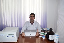 Seyyed Sohrab Rostamkalaei合作论文数Fac Pharm, Islamic Azad Univ17
 Mohammad Azadbakht合作论文数Faculty of Pharmacy, Mazandaran University of Medical Sciences15
 Seyyed Mobin Rahimnia合作论文数13
 Hamid Reza Kelidari合作论文数10
 Asare-Addo Kofi合作论文数Department of Pharmacy, University of Huddersfield10
 Seyed Jalal Hosseinimehr合作论文数Department of Radiopharmacy, Mazandaran University of Medical Sciences10
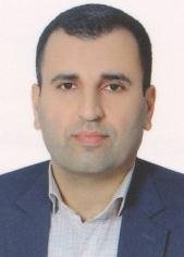 Ebrahim Salehifar合作论文数Faculty of Pharmacy, Mazandaran University of Medical Sciences9
 Mohammad Eghbali合作论文数Sch Nursing & Midwifery, Torbat Heydariyeh Univ Med Sci8
 Hadi Valizadeh合作论文数Faculty of Pharmacy and Drug Applied Research Center, Tabriz University of Medical Sciences8Zohreh Hajheydari合作论文数8
 Mohammad Seyedabadi合作论文数8
 Pedram Ebrahimnejad合作论文数Faculty of Pharmacy, Mazandaran University of Medical Sciences7
 Arash Ghasemi合作论文数Fac Med, Mazandaran Univ Med Sci6
 Fatemeh Ahmadi合作论文数6
 Soheil Azizi合作论文数6
 Ebrahim Nasiri .合作论文数6
 Ali Farmoudeh合作论文数Faculty of Pharmacy, Mazandaran University of Medical Sciences5
 Maryam Ghasemieshkaftaki合作论文数Faculty of Medicine, Mazandaran University of Medical Sciences5Saman Hosseinkhani合作论文数5Anahita Ghorbani合作论文数4Sahar Esfahani合作论文数4jafar m nezhad合作论文数4
 Maryam Moazeni合作论文数4
 Shidrokh Abootorabi合作论文数Mazandaran University of Medical Sciences4
 Adeleh Sahebnasagh合作论文数Dept Internal Med, North Khorasan Univ Med Sci4
 Reza Negarandeh合作论文数Nursing & Midwifery Care Res Ctr, Univ Tehran Med Sci4
 Mahmood Moosazadeh合作论文数Addiction Institute, Mazandaran University of Medical Sciences4
 Yousefi Ali合作论文数Department of Physical Chemistry, Tarbiat Modares University4
 Hedayatizadeh-Omran A合作论文数Molecular and Cell Biology Research Center, Mazandaran University of Medical Sciences4
 Ala Shahram合作论文数Faculty of Pharmacy, Mazandaran University of Medical Sciences4Tajbakhsh Mahgol合作论文数4
 Alipour Abbas合作论文数School of Medicine, Mazandaran University of Medical Sciences;School of Medicine, Mazandaran University of Medical Sciences4A. Heydari合作论文数a Chemistry Department, Tarbiat Modarres University, P. O. Box 14155-4838, Tehran, Iran4
 Mehdi Khalaj合作论文数Department of Chemistry, Islamic Azad University4
 Maryam Esfandyari合作论文数3Alireza Sanaee合作论文数3Seyed Mahmoud Mousavi-Safavi合作论文数3Behrouz Jafarkhani合作论文数3Shahram Ghasemi合作论文数3Amin Goodarzi合作论文数3
 Hamed Morad合作论文数These two authors contributed equally to this work.3Hossein Lashkardoost合作论文数Dept Epidemiol & Biostat, North Khorasan Univ Med Sci3




