Endemic coronaviruses circulate seasonally and, while typically mild, can produce robust inflammation and severe pneumonia. Immunological memory to coronaviruses can be cross-reactive for multiple coronaviruses including OC43 and SARS-CoV-2. In this study, we used mouse models to investigate immune responses elicited by pulmonary infection with OC43. OC43 caused a mild, self-limiting infection. Early after infection, there was acute influx of myeloid cells and lymphocytes to the lung as well as a type I interferon-dependent production of IFN-γ. However, blocking interferons did not enhance viral infection. After recovery, lungs contained resident memory lymphocytes, and both the blood and lungs contained antibodies against OC43. When infection was supplemented with proinflammatory stimuli to model more severe disease, OC43-elicited immune changes were bolstered, including increased virus-specific plasma IgG and lung IgA, enhanced lung B-cell class-switching, and increased antigen-specific Th1 and Th17 cytokine production from lung CD4+ T cells. The antibodies and CD4+ T cells in mice recovered from OC43 infection, even with immunostimulatory agents, reacted only with OC43 proteins and peptides and not with those from other coronaviruses. Therefore, OC43 infection in wild-type mice does not induce the heterotypic immunity observed in humans. In conclusion, OC43 induces a self-limiting lung infection in mice accompanied by homotypic lung-resident and systemic memory, amplified by proinflammatory stimuli during infection.
Establishing sub-phenotypes of pneumonia based on distinct host processes will be a step towards using host-directed therapies (to complement microbe-directed therapies) more rationally and precisely. Although pneumonia is a pulmonary pathophysiology, histological changes within the lungs have not been leveraged for sub-phenotyping. We addressed this by scoring 18 histopathology features (e.g., type 2 cell hyperplasia or necrosis) across rapid autopsy lung samples from 276 elderly subjects with pneumonia. Machine learning algorithms segregated subjects into seven different sub-phenotypes of pneumonia with distinct histopathology signatures. Quantitative immunofluorescence demonstrated associations of macrophages, neutrophils, T cells, and B cells with select histology features and pulmonary pathology sub-phenotypes. Mouse models revealed corollary sub-phenotypes, although some histology features observed in human lungs were never observed in mice. By illuminating this spectrum of histopathologies and discriminating discrete sub-phenotypes of pneumonia, a foundational framework emerges for developing and using host-directed therapies for subsets of pneumonia patients.
ABSTRACT Cytomegalovirus (CMV) has been implicated in glioblastoma (GBM) progression. Ongoing clinical trials are assessing therapeutic approaches targeting CMV in GBM but to date no new therapy has been approved outside the standard of care. Previous preclinical studies have highlighted the potential of the antiviral drug Cidofovir (CDV) in GBM; however, its clinical use is limited by dose-dependent nephrotoxicity and poor cellular uptake, necessitating high intravenous doses to achieve therapeutic activity. Brincidofovir (BCV), a lipid conjugate of CDV has been developed, which does not induce nephrotoxicity and has significantly greater cellular bioavailability. Here we examined the effects of BCV in a newly established CMV-driven GBM model (SB28) and in patient-derived tumor neurospheres. We show that BCV prolongs survival in vivo and exerts both CMV-dependent and independent antitumor effects. Mechanistically, BCV induces DNA damage and cell cycle dysregulation in GBM cells and inhibits proliferation of patient-derived neurospheres in a dose-dependent manner. These data identify BCV as a dual-action therapeutic that suppresses viral oncomodulation while directly targeting tumor cell viability.
Group 3 innate lymphoid cells (ILC3s) are key sensors of the intestinal environment, integrating dietary and microbial cues to maintain intestinal immunity. We found that intestinal ILC3s were reduced in overweight and obese humans and in high-fat diet (HFD)-fed mice. ILC3 loss occurred independently of caloric excess, weight gain, or glucose intolerance. Instead, impairment arose within hours of HFD consumption and was initiated by microbiota-driven intestinal barrier permeability and concomitant activation of inflammatory mononuclear phagocytes (MNPs). This response to inflammation impaired fatty acid oxidation in lipid-loaded ILC3s, resulting in mitochondrial damage and cell death. Intestinal ILC3 cell death was rescued by removal of excess fats from the diet. ILC3s from individuals with obesity also exhibited impaired fatty acid oxidation. Together, our findings define a malleable mechanism whereby dietary fats and microbial cues drive ILC3 maladaptation and death, with consequences for intestinal homeostasis.
BACKGROUND:Mechanisms driving aggressive meningiomas remain poorly understood. Given the pivotal role of the immune microenvironment in tumor progression, we developed a comprehensive atlas of the meningioma microenvironment, with a view toward identifying modifiable opportunities. METHODS:The immune microenvironment of 2,727 meningiomas was profiled using orthogonal methodologies, including 24 with mass cytometry, 24 single-cell RNAseq, 1,437 bulk RNAseq, 1,125 DNA methylation, 117 multiplex immunofluorescence, as well as that of 5 paired peripheral blood samples and 10 human meninges. Patient-derived organotypic tumor spheroids (PDOTS) were established to assess the effect of STING stimulation combined with anti-PD-1 treatment. RESULTS:We revealed a rich immune infiltration in meningioma, among the highest across 34 human cancer types (n = 12,188). Macrophages predominated in meningioma microenvironment, in contrast to the lymphoid dominance of peripheral blood, with meninges exhibiting an intermediate immune profile between meningiomas and peripheral blood. Cellular states and phenotypes of both immune and tumor cells shifted during tumor progression, with aggressive meningiomas possessing earlier-stage, immunosuppressive immune cells and proliferative tumor cells. Using ex vivo meningioma PDOTS, we demonstrated inducible responses to STING activation, marked by elevated cytokine release, which were synergistic when combined with PD-1 blockade. CONCLUSIONS:These findings offer an extensive resource on the cellular heterogeneity of the meningioma microenvironment and provide a framework for rational therapeutic modeling and strategy development.
T cell memory significantly alters the immune response and organ dysfunction induced by the murine cecal ligation and puncture (CLP) model of sepsis. Enhanced T cell memory activation promotes hepatic neutrophilic responses and induces hepatic dysfunction, which is a common complication of human sepsis associated with poor outcomes. We used a novel Immune Educated CLP sepsis mouse model to examine the role of memory T cell cytokine responses in driving innate immunity and organ dysfunction. Through this approach, we found that induced T cell memory prior to CLP led to higher serum levels of IL12, TNF, IL17, and IL1β – all dependent on memory CD8 T cell derived IFNγ following CLP. IFNγ induced activated hepatic IL12+ monocyte-derived dendritic cells. Increased neutrophilic responses occurred in Educated CLP which was found to depend on TNF, and were suppressed by IFNγ. Hepatic dysfunction in response to CLP was worsened by CD4 and CD8 T cell immune memory and prevented by IFNγ and IL17F blockade. These findings were recapitulated in naturalized outbred pet shop mice with natural immune memory to provide translational relevance to our Immune Educated CLP sepsis model. IL17F or IFNγ blockade may represent potential targets for treatment in sepsis with hepatic dysfunction.
Lupus is a complex autoimmune disease that involves dysregulation of the B cell compartment. Potential mechanisms for this dysregulation include abnormalities in immune dampening pathways, such as the programmed cell death 1 receptor (PD-1). Though PD-1 functions as a negative regulator in T cells, its role in B cells is not clear. We used comprehensive flow and mass cytometry to examine the expression of PD-1 on peripheral blood B cells from lupus and healthy controls in two large patient cohorts. PD-1 was increased on antigen experienced B cells including double negative, memory, and CD11c+ age-associated B cells. Heterogeneity in patient PD-1+ B cells was uncovered, including an enriched PD-1+CD11c+CXCR3+ B cell subset which we speculate may be pathogenic due to their potential to migrate to sites of inflammation, and a diminished PD-1+PD-L1+ B cell subset which we expect is an immunosuppressive phenotype. PD-1 expression was higher in patients with elevated anti-dsDNA, high disease activity and increased with in vitro stimulation (anti-BCR and TLR9). Surprisingly, PD-1+ B cells proliferated more than PD-1- B cells, and PD-1 blockade with an anti-PD-1 antibody further increased proliferation. Our results suggest that PD-1 function in B cells is different than its inhibitory signaling role in T cells. Further, the expansion of PD-1 expressing B cells in lupus, especially in patients with high disease activity, highlights a new potential pathogenic B cell population. Supported by T32ES007026; Public Health Sciences Pilot Award; NIH Accelerating Medicines Partnership (RA/SLE Network) Basic Autoimmunity (BA)
Lupus nephritis (LN) is a frequent manifestation of systemic lupus erythematosus, and fewer than half of patients achieve complete renal response with standard immunosuppressants. Identifying noninvasive, blood-based immune alterations associated with renal injury could aid therapeutic decisions. Here, we used mass cytometry immunophenotyping of peripheral blood mononuclear cells in 145 patients with biopsy-proven LN and 40 healthy controls to evaluate the heterogeneity of immune activation and identify correlates of renal parameters. Unbiased analysis identified 3 immunologically distinct groups of patients that were associated with different patterns of histopathology, renal cell infiltrates, urine proteomic profiles, and treatment response at 1 year. Patients with enriched circulating granzyme B+ T cells showed more active disease and increased numbers of activated CD8+ T cells in the kidney, yet they had the highest likelihood of treatment response. A second group characterized by a high type I interferon signature had a lower likelihood of response to therapy, while a third group appeared immunologically inactive but with chronic renal injuries. The major immunologic axes of variation could be distilled down to 5 simple cytometric parameters that recapitulate several clinical associations, highlighting the potential for blood immunoprofiling to translate to clinically useful noninvasive metrics to assess immune-mediated disease in LN.
Rheumatoid arthritis (RA) is a systemic autoimmune disease currently with no universally highly effective prevention strategies. Identifying pathogenic immune phenotypes in at-risk populations prior to clinical onset is crucial to establishing effective prevention strategies. Here, we applied multimodal single-cell technologies (mass cytometry and CITE-Seq) to characterize the immunophenotypes in blood from at-risk individuals (ARIs) identified through the presence of serum antibodies against citrullinated protein antigens (ACPAs) and/or first-degree relative (FDR) status, as compared with patients with established RA and people in a healthy control group. We identified significant cell expansions in ARIs compared with controls, including CCR2+CD4+ T cells, T peripheral helper (Tph) cells, type 1 T helper cells, and CXCR5+CD8+ T cells. We also found that CD15+ classical monocytes were specifically expanded in ACPA-negative FDRs, and an activated PAX5lo naive B cell population was expanded in ACPA-positive FDRs. Further, we uncovered the molecular phenotype of the CCR2+CD4+ T cells, expressing high levels of Th17- and Th22-related signature transcripts including CCR6, IL23R, KLRB1, CD96, and IL22. Our integrated study provides a promising approach to identify targets to improve prevention strategy development for RA.
OBJECTIVE:Ki-67 is a widely used marker of proliferation in meningiomas, influencing prognostic assessment and treatment decisions, including adjuvant radiation therapy. However, it is increasingly appreciated that some meningiomas are enriched with immune infiltration, which may confound Ki-67 interpretation as both immune and tumor cells exhibit proliferative potential. The authors aimed to dissect the cellular source and distribution of Ki-67 within the meningioma microenvironment and explore their clinical, genomic, and biological associations. METHODS:The cellular composition of 32 resected meningiomas, including tumor and immune lineages, was profiled with single-cell mass cytometry (cytometry by time of flight [CyTOF]) and single-cell RNA sequencing (scRNAseq). The Ki-67 index and mitotic count were assessed by immunohistochemistry. CDKN2A/B deletion and high-risk chromosome alterations were evaluated to establish a molecular Integrated Grade. An extrapolation cohort of 448 newly diagnosed meningiomas with gross-total resection was used for validation. RESULTS:Ki-67 is expressed by multiple cell lineages, both tumor and immune, as inferred by CyTOF on 77,498 cells and scRNAseq on 45,460 cells. The composition of cells contributing to Ki-67 expression changes from WHO grade 1 to grades 2 and 3, with Ki-67+ cells in WHO grade 1 tumors composed of mostly myeloid-lineage cells, while nonimmune tumor cells dominated Ki-67+ cells in grade 2 and 3 meningiomas. Ki-67 indices were markedly elevated in meningiomas from older patients (age > 70 years) and influenced by the timing of radiation exposure. The optimal Ki-67 threshold associated with future recurrence varied with time of follow-up. Furthermore, the authors highlight two scenarios of focally elevated Ki-67 expression, central infarction and extramedullary hematopoiesis, in which apparent proliferation does not correlate with tumor aggressiveness. CONCLUSIONS:These findings unveil the complexity of Ki-67 expression in meningiomas, emphasizing the need for a nuanced interpretation of proliferation indices. The Ki-67 index remains a reliable parameter for assessing the clinical, molecular, and prognostic characteristics of meningiomas on careful evaluation and consideration of potential confounding factors.
Lupus nephritis (LN) is a severe manifestation of systemic lupus erythematosus (SLE) with limited biomarkers for early detection. While neutrophils contribute to SLE pathogenesis, their phenotypic heterogeneity in disease remains poorly characterized. Here, we used mass cytometry to profile blood neutrophils from patients with biopsy-confirmed proliferative LN and healthy controls. We identified a distinct population of activated neutrophils, marked by surface expression of lysosomal-associated membrane protein 1 (LAMP1/CD107a), that was virtually absent in healthy individuals. We demonstrate that LAMP1 resides intracellularly in resting neutrophils and translocates to the cell surface upon activation. Transcriptomic analysis revealed no difference in LAMP1 mRNA expression between patients with SLE and controls, confirming that surface LAMP1 reflects neutrophil activation rather than increased transcription. Soluble LAMP1 was significantly elevated in serum from patients with SLE compared with controls, with the highest levels in proliferative LN. In a large cohort of 225 patients with LN, urinary LAMP1 correlated with glomerular filtration rate, proteinuria, and histological activity indices. Together, our findings reveal LAMP1 as a marker of neutrophil activation in SLE and identify serum and urinary LAMP1 as potential noninvasive biomarkers for proliferative LN.
Sepsis is a complex and life-threatening disease process related to a systemic response to severe infection. Due to the challenges of treating patients with sepsis, new therapies are being investigated, including cell-based approaches. Trophoblast stem cells (TSCs) are immune privileged cells with immunomodulatory properties. Thus, we proposed that TSCs may be beneficial in experimental models of sepsis to regulate the immune response and curtail organ injury. Sepsis was induced by experimental models in mice; cecal ligation and puncture (CLP) and lung infection with Streptococcus (S.) pneumoniae. TSCs were isolated from the chorionic villi of human (h) term placentas, and from mouse (m) placentas using anti-CD117 MicroBeads, and were administered intravenously 6 h after CLP or S. pneumoniae infection. We assessed mortality, bacterial clearance, organ injury, inflammatory response, and production of cytokines and chemokines. CD117+ hTSCs did not express human leukocyte antigen (HLA) I or II, and were clonogenic and self-renewing. CLP led to severe mortality by 7 days, and administration of either hTSCs or mTSCs resulted in markedly improved survival compared with control cells or vehicle. hTSCs promoted bacterial clearance and decreased organ injury in the liver, kidney, spleen, and bowel. The elevated innate immune response in the peritoneum, predominantly neutrophils, was attenuated by hTSCs. In addition, neutrophil infiltration into the spleen was less in mice receiving hTSCs, which corresponded with reduced plasma pro-inflammatory cytokines and chemokines. When assessing the lung response to S. pneumoniae infection, administration of hTSCs resulted in fewer bacteria in bronchoalveolar lavage fluid (BALF) and lung tissue, and less lung edema and injury. Neutrophils, which were markedly increased in BALF, were diminished and infiltration of neutrophils and macrophages into the lungs was decreased by hTSCs. BALF pro-inflammatory cytokines and chemokines were mitigated by hTSCs to levels of Sham mice, and systemic injury to the liver and spleen was attenuated. CD117+ hTSCs are immune privileged cells that when given after the onset of experimental models of infection/sepsis resulted in improved outcomes due to enhanced bacterial clearance, resolving inflammation, and less organ injury. These data support hTSCs as a potential cell-based therapy for sepsis.
Synovial tissue inflammation is the hallmark of rheumatoid arthritis (RA). Recent work has identified prominent pathogenic cell states in inflamed RA synovial tissue, such as T peripheral helper cells; however, the epigenetic regulation of these states has yet to be defined. We measured genome-wide open chromatin at single cell resolution from 30 synovial tissue samples, including 12 samples with transcriptional data in multimodal experiments. We identified 24 chromatin classes and predicted their associated transcription factors, including a CD8+ GZMK+ class associated with EOMES and a lining fibroblast class associated with AP-1. By integrating an RA tissue transcriptional atlas, we found that the chromatin classes represented 'superstates' corresponding to multiple transcriptional cell states. Finally, we demonstrated the utility of this RA tissue chromatin atlas through the associations between disease phenotypes and chromatin class abundance as well as the nomination of classes mediating the effects of putatively causal RA genetic variants.
 Joshua Keegan合作论文数56
 Malcolm P MacConmara合作论文数Brigham and Women’s Hospital, Harvard Medical School45Ja Mannick合作论文数43
 Deepak A Rao合作论文数Research Center for Genetic Medicine, Children's National Medical Center43
 John A Mannick合作论文数27
 Michael B. Brenner合作论文数Brigham And Women's Hospital;Department of Medicine, Harvard Medical School;Harvard Cancer Center27
 Soumya Raychaudhuri合作论文数The Raychaudhuri Lab, Harvard Medical School, Harvard University;Center for Data Sciences, Brigham and Women’s Hospital;Broad Institute26
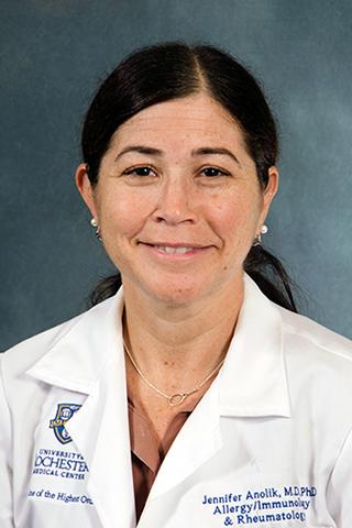 Jennifer Howitt Anolik合作论文数Anolik Lab, University of Rochester Medical Center24Alec Griffith合作论文数Dept Surg, Brigham & Womens Hosp22Wei Kevin合作论文数Div Rheumatol Immunol & Allergy, Brigham & Womens Hosp19
 Goro Tajima合作论文数19
 Nir Hacohen合作论文数Center for Cancer Immunology, Massachusetts General Hospital;Mass General Research Institute;Broad Institute19Laura A Cahill合作论文数17
 Laura Donlin合作论文数HSS Research Institute;Hospital for Special Surgery, Weill Cornell Medicine17Bykerk Vivian P合作论文数17
 Judith A. James合作论文数Oklahoma Medical Research Foundation;University of Oklahoma Health Sciences Center17
 Fei Guo (郭菲)合作论文数Beijing Association for Science Training Program17
 Fionnuala O’Leary合作论文数BWH/Harvard Medical School17Adam Delisle合作论文数17
 Zhang Fan合作论文数Worcester Polytechnic Institute, University of Massachusetts16
 Andrew Filer合作论文数Institute of Inflammation and Ageing, College of Medical and Dental Sciences, University of Birmingham16
 Joel Guthridge合作论文数Oklahoma Medical Research Foundation15
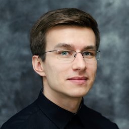 Slowikowski Kamil合作论文数Division of Rheumatology, Immunology, Allergy, Brigham and Women's Hospital and Harvard Medical School, Boston, MA, USA.15
 Susan M. Goodman合作论文数Department of Rheumatology, Hospital for Special Surgery;Weill Cornell Medical College15Kaitlyn Howard合作论文数14Seshadri Anupamaa合作论文数Brigham and Women's Hospital14John a Mannick合作论文数14
 V. Michael Holers合作论文数Department of Medicine-Rheumatology, School of Medicine, University of Colorado14Akinori Osuka合作论文数Aichi Children’s Health and Medical Center Pediatric Surgery14William Apruzzese合作论文数Department of Orthopedics (WA, GSP),, Massachusetts General Hospital14Yasutaka Nakahori合作论文数Harvard Med Sch, Brigham & Womens Hosp, Boston, MA USA13
 Jennifer L Nguyen合作论文数13Frank Borriello合作论文数13
 Adrian Maung合作论文数Yale Department of Surgery, Yale School of Medicine, Yale University13
 Costantino Pitzalis合作论文数William Harvey Research Institute, Barts and The London School of Medicine and Dentistry, Queen Mary University of London13
 Irshad Chaudry合作论文数Department of Surgery, Heersink School of Medicine, University of Alabama at Birmingham13Mark A. Perrella合作论文数Pulmonary and Critical Care Division, Brigham and Women's Hospital13Satoshi Fujimi合作论文数13
 Ann Mckenna合作论文数Brigham and Women’s Hospital, Harvard Medical School13
 Larry Moreland合作论文数Division of Rheumatology, School of Medicine, University of Colorado13S Fujimi合作论文数12
 Peter Nigrovic合作论文数Division of Medical Sciences, Harvard Medical School, Harvard University;Division of Immunology, Boston Children's Hospital12
 Gary S. Firestein合作论文数Department of Medicine, School of Medicine, University of California12Julie Ng合作论文数12
 Michael B. Yaffe合作论文数Koch Institute for Integrative Cancer Research, Massachusetts Institute of Technology;Beth Israel Deaconess Medical Center12
 Betty Diamond合作论文数Center for Autoimmune, Musculoskeletal and Hematopoietic Diseases, The Feinstein Institutes for Medical Research;Donald and Barbara Zucker School of Medicine at Hofstra/Northwell12Nida Meednu合作论文数12Peter H. Lapchak合作论文数Brigham and Women’s Hospital, Harvard Medical School12Alain Delisle合作论文数12




