Primary pulmonary non-Hodgkin lymphoma (PPL) is a rare malignancy most commonly presented as indolent marginal zone B-cell lymphoma of mucosa-associated lymphoid tissue (MALT) or bronchus-associated lymphoid tissue (BALT). Aggressive subtypes of PPL, such as diffuse large B-cell lymphoma (DLBCL), occur less frequently. Diagnosis and workup require tissue biopsy, immunophenotyping, and cross-sectional imaging, often supplemented by positron emission tomography (PET)/computed tomography (CT) for staging and treatment planning. Radiation therapy or radiotherapy (RT) is a cornerstone in managing localized, indolent PPL, offering durable local control with minimal toxicity. Modern involved-site RT (ISRT) techniques deliver radiation in conventional fractionation schedules, precisely targeting pulmonary lesions while sparing surrounding lung and mediastinal structures. RT can be used alone for early-stage disease or following systemic therapy for residual or refractory lesions. The cases in this report highlight the importance of histology-driven, individualized treatment planning and the pivotal role of RT in achieving optimal outcomes in this rare malignancy.
Stereotactic ablative radiotherapy (SABR) is standard for early stage NSCLC, but strategies to enhance its systemic antitumor immune effects remain limited. Preclinical data suggest that severe acute respiratory syndrome coronavirus 2 mRNA vaccines enhance innate immunity and antigen presentation, raising the possibility that vaccination could augment radiotherapy-induced immune priming. We evaluated the clinical impact of peri-SABR vaccination (defined as within 3 mo before or after SABR) in 413 real-world patients with T1 to 3N0M0 NSCLC who were treated during the pandemic era, including 144 patients who received vaccination within this window. In time-varying analyses, vaccination was associated with improved recurrence-free survival (hazard ratio [HR] = 0.69, 95% confidence interval [CI]: 0.49-0.98, p = 0.039) and a trend toward improved overall survival (HR = 0.66, 95% CI: 0.42-1.03, p = 0.069). In propensity score-matched time-dependent analyses, vaccination remained associated with improved recurrence-free survival (HR = 0.61, 95% CI: 0.40-0.91, p = 0.016) and overall survival (HR = 0.57, 95% CI: 0.34-0.94, p = 0.029), with directionally consistent findings observed after multivariable adjustment. These findings support further investigation into potential interactions between severe acute respiratory syndrome coronavirus 2 mRNA vaccination and SABR-induced antitumor immunity.
PURPOSE:NRG Oncology/Alliance LU005 (ClinicalTrials.gov identifier: NCT03811002) tested the addition of atezolizumab to concurrent chemoradiation (CRT) in this open-label, phase III international trial. METHODS:Patients with limited-stage small cell lung cancer (LS-SCLC), stage Tx-IV, N0-3, and M0 with Eastern Cooperative Group performance status (PS) 0-2 received one cycle of chemotherapy (platinum/etoposide) before study registration and were randomly assigned to CRT alone versus CRT plus concurrent and adjuvant atezolizumab, 1,200 mg once daily, every 3 weeks until investigator-assessed progression or intolerable side effects for a maximum of 17 cycles. Patients were stratified by choice of chemotherapy (cisplatin v carboplatin), radiation fractionation schedule (66 Gy once daily v 45 Gy twice daily), sex, and PS (0/1 v 2). The primary end point was overall survival (OS). Secondary end points included investigator-assessed progression-free survival (PFS), objective response rate, local control, and distant-metastasis-free survival (DMFS). RESULTS:patients were randomly assigned from May 2019 to December 2023. The median OS was 36.1 months (95% CI, 28.1 to 42.5) for the CRT-alone arm and 31.1 months (95% CI, 28.5 to 44.7) for the CRT + atezolizumab arm, respectively (hazard ratio [HR], 1.03 [95% CI, 0.80 to 1.32]). The median PFS was 11.4 months (95% CI, 10.3 to 13.2) for the CRT-alone arm and 12.1 months (95% CI, 10.9 to 15.2) for the CRT + atezolizumab arm, respectively (HR, 0.98 [95% CI, 0.79 to 1.22]). The median DMFS was 13.0 months (95% CI, 11.3 to 18.2) for the CRT-alone arm and 16.8 months (95% CI, 12.1 to 21.6) for the CRT + atezolizumab arm (HR, 0.96 [95% CI, 0.76 to 1.21]). No unexpected safety signals with concurrent atezolizumab were observed. CONCLUSION:Concurrent and adjuvant atezolizumab with chemoradiation did not improve survival in patients with LS-SCLC.
When identifying electrical, mechanical, or biological systems, parametric continuous-time identification methods can lead to interpretable and parsimonious models when the model structure aligns with the physical properties of the system. Traditional linear system identification may not consider the most parsimonious model when relying solely on unfactored transfer functions, which typically result from standard direct approaches. This paper presents a novel identification method that delivers additive models for both open and closed-loop setups. The estimators that are derived are shown to be generically consistent, and can admit the identification of marginally stable additive systems. Numerical simulations show the efficacy of the proposed approach, and its performance in identifying a modal representation of a flexible beam is verified using experimental data.
Cancer creates an immunosuppressive environment that hampers immune responses, allowing tumors to grow and resist therapy. One way the immune system fights back is by inducing ferroptosis, a type of cell death, in tumor cells through CD8 + T cells. This involves lipid peroxidation and enzymes like lysophosphatidylcholine acyltransferase 3 (Lpcat3), which makes cells more prone to ferroptosis. However, the mechanisms by which cancer cells avoid immunotherapy-mediated ferroptosis are unclear. Our study reveals how cancer cells evade ferroptosis and anti-tumor immunity through the upregulation of fatty acid-binding protein 7 (Fabp7). To explore how cancer cells resist immune cell-mediated ferroptosis, we used a comprehensive range of techniques. We worked with cell lines including PD1-sensitive, PD1-resistant, B16F10, and QPP7 glioblastoma cells, and conducted in vivo studies in syngeneic 129 Sv/Ev, C57BL/6, and conditional knockout mice with Rora deletion specifically in CD8+ T cells, Cd8 cre;Rorafl mice. Methods included mass spectrometry-based lipidomics, targeted lipidomics, Oil Red O staining, Seahorse analysis, quantitative PCR, immunohistochemistry, PPARγ transcription factor assays, ChIP-seq, untargeted lipidomic analysis, ROS assay, ex vivo co-culture of CD8+ T cells with cancer cells, ATAC-seq, RNA-seq, Western blotting, co-immunoprecipitation assay, flow cytometry and Imaging Mass Cytometry. PD1-resistant tumors upregulate Fabp7, driving protective metabolic changes that shield cells from ferroptosis and evade anti-tumor immunity. Fabp7 decreases the transcription of ferroptosis-inducing genes like Lpcat3 and increases the transcription of ferroptosis-protective genes such as Bmal1 through epigenetic reprogramming. Lipidomic profiling revealed that Fabp7 increases triglycerides and monounsaturated fatty acids (MUFAs), which impede lipid peroxidation and ROS generation. Fabp7 also improves mitochondrial function and fatty acid oxidation (FAO), enhancing cancer cell survival. Furthermore, cancer cells increase Fabp7 expression in CD8+ T cells, disrupting circadian clock gene expression and triggering apoptosis through p53 stabilization. Clinical trial data revealed that higher FABP7 expression correlates with poorer overall survival and progression-free survival in patients undergoing immunotherapy. Our study uncovers a novel mechanism by which cancer cells evade immune-mediated ferroptosis through Fabp7 upregulation. This protein reprograms lipid metabolism and disrupts circadian regulation in immune cells, promoting tumor survival and resistance to immunotherapy. Targeting Fabp7 could enhance immunotherapy effectiveness by re-sensitizing resistant tumors to ferroptosis.
The Publisher regrets that this article is an accidental duplication of an article that has already been published, http://dx.doi.org/10.1016/j.clbc.2025.09.009. The duplicate article has therefore been withdrawn. The full Elsevier Policy on Article Withdrawal can be found at https://www.elsevier.com/about/policies-and-standards/article-withdrawal.
419 Background: A third of patients with biochemical recurrence after radiation (RT) have intraprostatic radiorecurrence (IPR) on PSMA PET/CT. Patients with IPR have worse metastasis-free survival - a surrogate for progression to lethal prostate cancer (PCa). We previously reported the results from our F-SHARP clinical trial that demonstrated salvage reirradiation using focal dose-escalated high dose rate (HDR) brachytherapy is safe and effective. Here, we examine the Decipher score to determine if it could be a tool to risk stratify patients with IPR. Methods: F-SHARP (NCT03312972) is a multi-institutional phase I/II trial of focal dose-escalated salvage HDR for IPR. Patients were recruited from 2017-2023 at 3 centers. Eligibility criteria included a history of localized PCa treated with any form of definitive RT and biopsy-proven IPR with no regional or distant metastasis. Of the 62 participants, 37 consented for the biomarker correlative study and 31 (50%) had sample data passing quality control for Decipher analysis (Veracyte, San Diego, CA). De-identified data from 146,940 patients tested (2016-2024) with the Decipher prostate genomic classifier were retrieved from the GRID registry (NCT02609269) and used to create a matched cohort based on NCCN risk at diagnosis. Univariable Cox proportional hazards models were used to compare oncologic, CTCAE v4.03 toxicity, and EPIC-26 hrQoL events by Decipher risk group. Results: The biomarker cohort had similar baseline characteristics to the overall trial cohort. 30% received ADT with initial RT (73% external beam, 27% LDR brachytherapy). At recurrence, 71% had high Decipher risk (median score 0.67) as compared to only 35% (median score 0.48) in the matched GRID cases (n=130,760). Median time from initial RT to enrollment in Decipher low (<0.45) was 16.9 years, compared to 8.0 and 7.4 years in the intermediate (0.45-0.6) and high (>0.6) score patients. Median follow up was 32.3 months. Decipher score was not associated with toxicity or quality of life post-HDR (all p>0.05). As depicted in the table, higher Decipher score was associated with an increased risk of biochemical progression-free survival (bFS; p=0.03), local recurrence-free survival (LRFS; p=0.04), and radiographic progression-free survival (rPFS; p=0.01). At 3 years, bPFS was 43% vs. 75%, LRFS was 58% vs. 100%, and rPFS was 42% vs. 100% for Decipher high vs. lower risk (<0.6). Conclusions: Salvage reirradiation is a growing indication for RT in PCa. This is the first use of genomic risk stratification in this setting. Nearly a third of patients with IPR have a lower Decipher risk score, and our data suggest especially favorable outcomes with salvage HDR. Future studies to determine how Decipher risk stratification can be used to tailor further treatment intensification with systemic therapy for those most at risk of reirradiation failure are warranted. Clinical trial information: NCT03312972 . Endpoint Hazard Ratio per 0.1 unit (95% CI) bPFS 1.70 (1.05-2.75) LRFS 2.30 (1.02-5.18) rPFS 2.47 (1.23-4.97)
Recent advances in oncology research have highlighted the promising synergy between low-dose radiation therapy (LDRT) and immunotherapies, with growing evidence highlighting the unique benefits of the combination. LDRT has emerged as a potent tool for stimulating the immune system, triggering systemic antitumor effects by remodeling the tumor microenvironment. Notably, LDRT demonstrates remarkable efficacy even in challenging metastatic sites such as the liver (uveal) and brain (cutaneous), particularly in advanced melanoma stages. The increasing interest in utilizing LDRT for secondary metastatic sites of uveal, mucosal, or cutaneous melanomas underscores its potential efficacy in combination with various immunotherapies. This comprehensive review traverses the journey from laboratory research to clinical applications, elucidating LDRT’s immunomodulatory role on the tumor immune microenvironment (TIME) and systemic immune responses. We meticulously examine the preclinical evidence and ongoing clinical trials, throwing light on the promising prospects of LDRT as a complementary therapy in melanoma treatment. Furthermore, we explore the challenges associated with LDRT’s integration into combination therapies, addressing crucial factors such as optimal dosage, fractionation, treatment frequency, and synergy with other pharmacological agents. Considering its low toxicity profile, LDRT presents a compelling case for application across multiple lesions, augmenting the antitumor immune response in poly-metastatic disease scenarios. The convergence of LDRT with other disciplines holds immense potential for developing novel radiotherapy-combined modalities, paving the way for more effective and personalized treatment strategies in melanoma and beyond. Moreover, the dose-related toxicities of immunotherapies may be reduced by synergistic amplification of antitumor efficacy with LDRT.
The relationship between breast size and breast cancer risk is complex and not fully understood. This study investigates how breast size, categorized by cup size, correlates with age-standardized rates (ASR) of breast cancer incidence.Data were collected from two sources: breast cancer incidence rates from the Global Cancer Observatory (GCO) and breast size data from “Data Pandas,” an open-access database. This allowed for a cross-country analysis of breast cancer incidence and breast size characteristics. Descriptive statistics indicated that ASR increased with larger cup sizes, ranging from 34.72 (AA) to 90.17 (C). An ANOVA test revealed significant differences in mean ASR among cup size groups (F=14.416, P<0.001), with Bonferroni comparisons showing distinct clusters: smaller sizes (AA, AA-A, A) differed significantly from larger sizes (A-B, B, B-C, C).The largest mean ASR difference was between groups A and C (-42.93, P=0.001), highlighting higher ASR in larger cup sizes. This suggests a significant association between breast cup size and breast cancer ASR, potentially linked to physiological or hormonal factors.Despite limitations, these findings prompt further investigation. The next phase will focus on breast cancer patients, addressing relevant risk factors for a more comprehensive understanding of the associations observed.
PURPOSE:Up to 50% of clinical recurrences after curative-intent prostate cancer radiation are intraprostatic radiorecurrences (IPRRs). Salvage local therapy (SLT) is increasingly offered, particularly as focal SLT, to reduce toxicity due to prior radiation. Limited data exist on the relative value of magnetic resonance imaging (MRI), positron emission tomography/computed tomography (PET/CT), and biopsy on SLT target delineation. We compared MRI, PET/CT, and biopsy in patients with IPRRs and the impact each modality has on identifying IPRRs and defining the extent of prostatic involvement. METHODS AND MATERIALS:We performed a secondary analysis of 62 patients enrolled in a phase 1/2 clinical trial of salvage high-dose-rate brachytherapy. The IPRR was delineated using each imaging modality and by defining the involved regions of the prostate on biopsy. The exact binomial distribution was used to estimate the sensitivity of MRI and PET/CT to detect the IPRR. Exact conditional logistic regression was used to compare the tumor identified by MRI and PET/CT with the areas of biopsy involvement (gold standard) and estimate the proportion of patients with prostatic involvement outside of the image-defined targets. RESULTS:The sensitivity for detecting the IPRR was 91.8% for MRI and 85.5% for PET/CT. Most patients had biopsy-proven cancer outside of the MRI-defined (70.5%) and PET/CT-defined (73.8%) target. Delineating the brachytherapy target using imaging only would have missed the full extent of recurrence in 63.9%. CONCLUSIONS:Although MRI and PET/CT are valuable, a thorough biopsy is a mandatory tool to avoid missing areas of imaging-occult prostatic involvement when delivering focal SLT.
Radiotherapy remains a cornerstone in cancer treatment, used in over 50% of cases. It employs ionizing radiation, primarily X-rays, to target and destroy tumors through direct DNA damage and indirect effects via reactive oxygen species. Despite technological advancements improving precision of the delivered dose to the tumor, radiotherapy faces critical challenges, particularly damage to healthy tissues, which limits the maximum safe dose. Recent years have seen significant improvements in radiation delivery, including advanced imaging for real-time tumor tracking and combinations with immunotherapy. However, the need for innovative strategies to enhance radiotherapy's therapeutic index remains essential. The radioenhancer NBTXR3 could represent a solution in addressing these limitations. This nanotechnology has been designed to amplify radiotherapy's effects within tumors without increasing toxicity in non-injected adjacent healthy tissues. Beyond better cancer cell destruction and tumor control, radiotherapy-activated NBTXR3 nanoparticles can also stimulate systemic antitumor immune responses in preclinical models. This review aims to provide a comprehensive analysis of preclinical research on NBTXR3, focusing on its mechanism of action and role in initiating and enhancing antitumor immune responses.
Objectives Radiation Adaptive Response (AR) is a biological phenomenon in which exposure to low-dose radiation (LDR) enhances an organism’s ability to withstand subsequent higher doses. This scoping review explores AR across multiple disciplines, summarizing evidence, identifying research gaps, and evaluating potential applications in cancer therapy, neurodegenerative disease management, space medicine, and pandemic response. Methods A comprehensive review of experimental/clinical studies on AR was conducted, focusing on molecular mechanisms, biological implications, biophysical modeling, and translational applications. Results In oncology, AR has shown promise in selectively protecting normal tissues during radiotherapy while sensitizing tumor cells, yet its effects remain cell-type dependent. LDR may manage neurodegenerative diseases by modulating oxidative stress and inflammation. In space medicine, AR-based astronaut selection has been proposed as a novel strategy to mitigate radiation risks during long-term space missions, although empirical validation is lacking. LDR therapy for managing COVID-19 pneumonia has been explored, but ethical concerns and long-term safety risks require further investigation. Conclusion Despite AR’s potential, its clinical and spaceflight implementation requires mechanistic elucidation, standardized protocols, and rigorous studies. The risks of tumorigenesis, individual variability in AR, and potential immunomodulatory effects must be evaluated before widespread application. Moreover, inconsistent AR appearance complicates its study and clinical use.
This mini-review explores adaptive responses in organisms exposed to high radiation levels, drawing comparisons between Chernobyl’s wildlife—specifically its darker-pigmented frogs—and residents of Ramsar, Iran, a region with high natural background radiation. Chernobyl’s wildlife adaptations are not surprising, as substantial evidence in humans, demonstrates similar adaptation to high radiation levels. Studies reveal that mechanisms such as increased melanin production in frogs and enhanced DNA repair capabilities in Ramsar residents help mitigate radiation damage. These adaptations provide a framework for understanding resilience to environmental stressors and contribute to broader discussions on evolutionary survival mechanisms in extreme environments. By examining ecological and physiological responses across species, this review sheds light on radiation’s role in natural selection and potential applications for environmental and radiobiological research.
Challenging the Mutation-Only Paradigm: Evidence from Ramsar High Background Radiation AreasChallenging the Mutation-Only Paradigm: Evidence from Ramsar High Background Radiation AreasChallenging the Mutation-Only Paradigm: Evidence from Ramsar High Background Radiation AreasChallenging the Mutation-Only Paradigm: Evidence from Ramsar High Background Radiation AreasChallenging the Mutation-Only Paradigm: Evidence from Ramsar High Background Radiation AreasChallenging the Mutation-Only Paradigm: Evidence from Ramsar High Background Radiation AreasChallenging the Mutation-Only Paradigm: Evidence from Ramsar High Background Radiation AreasChallenging the Mutation-Only Paradigm: Evidence from Ramsar High Background Radiation AreasChallenging the Mutation-Only Paradigm: Evidence from Ramsar High Background Radiation Areas
Integrating cellular sarcoplasmic reticulum (SR) Ca2+ release with the known Ca2+ activation properties of RyR2s remains challenging. The sharp increase in SR Ca2+ permeability above a threshold SR luminal [Ca2+] is not reflected in RyR2 kinetics from single-channel studies. Additionally, the current paradigm that global Ca2+ release (Ca2+ waves) arises from interacting local events (Ca2+ sparks) faces a key issue that these events rarely activate neighboring sites. We present a multiscale model that reproduces Ca2+ sparks and waves in skinned ventricular myocytes using experimentally validated RyR2 kinetics. The model spans spatial domains from 10-8 to 10-4 m and timescales from 10-6 to 10 s. Ca2+ release sites are distributed in cubic voxels (0.25-µm sides) informed by super-resolution micrographs. We use parallel computing to calculate Ca2+ transport, diffusion, and buffering. Substantial increases in SR Ca2+ release occur, and Ca2+ waves initiate when Ca2+ sparks become prolonged above a threshold SR [Ca2+]. These prolonged events (Ca2+ embers) are much more likely than Ca2+ sparks to activate release from neighboring sites and accumulate increases in cytoplasmic [Ca2+] along with an associated fall in Ca2+ buffering power. This primes the cytoplasm for Ca2+-induced Ca2+ release (CICR) that produces Ca2+ waves. Thus, Ca2+ ember formation and CICR are both essential for initiation and propagation of Ca2+ waves. Cell architecture, along with the differential effects of RyR2 opening and closing rates, collectively determines the SR [Ca2+] threshold for Ca2+ embers, waves, and the phenomenon of store overload-induced Ca2+ release.
Background: Dual primary malignancies, including colorectal (CRC) and gastric cancers (GC), are complicated cases due to the complexity of managing patients. Case Report: This case report presents a 62-year-old male patient with rectal and gastric adenocarcinomas. Initially, rectal adenocarcinoma after a complaint of hematochezia was diagnosed by prognostic modalities. The patient received total neoadjuvant therapy with FOLFOX chemotherapy and chemoradiotherapy. After surgery, a complete pathological response was obtained. A few months later, gastric adenocarcinoma with persistent heartburn was detected through esophagogastroduodenoscopy (EGD). total neoadjuvant therapy with FOLFOX chemotherapy and chemoradiotherapy followed by total gastrectomy were prescribed. After gastrectomy, a complete pathological response was obtained. Conclusion: This case of synchronous CRC and GC, diagnosed 5 months apart, underscores the pivotal role of early detection and multidisciplinary management in achieving favorable outcomes. Complete pathologic responses in both malignancies following tailored TNT with FOLFOX and FLOT regimens, combined with surgical interventions, highlight the efficacy of personalized treatment strategies, even in resource-constrained settings. Continued research is essential to optimize diagnostic protocols, refine therapeutic approaches, and improve access to genetic testing for synchronous and metachronous malignancies, promoting equitable cancer care globally.
Purpose:This study aimed to compare the outcomes of proton radiotherapy alone versus its combination with immuno-oncology agents (Proton-IO) or tyrosine kinase inhibitors (Proton-TKI) in patients with intermediate- to advanced-stage hepatocellular carcinoma (HCC). Methods:We retrospectively reviewed the medical records of 137 patients with Barcelona Clinic Liver Cancer (BCLC) stage B or C HCC treated with proton radiotherapy at Linkou Chang Gung Memorial Hospital between 2020 and 2023. Patients were stratified into three groups: proton radiotherapy alone (n = 64), Proton-IO (n = 44), and Proton-TKI (n = 29). The most frequently used immuno-oncology agents were atezolizumab-bevacizumab (n = 33) and pembrolizumab (n = 5). Tyrosine kinase inhibitors (TKIs) included lenvatinib (n = 16) and sorafenib (n = 13). Results:With a median follow-up of 30 months, patients in the Proton-IO group were significantly associated with higher 2-year overall survival (OS) rates compared with those receiving Proton-TKI or proton radiotherapy alone (77.0% vs. 47.2% vs. 52.7%; p = 0.002). Proton-IO was also associated with significantly longer time to progression (TTP) and distant metastasis-free survival (DMFS) (2-year TTP: 50.5% vs. 28.1% vs. 24.2%, p = 0.003; 2-year DMFS: 83.4% vs. 61.1% vs. 67.2%, p = 0.027). No significant differences in 2-year local control rates were observed among the treatment groups (97.7% vs. 92.9% vs. 86.8%; p = 0.230). Multivariate analysis identified Proton-IO as an independent predictor of improved OS (p < 0.001), TTP (p < 0.001), and DMFS (p = 0.004). Grade 3-4 upper gastrointestinal (UGI) bleeding was observed in 2 (1.5%) patients (proton monotherapy, n = 1; Proton-IO, n = 1). There were no significant differences among the groups in the incidence of grade ≥3 UGI bleeding, liver toxicity, colitis, rib fractures, or hematologic adverse events. Conclusion:In BCLC stage B/C HCC, proton radiotherapy combined with immunotherapy was significantly associated with higher OS, TTP, and DMFS without an increase in grade ≥3 toxicity compared with proton radiotherapy alone or Proton-TKIs.
Among all the treatment modalities, despite the development of targeted treatments, radiotherapy is still considered one of the most effective treatment approaches. While this modality is highly effective in tumor control, it still faces resistance in cancer cells through complex mechanisms. Hypoxia has been considered one of the important characteristics of solid tumors, effectively impacting therapeutic response and a challenge for treatment because of radiation resistance, poor prognosis, and reduced effectiveness of radiation therapy. Hypoxia may facilitate a series of adaptive responses by which tumors survive and progress despite radiation therapy, acting through several pathways: stabilizing hypoxia-inducible factors, metabolic reprogramming, disruption of DNA damage signaling, genomic instability, and cell cycle arrest. More specifically, hypoxia PET, MRI-based oxygenation measurements, and the integration of artificial intelligence with imaging approaches have considerably given valuable information on tumor oxygenation and related mechanisms of radiation resistance, supplemented by HIF-1 alpha, CAIX, VEGF, GLUT1, and some hypoxia-associated microRNAs. This review discusses the key molecular and cellular pathways taking part in the processes leading to radioresistance brought about by hypoxia and highlights biomarkers able to predict radiation response. A deep understanding of these mechanisms may enable the development of novel therapeutic approaches to overcome hypoxia-induced radioresistance, thereby improving personalized cancer therapy.
The infiltration of T cells and chimeric antigen receptor T (CAR-T) cells into solid tumors is essential for robust antitumor response and therapeutic outcomes. While cell therapies succeeded in hematologic malignancies, their efficacy in solid tumors is limited due to poor tumor penetration and the immunosuppressive tumor immune microenvironment (TIME). Our previous studies showed that combining low-dose radiotherapy (LDRT) with T-cell therapy synergistically enhanced tumor control and survival. Nanostring metabolic gene analysis indicated that LDRT modulates T-cell metabolism within TIME. Therefore, we hypothesized that LDRT enhances T-cell metabolism, favoring anticancer potency. To investigate the role of LDRT in T-cell metabolism, we established a TIME using co-culture of cancer cells and PBMCs in xenograft NSG mice, followed by LDRT. Using nCounter Metabolic Pathways, we quantified the expression of genes associated with core metabolic and immune metabolic processes in intratumoral T cells. Nanostring analysis revealed key pathways and genes that regulate T-cell effector functions, further underscoring LDRT's impact on T-cell metabolism. Seahorse assays highlighted that LDRT positively influenced the oxygen consumption rate (OCR) and extracellular acidification rate (ECAR), favoring improved metabolic and effector functions in T/ CAR-T cells. Mitotracker analysis revealed that LDRT-treated T/ CAR-T cells exhibited enhanced mitochondrial membrane potential (Mitotracker Deep Red) and mitochondrial mass (Mitotracker Green), suggesting robust mitochondrial health and functionality. Transmission electron microscopy (TEM) showed improved mitochondrial morphology of T cells following LDRT. Additionally, Incucyte live-cell analysis demonstrated that CAR-T cells treated with LDRT exhibited significantly enhanced cancer cell killing (P ≤ 0.001) compared to controls. CAR-T cells conditioned with LDRT showed increased tumor cell killing in vivo, further validating the potentiating effect of LDRT on cell-based therapies. These findings collectively highlight the pivotal role of LDRT in modulating T-cell metabolism and enhancing antitumor immune responses. The study highlights the translational potential of combining LDRT with cell therapies in solid tumors. By reprogramming T-cell metabolism within TIME, LDRT significantly enhanced tumor control and improved survival outcomes, establishing a promising foundation for metabolism-targeted therapies. These findings provide compelling evidence to support further exploration and clinical investigation of this synergistic approach, aiming to optimize the efficacy of cell-based immunotherapies and improve outcomes for patients with challenging solid tumor malignancies. Zahid Rafiq, Puebla Nahum Osorio, Weiqin Lu, James Welsh. Low dose radiation enhanced antitumor immune response by reprogramming T cell and CAR-T cell metabolism in a tumor immune microenvironment [abstract]. In: Proceedings of the American Association for Cancer Research Annual Meeting 2025; Part 1 (Regular Abstracts); 2025 Apr 25-30; Chicago, IL. Philadelphia (PA): AACR; Cancer Res 2025;85(8_Suppl_1):Abstract nr 4543.
 Joe Y. Chang合作论文数Division of Radiation Oncology, University of Texas MD Anderson Cancer Center;Department of Radiation Oncology, University of Texas MD Anderson Cancer Center150
 Ritsuko Komaki合作论文数University of Texas MD Anderson Cancer Center;Department of Radiation Oncology, Baylor College of Medicine;Department of Scientific Research, Baylor College of Medicine149
 Maria Angelica Cortez合作论文数Medical School, The University of Texas Southwestern Medical Center;Children's Medical Center Research Institute, The University of Texas Southwestern Medical Center112
 Daniel Gomez合作论文数101
 Zhongxing Liao合作论文数Department of Radiation Oncology, Division of Radiation Oncology, The University of Texas MD Anderson Cancer Center94Hampartsoum B Barsoumian合作论文数Univ Texas MD Anderson Canc Ctr, Dept Radiat Oncol, Houston, TX 77030 USA91
 John V. Heymach合作论文数Department of Thoracic-Head & Neck Med Onc, Division of Cancer Medicine, The University of Texas MD Anderson Cancer Center;Graduate School of Biomedical Sciences, The University of Texas MD Anderson Cancer Center86
 Steven H. Lin合作论文数Department of Experimental Radiation Oncology, The University of Texas MD Anderson Cancer Center/Department of Radiation Oncology, The University of Texas MD Anderson Cancer Center80
 Chad Tang合作论文数Department of Radiation Oncology, Division of Radiation Oncology, The University of Texas MD Anderson Cancer Center65Pamela K. Allen合作论文数Division of Radiation Oncology, University of Texas MD Anderson Cancer Center59Verma Vivek合作论文数Allegheny General Hospital;Mercy Health - St. Elizabeth Boardman Hospital51
 David S Hong合作论文数Department of Investigational Cancer Therapeutics, Division of Cancer Medicine, The University of Texas MD Anderson Cancer Center42
 Jonathan E Schoenhals合作论文数Department of Radiation Oncology, University of Texas MD Anderson Cancer Center41
 Quynh-Nhu Nguyen合作论文数38
 Stephen G. Swisher合作论文数Department of Thoracic and Cardiovascular Surgery, Division of Surgery, The University of Texas MD Anderson Cancer Center;Lung Cancer35
 Taylor R Cushman合作论文数Dept Radiat Oncol, Univ Texas MD Anderson Canc Ctr34
 Wayne L. Hofstetter合作论文数Department of Thoracic and Cardiovascular Surgery, Division of Surgery, The University of Texas MD Anderson Cancer Center34
 Stephen Hahn合作论文数33
 Jaffer A. Ajani合作论文数Department of Gastrointestinal Medical Oncology, Division of Cancer Medicine, The University of Texas MD Anderson Cancer Center33
 James D. Cox合作论文数Division of Radiation Oncology, The University of Texas MD Anderson Cancer Center32
 Yun Hu合作论文数31
 Hari Menon合作论文数Penn State Geisinger Medical Center30
 Nahum Puebla-Osorio合作论文数30Masrorpour Fatemeh合作论文数29
 Dawei Chen (陈大卫)合作论文数Department of Radiation Oncology Shandong Cancer Hospital and Institute, Shandong First Medical University28
 Ignacio I. Wistuba合作论文数Department of Translational Molecular Pathology, The University of Texas MD Anderson Cancer Center;Department of Pathology, The University of Texas MD Anderson Cancer Center28
 Stephen G. Chun合作论文数Department of Radiation Oncology, Division of Radiation Oncology, The University of Texas MD Anderson Cancer Center27Ailin Li合作论文数26
 Lauren Averett Byers合作论文数Department of Thoracic-Head and Neck Medical Oncology, Division of Cancer Medicine, The University of Texas MD Anderson Cancer Center26
 Reza J. Mehran合作论文数Department of Thoracic and Cardiovascular Surgery, Division of Surgery, The University of Texas MD Anderson Cancer Center26
 Manoop S. Bhutani合作论文数Department of Gastroenterology Hepatology and Nutrition, Division of Internal Medicine, The University of Texas MD Anderson Cancer Center;Department of Radiation Oncology, Division of Radiation Oncology, The University of Texas MD Anderson Cancer Center;Department of Endoscopic Research and Development, The University of Texas MD Anderson Cancer Center25Blumenschein George R合作论文数24
 Jeffrey H. Lee合作论文数Department of Gastroenterology Hepatology and Nutrition, Division of Internal Medicine, The University of Texas MD Anderson Cancer Center23Saumil J. Gandhi合作论文数22
 Peter A. Balter合作论文数Department of Radiation Physics, Division of Radiation Oncology, The University of Texas MD Anderson Cancer Center21Jeremy J. Erasmus合作论文数21
 Clifford G. Robinson合作论文数Division of Clinical Research, Department of Radiation Oncology, Washington University School of Medicine in St. Louis;Center for Advanced Medicine Siteman Cancer Center, Washington University;Barnes-Jewish Hospital;Barnes-Jewish West County Hospital20Samir Narayan合作论文数20Melenda Jeter合作论文数20
 Kewen He合作论文数19
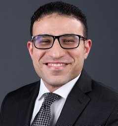 Ahmed I Younes合作论文数Section of Lymphoma, University of Texas M. D. Anderson Cancer Center19
 Arya Amini合作论文数Department of Radiation Oncology, City of Hope National Medical Center19Jeffrey D. Bradley合作论文数19Q.N. Nguyen合作论文数18
 Duygu Sezen合作论文数18Joseph J. Bevelacqua合作论文数18
 Sebastien Paris合作论文数18
 Rishab Ramapriyan合作论文数Univ Texas MD Anderson Canc Ctr, Dept Radiat Oncol, Houston, TX 77030 USA18
 Yolanda I. Garces合作论文数Department of Radiation Oncology, Mayo Clinic;Mayo Clinic Comprehensive Cancer Center18




