Currently, there is substantial ongoing discussion around the functional role of the thalamus in consciousness. What is missing in the literature, however, is a systematic investigation of the relevance of specific thalamic nuclei in pharmacologically and pathologically altered states of consciousness in humans. Using resting-state functional magnetic resonance imaging in both healthy anaesthetized volunteers (n = 16) and patients with disorders of consciousness (n = 22), we sought to identify which specific thalamic subregions in both cohorts may be differentially significant for loss of consciousness. Our findings revealed that, among all nuclei, the pulvinar was found to have the strongest functional connectivity change with loss of consciousness following anaesthesia, while demonstrating distinct functional connectivity patterns related to higher-order default mode and executive control networks. Remarkably, in loss of consciousness in disorders of consciousness patients, the ventral-latero-ventral was found to have the strongest connectivity change in comparison with healthy controls, while exhibiting discrete functional connectivity patterns related to higher-order default mode, executive control and frontoparietal networks. Furthermore, we provide evidence that this neural connectivity biomarker in patients also mirrored the changes observed at the behavioural level, which could have clinical implications for targeted deep brain stimulation in therapy for disorders of consciousness.
Consciousness and cognition arise from the ongoing interactions between brain regions. Synchronous fluctuations of fMRI signals may indicate that two brain regions perform similar cognitive functions, but neural interactions are also constrained by anatomical connectivity and regions' molecular, cytoarchitectonic, and metabolic profiles. Here we disentangle the respective contributions of ongoing cognition and multimodal neurobiological constraints in shaping functional connectivity. We jointly contextualise haemodynamic FC against eight distinct multimodal representations of the human connectome: (i) structural connectivity from diffusion tractography; (ii) spatial embedding; (iii) similarity of transcriptional profiles from gene expression; (iv) similarity of receptor profiles from Positron Emission Tomography; (v) laminar profile similarity from histology; (vi) correlated electrophysiological activity from magnetoencephalography; (vii) correlated metabolic activity from PET glucose uptake; (viii) coordinated activation across 123 cognitive operations from the NeuroSynth meta-analytic engine. We demonstrate that cognitive co-activation is the dominant predictor of inter-regional fMRI synchrony in the awake human brain, even when quantified using intracranial electrical stimulation. Crucially, this predominance of cognitive co-activation for shaping functional connectivity is systematically obliterated across five datasets of pharmacological and pathological perturbations of consciousness (chronic disorders of consciousness; anaesthesia with sevoflurane, propofol, or ketamine) when cognition is disconnected from the environment or altogether abolished. Altogether, we show that multimodal predictors of functional architecture shift away from cognitive co-activation and toward anatomical-molecular constraints during pharmacological and pathological perturbations of consciousness.
BACKGROUND AND OBJECTIVES:The clinical characteristics and natural history of post-traumatic cerebral venous outflow compromise (VOC) are poorly characterized. We aimed to determine the prevalence of VOC in at-risk traumatic brain injury patients, assess its effect on long-term mortality, and describe our management practices. METHODS:We conducted a retrospective single-center case-control study. We included patients admitted to the neurocritical care unit after traumatic brain injury who had been investigated with computed tomography venography because of clinical suspicion of VOC between 2008 and 2015. RESULTS:A total of 89 patients underwent computed tomography venography during their neurocritical care unit admission. A total of 43 patients (48%, 32 male [74%], mean age 40 years [SD 16.2]) had evidence of VOC. Of these, 28 (65%) were due to an intraluminal dural venous sinus thrombosis (DVST) and 15 (35%) due to an extraluminal cause. Twelve (43%) of the DVSTs were occlusive, and 16 (57%) were nonocclusive. A total of 24 patients (27%) underwent decompressive craniectomy, which was more commonly performed for patients with an occlusive thrombosis than those with partial or no VOC (67% vs 23% vs 20%, P < .01). A total of 4 patients (14%) with an intraluminal thrombosis received antithrombotic therapy. Mortality rate of those with VOC may have been higher compared with those without VOC at 14 days (14% vs 7%, P = .31) but was similar at 5 years (21% vs 22%, P = .93). Patients with a midline or bilateral thrombosis had higher overall mortality (83% vs 18%, P = .01) than those with a thrombosis located elsewhere. CONCLUSION:Among those at risk, patients with evidence of post-traumatic VOC may have had higher short-term mortality, but VOCs did not increase long-term mortality rates compared with those without VOC. Patients with an occlusive thrombosis were more likely to undergo decompressive craniectomy. Most patients with a DVST received prophylactic rather than treatment-dose antithrombosis. Further studies are required to determine the optimal management of post-traumatic VOC.
How is the information-processing architecture of the human brain organised, and how does its organisation support consciousness? Here, we combine network science and a rigorous information-theoretic notion of synergy to delineate a ‘synergistic global workspace’, comprising gateway regions that gather synergistic information from specialised modules across the human brain. This information is then integrated within the workspace and widely distributed via broadcaster regions. Through functional MRI analysis, we show that gateway regions of the synergistic workspace correspond to the human brain’s default mode network, whereas broadcasters coincide with the executive control network. We find that loss of consciousness due to general anaesthesia or disorders of consciousness corresponds to diminished ability of the synergistic workspace to integrate information, which is restored upon recovery. Thus, loss of consciousness coincides with a breakdown of information integration within the synergistic workspace of the human brain. This work contributes to conceptual and empirical reconciliation between two prominent scientific theories of consciousness, the Global Neuronal Workspace and Integrated Information Theory, while also advancing our understanding of how the human brain supports consciousness through the synergistic integration of information.
BACKGROUND:Patients with brain injury who are unresponsive to commands may perform cognitive tasks that are detected on functional magnetic resonance imaging (fMRI) and electroencephalography (EEG). This phenomenon, known as cognitive motor dissociation, has not been systematically studied in a large cohort of persons with disorders of consciousness. METHODS:In this prospective cohort study conducted at six international centers, we collected clinical, behavioral, and task-based fMRI and EEG data from a convenience sample of 353 adults with disorders of consciousness. We assessed the response to commands on task-based fMRI or EEG in participants without an observable response to verbal commands (i.e., those with a behavioral diagnosis of coma, vegetative state, or minimally conscious state-minus) and in participants with an observable response to verbal commands. The presence or absence of an observable response to commands was assessed with the use of the Coma Recovery Scale-Revised (CRS-R). RESULTS:Data from fMRI only or EEG only were available for 65% of the participants, and data from both fMRI and EEG were available for 35%. The median age of the participants was 37.9 years, the median time between brain injury and assessment with the CRS-R was 7.9 months (25% of the participants were assessed with the CRS-R within 28 days after injury), and brain trauma was an etiologic factor in 50%. We detected cognitive motor dissociation in 60 of the 241 participants (25%) without an observable response to commands, of whom 11 had been assessed with the use of fMRI only, 13 with the use of EEG only, and 36 with the use of both techniques. Cognitive motor dissociation was associated with younger age, longer time since injury, and brain trauma as an etiologic factor. In contrast, responses on task-based fMRI or EEG occurred in 43 of 112 participants (38%) with an observable response to verbal commands. CONCLUSIONS:Approximately one in four participants without an observable response to commands performed a cognitive task on fMRI or EEG as compared with one in three participants with an observable response to commands. (Funded by the James S. McDonnell Foundation and others.).
A central question in neuroscience is how consciousness arises from the dynamic interplay of brain structure and function. Here we decompose functional MRI signals from pathological and pharmacologically-induced perturbations of consciousness into distributed patterns of structure-function dependence across scales: the harmonic modes of the human structural connectome. We show that structure-function coupling is a generalisable indicator of consciousness that is under bi-directional neuromodulatory control. We find increased structure-function coupling across scales during loss of consciousness, whether due to anaesthesia or brain injury, capable of discriminating between behaviourally indistinguishable sub-categories of brain-injured patients, tracking the presence of covert consciousness. The opposite harmonic signature characterises the altered state induced by LSD or ketamine, reflecting psychedelic-induced decoupling of brain function from structure and correlating with physiological and subjective scores. Overall, connectome harmonic decomposition reveals how neuromodulation and the network architecture of the human connectome jointly shape consciousness and distributed functional activation across scales.
This scientific commentary refers to ‘Physiological alterations of pineal recess crowding in symptomatic non-hydrocephalic pineal cysts’ by Eide et al. (https://doi.org/10.1093/braincomms/fcad078).
High-level brain functions are widely believed to emerge from the orchestrated activity of multiple neural systems. However, lacking a formal definition and practical quantification of emergence for experimental data, neuroscientists have been unable to empirically test this long-standing conjecture. Here we investigate this fundamental question by leveraging a recently proposed framework known as "Integrated Information Decomposition," which establishes a principled information-theoretic approach to operationalise and quantify emergence in dynamical systems — including the human brain. By analysing functional MRI data, our results show that the emergent and hierarchical character of neural dynamics is significantly diminished in chronically unresponsive patients suffering from severe brain injury. At a functional level, we demonstrate that emergence capacity is positively correlated with the extent of hierarchical organisation in brain activity. Furthermore, by combining computational approaches from network control theory and whole-brain biophysical modelling, we show that the reduced capacity for emergent and hierarchical dynamics in severely brain-injured patients can be mechanistically explained by disruptions in the patients' structural connectome. Overall, our results suggest that chronic unresponsiveness resulting from severe brain injury may be related to structural impairment of the fundamental neural infrastructures required for brain dynamics to support emergence.
OBJECTIVE:Meningiomas invading the intracranial venous sinuses may cause intracranial venous hypertension, papilledema, and visual compromise. Sinus resection and graft reconstructions, however, add significant complexity to tumor surgery, with the potential for increased morbidity. In this study, the authors explored whether venous sinus stenting might provide an alternative means of controlling venous hypertension that would be sustainable over the long term. METHODS:The authors performed a retrospective review of all 16 patients with intracranial meningiomas who underwent stenting at their institution for venous sinus compromise. At presentation, all had headache and 9 had papilledema. Thirteen patients had 1 meningioma and 3 had 2 or more. Three patients had had previous tumor resection and radiotherapy. One patient had been treated with a lumboperitoneal shunt and radiotherapy. The median length of clinical follow-up was 8 years (range 4 months-18 years). RESULTS:Venous sinus narrowing was often not confined to the site of meningioma, and bilateral transverse sinus narrowing, reminiscent of that seen in idiopathic intracranial hypertension, was present in 7 patients with sagittal sinus meningiomas. Eleven patients had stents placed solely across sinus narrowing caused by meningioma. Five patients had additional stents placed at other sites of venous narrowing at the same time: in one of these patients, a stent was placed across a defect in the sagittal sinus caused by previous surgery, and in the 4 other patients, stents were placed across nontumor narrowings of the transverse sinuses. In 1 patient, the jugular vein was also stented. Nine patients developed symptomatic in-stent restenosis at the meningioma site. Eight had further stenting procedures with variable success in restoring the in-stent lumen. The remaining patient, with a late partial relapse, is being reinvestigated. Papilledema resolved in all patients after stenting. Six patients experienced prolonged and very substantial relief of all symptoms. Five patients had persistent headache despite restoration of the sinus lumen. Five had persistent symptoms associated with resistant in-stent stenosis. There were no significant complications from any of the diagnostic or therapeutic procedures. CONCLUSIONS:In patients who are symptomatic with meningiomas obstructing the venous sinuses, successful stenting of the affected segment can give a good outcome, especially in terms of relieving papilledema. However, further procedures are often necessary to maintain stent patency, other areas of venous compromise frequently coexist, and some patients remain symptomatic despite apparently successful treatment of the index lesion. Long-term surveillance is a requirement.
Background:The aim of this study was to create a simplistic taxonomy to improve transparency and consistency in, and reduce complexity of, interpreting diffusion tensor imaging (DTI) profiles in white matter disruption. Using a novel strategy of a periodic table of DTI elements, we examined if DTI profiles could demonstrate neural properties of disruption sufficient to characterize white matter changes specific for hydrocephalus vs. non-hydrocephalus, and to distinguish between cohorts of neural injury by their differing potential for reversibility.Methods:DTI datasets from three clinical cohorts representing pathological milestones from reversible to irreversible brain injury were compared to those of healthy controls at baseline, over time and with interventions. The final dataset comprised patients vs. controls in the following groupings: mild traumatic brain injury (mTBI), n = 24 vs. 27, normal pressure hydrocephalus (NPH), n = 16 vs. 9 and Alzheimer's disease (AD), n = 27 vs. 47. We generated DTI profiles from fractional anisotropy (FA) and mean, axial and radial diffusivity measures (MD, L1 and L2 and 3 respectively), and constructed an algorithm to map changes consistently to a periodic table of elements, which fully described their diffusivity and neural properties.Results:Mapping tissue signatures to a periodic table of DTI elements rapidly characterized cohorts by their differing patterns of injury. At baseline, patients with mTBI displayed the most preserved tracts. In NPH, the magnitude of changes was dependent on "familial" DTI neuroanatomy, i.e., potential for neural distortion from risk of ventriculomegaly. With time, patients with Alzheimer's disease were significantly different to controls across multiple measures. By contrast, patients with mTBI showed both loss of integrity and pathophysiological processes of neural repair. In NPH, some patterns of injury, such as "stretch/compression" and "compression" were more reversible following intervention than others; these neural profile properties suggested "microstructural resilience" to injury.Conclusion:Using the novel strategy of a periodic table of DTI elements, our study has demonstrated it is possible to distinguish between different cohorts along the spectrum of brain injury by describing neural profile properties of white matter disruption. Further work to contribute datasets of disease toward this proposed taxonomic framework would enhance the translatability of DTI profiles to the clinical-research interface.
Background In 2020, The London Royal College of Physicians published "Prolonged disorders of consciousness following sudden-onset brain injury: national clinical guidelines". In 2021, in the journal Brain, Scolding et al. published "a critical evaluation of the new UK guidelines". This evaluation focussed on one of the 73 recommendations in the National Clinical Guidelines. They also alleged that the guidelines were unethical. Criticisms They criticised our recommendation not to use activation protocols using fMRI, electroencephalography, or Positron Emission Tomography. They claim these tests can (a) detect 'covert consciousness', (b) add predictive value and (c) should be part of routine clinical care. They also suggest that our guideline was driven by cost considerations, leading to clinicians deciding to withdraw treatment at 72 h. Evidence Our detailed review of the evidence confirms the American Academy of Neurology Practise Guideline (2018) and the European Academy of Neurology Guideline (2020), which agree that insufficient evidence supports their approach. Ethics The ethical objections are based on unwarranted assumptions. Our guideline does not make any recommendations about management until at least four weeks have passed. We explicitly recommend that expert assessors undertake ongoing surveillance and monitoring; we do not suggest that patients be abandoned. Our recommendation will increase the cost We had ethicists in the working party. Conclusion We conclude the "critical evaluation" fails to provide evidence for their criticism and that the ethical objections arise from incorrect assumptions and unsupported interpretations of evidence and our guideline. The 2020 UK national guidelines remain valid.
Typical consciousness can be defined as an individual-specific stream of experiences. Modern consciousness research on dynamic functional connectivity uses clustering techniques to create common bases on which to compare different individuals. We propose an alternative approach by combining modern theories of consciousness and insights arising from phenomenology and dynamical systems theory. This approach enables a representation of an individual's connectivity dynamics in an intrinsically-defined, individual-specific landscape. Given the wealth of evidence relating functional connectivity to experiential states, we assume this landscape is a proxy measure of an individual's stream of consciousness. By investigating the properties of this landscape in individuals in different states of consciousness, we show that consciousness is associated with short term transitions that are less predictable, quicker, but, on average, more constant. We also show that temporally-specific connectivity states are less easily describable by network patterns that are distant in time, suggesting a richer space of possible states. We show that the cortex, cerebellum and subcortex all display consciousness-relevant dynamics and discuss the implication of our results in forming a point of contact between dynamical systems interpretations and phenomenology.
Small world topologies are thought to provide a valuable insight into human brain organisation and consciousness. However, functional magnetic resonance imaging studies in consciousness have not yielded consistent results. Given the importance of dynamics for both consciousness and cognition, here we investigate how the diversity of small world dynamics (quantified by sample entropy; dSW-E1) scales with decreasing levels of awareness (i.e., sedation and disorders of consciousness). Paying particular attention to result reproducibility, we show that dSW-E is a consistent predictor of levels of awareness even when controlling for the underlying functional connectivity dynamics. We find that dSW-E of subcortical, and cortical areas are predictive, with the former showing higher and more robust effect sizes across analyses. We find that the network dynamics of intermodular communication in the cerebellum also have unique predictive power for levels of awareness. Consequently, we propose that the dynamic reorganisation of the functional information architecture, in particular of the subcortex, is a characteristic that emerges with awareness and has explanatory power beyond that of the complexity of dynamic functional connectivity.
The human brain entertains rich spatiotemporal dynamics, which are drastically reconfigured when consciousness is lost due to anaesthesia or disorders of consciousness (DOC). Here, we sought to identify the neurobiological mechanisms that explain how transient pharmacological intervention and chronic neuroanatomical injury can lead to common reconfigurations of neural activity. We developed and systematically perturbed a neurobiologically realistic model of whole-brain haemodynamic signals. By incorporating PET data about the cortical distribution of GABA receptors, our computational model reveals a key role of spatially-specific local inhibition for reproducing the functional MRI activity observed during anaesthesia with the GABA-ergic agent propofol. Additionally, incorporating diffusion MRI data obtained from DOC patients reveals that the dynamics that characterise loss of consciousness can also emerge from randomised neuroanatomical connectivity. Our results generalise between anaesthesia and DOC datasets, demonstrating how increased inhibition and connectome perturbation represent distinct neurobiological paths towards the characteristic activity of the unconscious brain.
BACKGROUND: The provision of equitable and affordable health care has become increasingly challenging as advanced technology is introduced, particularly in developing countries. We explored the hypothesis that focused, small-scale mini-public-private partnerships have a potential role in providing equitable and affordable access to advanced technology for the benefit of all patients in developing nations, particularly middle-income countries. METHODS: A clinician-led financial plan was developed at the University of Malaya to create the Centre for Image Guidance and Minimally Invasive Therapy (CIGMIT) to provide an integrated platform for high-end care for Malaysian patients of all ages, both public and private, requiring complex neurosurgical and spinal procedures and stereotactic and intensity-modulated radiotherapy. The challenges faced during development of the plan were documented together with an audit of patient throughput and analyses of financial risk and return. RESULTS: CIGMIT opened in 2015. Patient throughput, both public and private, progressively increased in all facilities. In 2015-2019, 37,724 patients used the Centre's facilities. CIGMIT has become progressively more profitable for the University of Malaya, the public and private hospitals, and the investor. CIGMIT has weathered the challenges posed by coronavirus disease 19. CONCLUSIONS: Focused, small-scale mini-public -private partnerships have a potential role in providing advanced technology for the benefit of patients in developing nations, particularly middle-income countries, subject to an approach that balances equity of access between public and private health care systems with fair reward.
 Marek Czosnyka合作论文数Neurosurgical Unit, University of Cambridge303
 David Menon合作论文数Division of Anaesthesia, Department of Medicine, Queens' College, University of Cambridge169
 Peter Smielewski合作论文数Division of Neurosurgery, University of Cambridge158
 Peter J A Hutchinson合作论文数Department of Clinical Neurosciences, Robinson College, University of Cambridge151
 Zofia Czosnyka合作论文数Division of Neurosurgery, University of Cambridge100
 PJ Kirkpatrick合作论文数63A. T. C. Carpenter合作论文数57Tim Fryer合作论文数Wolfson Brain Imaging Centre, Department of Clinical Neurosciences, University of Cambridge56
 Peter Kirkpatrick合作论文数Neurosurgery Unit, University of Cambridge;Addenbrookes Hospital52
 Jonathan Coles合作论文数Department of Medicine, University of Cambridge49Hugh Richards合作论文数49
 Adrian M. Owen合作论文数Centre for Brain and Mind, Western University;Institute for Neuroscience, Western University45
 Ivan Sergeevich Timofeev合作论文数University of Cambridge;Department of Neurosurgery;Addenbrooke ' s Hospital;Addenbrooke ' s Hospital, University of Cambridge37HK RICHARDS合作论文数36
 Magdalena Kasprowicz合作论文数Institute of Biomedical Engineering and Instrumentation, Wroclaw University of Technology35Judith Allanson合作论文数Department of Public Health and Primary Care, University of Cambridge34
 Barbara Sahakian合作论文数Department of Psychiatry, University of Cambridge34John C. Clark合作论文数32Guy B. Williams合作论文数Addenbrooke's Hospital, Cambridge University32
 Jonathan Gillard合作论文数Department of Radiology, University of Cambridge31
 Franklin Aigbirhio合作论文数Yusuf Hamied Department of Chemistry, University of Cambridge;Molecular Imaging Chemistry Laboratory, University of Cambridge29
 Budohoski Karol P合作论文数Department of Neurosurgery, University of Cambridge28
 Andrea Lavinio合作论文数Department of Anaesthesia Spedali Civili, University of Brescia28
 Stefan K Piechnik合作论文数Radcliffe Department of Medicine, Medical Sciences Division, University of Oxford28Martin R. Coleman合作论文数27
 Neil Burnet合作论文数Manchester Cancer Research Centre, The University of Manchester26
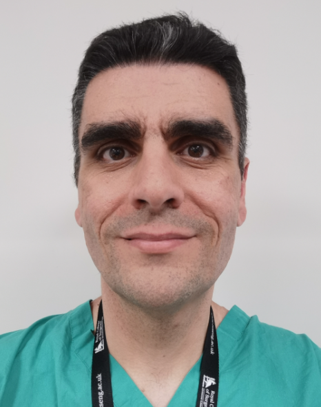 Angelos Kolias合作论文数Department of Clinical Neuroscience, Addenbrooke's Hospital;CRUK Cambridge Centre, Cancer Research UK Cambridge Institute, University of Cambridge24Da Chatfield合作论文数24Keri L. H. Carpenter合作论文数Division of Neurosurgery, University of Cambridge23
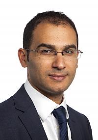 Adel Helmy合作论文数Division of Neurosurgery, University of Cambridge23
 Eric Schmidt合作论文数22
 Keong Nicole Chwee Har合作论文数Department of Neurosurgery, National Neuroscience Institute;Singapore General Hospital;Duke-NUS Graduate Medical School, National University of Singapore22
 Anthony Davenport合作论文数Department of Medicine, University of Cambridge21
 Helen Seeley合作论文数Neurosurgery Unit, UK Shunt Registry21
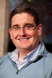 Stephen Price合作论文数Queens' College, University of Cambridge21M T O'Connell合作论文数20A Pena合作论文数19
 Paola Finoia合作论文数Department of Medicine, Division of Anaesthesia, University of Cambridge;Department of Clinical Neurosciences, University of Cambridge19Piotr Smielewski合作论文数19Peter Johnström合作论文数19
 Arun Gupta合作论文数Department of Radiodiagnosis, All India Institute of Medical Sciences19Nicholas Higgins合作论文数Addenbrooke’s Hospital18Brian K. Owler合作论文数18PG Al-Rawi合作论文数17
 Mark Gurnell合作论文数Metabolic Research Laboratories, University of Cambridge/Department of Medicine, University of Cambridge/Wellcome-MRC Institute of Metabolic Science, University of Cambridge16
 Luzius A. Steiner合作论文数16Matthew R Garnett合作论文数15Corteen Elizabeth A合作论文数15
 Neil Harris合作论文数Brain Research Institute, University of California, Los Angeles;Department of Neurosurgery, David Geffen School of Medicine, University of California, Los Angeles14




