Macropinocytosis is a large-scale endocytosis process primarily observed in phagocytes as part of their cellular function to ingest antigens. Once phagocytes encounter gram-negative bacteria, the receptor proteins identify lipopolysaccharides (LPSs), which trigger radical membrane ruffles that gradually change to cup-like structures. The open area of the cups closes to generate vesicles called macropinosomes. The target bacteria are isolated by the cups and engulfed by the cells as the cups close. In addition to its ingestion function, macropinocytosis also regulates the AKT pathway in macrophages. In the current study, we report that macropinocytic cups are critical for LPS-induced AKT phosphorylation (pAKT) and cytokine expression in macrophages. High-resolution scanning electron microscope observations detailed the macropinocytic cup structures induced by LPS stimulation. Confocal microscopy revealed that AKT and the kinase molecule mTORC2 were localized in the cups. The biochemical analysis showed that macropinocytosis inhibition blocked LPS-induced pAKT. RNA sequencing, quantitative polymerase chain reaction, and enzyme-linked immunosorbent assay analyses revealed that the inhibition of macropinocytosis or the AKT pathway causes a decrease in the expression of proinflammatory cytokines interlukin-6 and interlukin-1α. Moreover, activation of the transcription factor nuclear factor κB, which regulates the cytokine expression downstream of the AKT/IκB pathway, was hindered when macropinocytosis or AKT was inhibited. These results indicate that LPS-induced macropinocytic cups function as signal platforms for the AKT pathway to regulate the cytokine expression by modulating nuclear factor κB activity in LPS-stimulated macrophages. Based on these findings, we propose that macropinocytosis may be a good therapeutic target for controlling cytokine expression.
The human organic cation transporter 1 (hOCT1), also known as SLC22A1, is integral to hepatic uptake of structurally diversified endogenous and exogenous organic cations, influencing both metabolism and drug pharmacokinetics. hOCT1 has been implicated in the therapeutic dynamics of many drugs, making interactions with hOCT1 a key consideration in novel drug development and drug–drug interactions. Notably, metformin, the frontline medication for type 2 diabetes, is a prominent hOCT1 substrate. Conversely, hOCT1 can be inhibited by agents such as spironolactone, a steroid analog inhibitor of the aldosterone receptor, necessitating a deep understanding of hOCT1–drug interactions in the development of new pharmacological treatments. Despite extensive study, specifics of hOCT1 transport and inhibition mechanisms remain elusive at the molecular level. Here, we present cryo-electron microscopy structures of the hOCT1-metformin complex in three distinct conformational states — outward open, outward occluded, and inward occluded as well as substrate-free hOCT1 in both partially and fully open states. We also present hOCT1 in complex with spironolactone in both outward and inward facing conformations. These structures provide atomic-level insights into the dynamic metformin transfer process via hOCT1 and the mechanism by which spironolactone inhibits it. Additionally, we identify a ‘YER’ motif critical for the conformational flexibility of hOCT1 and likely other SLC22 family transporters. Our findings significantly advance the understanding of hOCT1 molecular function and offer a foundational framework for the design of new therapeutic agents targeting this transporter.
TACAN is an ion channel-like protein that may be involved in sensing mechanical pain. Here, we present the cryo-electron microscopic structure of human TACAN (hTACAN). hTACAN forms a dimer in which each protomer consists of a transmembrane globular domain (TMD) containing six helices and an intracellular domain (ICD) containing two helices. Molecular dynamic simulations suggest that each protomer contains a putative ion conduction pore. A single-point mutation of the key residue Met207 greatly increases membrane pressure-activated currents. In addition, each hTACAN subunit binds one cholesterol molecule. Our data show the molecular assembly of hTACAN and suggest that wild-type hTACAN is in a closed state.
A major component of bacterial biofilms is curli amyloid fibrils secreted by the curli biogenesis system. Understanding the curli biogenesis mechanism is critical for developing therapeutic agents for biofilm-related infections. Here we report a systematic study of the curli biogenesis system, highlighted by structural, biochemical and functional analysis of the secretion channel complexes (CsgF-CsgG) with and without the curli substrate. The dual-pore architecture of the CsgF-CsgG complex was observed and used to develop an approach to inhibit the curli secretion by physically reducing the size of the CsgF pore. We further elucidated the assembly of the CsgFG complex with curli components (CsgA and CsgB) and curli-cell association through CsgF. Importantly, the recognition of the CsgA substrate by CsgG was uncovered. Nine crevices outside of the CsgG channel provide specific and highly-conserved recognition sites for CsgA N-terminus. Together with analysis of CsgE, our study provides comprehensive insights into curli biogenesis.
 Kaixuan Gao (高凯旋)合作论文数1
 Angqi Zhu合作论文数Newstead Lab, Department of Biochemistry, University of Oxford1Xinhe Yu合作论文数1Jinzi Wei合作论文数1Baoliang Lan合作论文数1Li Wang合作论文数1Fang Kong合作论文数School of Life Science, Westlake University1
 Xiaowei Sun合作论文数1Tianlei Wen合作论文数1Ming Li合作论文数1
 Yanan Li (李亚楠)合作论文数1Xuhang Lu合作论文数1Yaojie Wang合作论文数1Shenghai Chang合作论文数Zhejiang Univ, Sch Med, Dept Biophys, Hangzhou 310058, Zhejiang, Peoples R China1Xiaoou Sun合作论文数1Ning Liu合作论文数1Lili Cheng合作论文数1
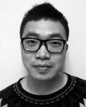 Yang Li合作论文数Univ Michigan, Dept Computat Med & Bioinformat, Ann Arbor, MI 48109 USA1Xiaozhe Chen合作论文数1Shuhao Zhang合作论文数1
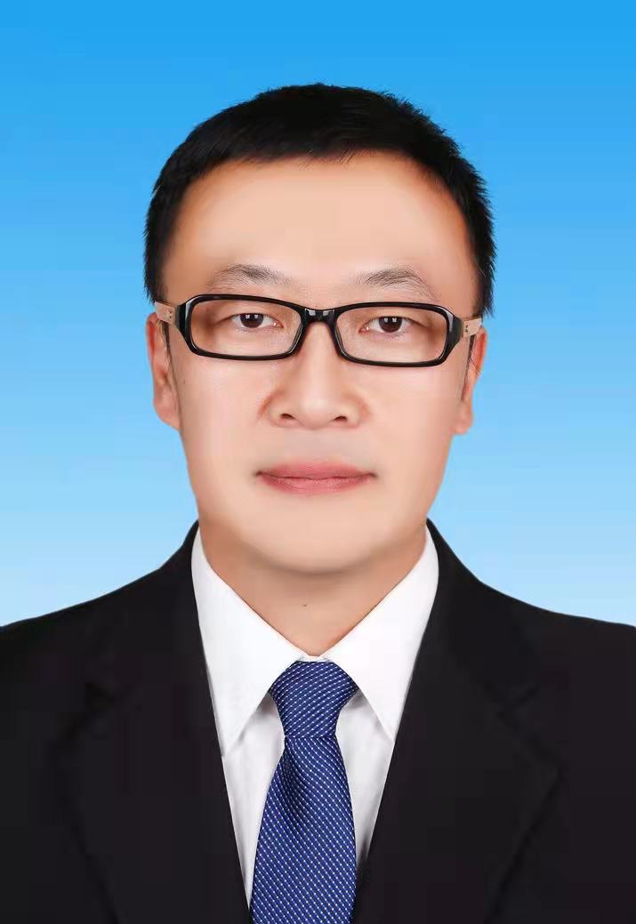 Xiangyu Liu (刘翔宇)合作论文数School of Chemistry and Chemical Engineering, Ningxia University1
 Ligong Chen (陈立功)合作论文数School of Pharmaceutical Sciences, Tsinghua University1
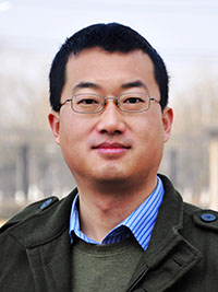 Xueming Li (李雪明)合作论文数School of Life Sciences, Tsinghua University1
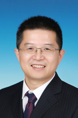 Yuequan Shen (沈月全)合作论文数College of Life Sciences, Nankai University;State Key Laboratory of Medicinal Chemical Biology, Nankai University1
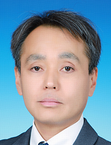 Sei Yoshida (吉田整)合作论文数College of Life Sciences, Nankai University;State Key Laboratory of Medicinal Chemical Biology, Nankai University1
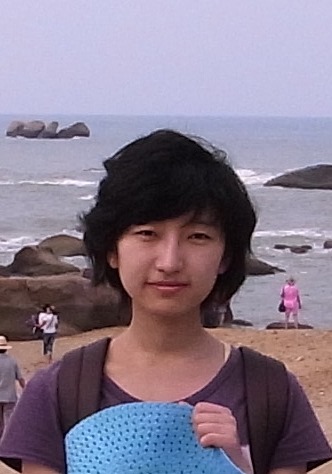 Xue Yang (杨雪)合作论文数College of Pharmacy, Nankai University1
 Xing Zhang (张兴)合作论文数Sir Run Run Shaw Hospital, School of Medicine, Zhejiang University;Center of Cryo Electron Microscopy, School of Medicine, Zhejiang University1Meng Yin (尹孟)合作论文数1Zhaofeng Yan (严照峰)合作论文数1Chuangye Yan (闫创业)合作论文数1




