Video capsule endoscopy has become increasingly important for investigating the small intestine within the gastrointestinal tract. However, a persistent challenge remains the short battery lifetime of such compact sensor edge devices. Integrating artificial intelligence can help overcome this limitation by enabling intelligent real-time decision- making, thereby reducing the energy consumption and prolonging the battery life. However, this remains challenging due to data sparsity and the limited resources of the device restricting the overall model size. In this work, we introduce a multi-task neural network that combines the functionalities of precise self-localization within the gastrointestinal tract with the ability to detect anomalies in the small intestine within a single model. Throughout the development process, we consistently restricted the total number of parameters to ensure the feasibility to deploy such model in a small capsule. We report the first multi-task results using the recently published Galar dataset, integrating established multi-task methods and Viterbi decoding for subsequent time-series analysis. This outperforms current single-task models and represents a significant ad- vance in AI-based approaches in this field. Our model achieves an accu- racy of 93.63
The classification performance of deep neural networks relies strongly on access to large, accurately annotated datasets. In medical imaging, however, obtaining such datasets is particularly challenging since annotations must be provided by specialized physicians, which severely limits the pool of annotators. Furthermore, class boundaries can often be ambiguous or difficult to define which further complicates machine learning-based classification. In this paper, we want to address this problem and introduce a framework for mislabel detection in medical datasets. This is validated on the two largest, publicly available datasets for Video Capsule Endoscopy, an important imaging procedure for examining the gastrointestinal tract based on a video stream of lowresolution images. In addition, potentially mislabeled samples identified by our pipeline were reviewed and re-annotated by three experienced gastroenterologists. Our results show that the proposed framework successfully detects incorrectly labeled data and results in an improved anomaly detection performance after cleaning the datasets compared to current baselines.
The differentiation between benign and malignant biliary strictures remains a significant clinical challenge. Recent studies have suggested that next generation sequencing (NGS) can improve the diagnostic accuracy. However, evidence on its performance in routine clinical practice is limited. Therefore, validation of the diagnostic value of NGS using real-world clinical data is warranted. We compared the performance of NGS analysis of cholangiocellular carcinoma (CCC)-associated mutations in endoscopic biopsies with that of histopathology, CA19-9 and cross-sectional imaging. NGS showed higher sensitivity for malignancy (65%) compared to histopathology (52%) at similar specificity (96% vs. 100%). The combination of NGS and histopathology further increased sensitivity for malignancy to 78% (p = 0.03) at similar specificity (96%). Our data provide support for the use of NGS in the diagnostic workup of biliary strictures.
Abstract:Metabolic dysfunction-associated steatotic liver disease (MASLD) is the most prevalent chronic liver disease worldwide, affecting around 30% of the population. It represents the hepatic manifestation of the metabolic syndrome and can progress to severe complications such as cirrhosis and hepatocellular carcinoma, with fibrosis as the key determinant of outcome. Current treatments, including the first liver-targeted pharmacotherapies, show limited efficacy in reversing fibrosis. The multidisciplinary consortium of the newly established transregional collaborative research center TRR 412 at the Charité Berlin and Dresden University of Technology aims to achieve a deeper mechanistic understanding of MASLD, promoting a holistic view that integrates pathogenic pathways of metabolic injury, inflammation, and fibrosis. MASLD is a complex and heterogeneous disease involving multiple liver cell types, and pathomechanisms are not (always) hierarchical, but intertwined across disease stages. In depth definition of the liver-specific molecular and cellular mechanisms driving MASLD progression will enable the development of innovative, mechanism-based therapies as well as improved patient stratification and personalized treatment approaches.
Various gastrointestinal disorders have been linked to gut microbiome dysbiosis, as it plays a critical role in immune regulation, metabolism, nutrient digestion, and pathogen suppression. However, the microbiome’s spatial variability across gastrointestinal segments and its intra- and interindividual differences complicate its study and clinical interpretation. While fecal DNA analysis is commonly used, stool samples only capture an accumulated signal and miss the spatial dynamics of microbial populations. To address this, we propose a modular sampling capsule capable of wirelessly collecting liquid. The capsule consists of two main modules: (i) an actuator module integrating a polymer-based microfluidic system with meltable wax-based opening valve, screen-printed microheater, cellulose membrane-based closing valve, evacuated sampling chamber with dried sample preservative material, filter membrane (size exclusion 150 μm), and sample extraction channel; and (ii) a control electronic module with communication, localization, and power supply units. The actuator module was validated in vitro using a diluted stool simulant (330 mg/mL) and an uncleaned porcine intestine. The opening valve activated within 3.6 ± 0.5 s at 120 ± 10 mA and 0.8 V. The sample was then filtered and aspirated into the sampling chamber within 1–2 s, and the closing valve sealed the inlet completely within 10 min. We overcame design, material, and fabrication challenges to construct an actuator module that functions effectively in liquids with variable physicochemical conditions (pH, chemical composition, viscosity, and particle size). These results demonstrate the feasibility of a controlled, segment-specific intestinal sampling capsule, representing a step towards precise and accurate microbiome profiling.
Supplementary Table S4 lists the 140 colorectal-cancer-associated loci and associations with colorectal cancer in European-ancestry population.
Supplementary Figure S6 shows the calibration on relative risk of PRS stratified by PRS with 7 bins in groups of different ancestry in the GERA cohort.
Supplementary Table S2 shows the descriptive statistics of GERA study participants by racial/ethnic groups and sex.
Handheld ultrasound (H-US) offers a widely accessible and cost-effective option for future medicine. Quantitative US methods, such as H-Scan, could broaden its impact by leveraging the enormous potential of radiofrequency (RF) ultrasound data. H-US derived steatosis and fibrosis assessments would reduce the need for expensive FibroScan (R) devices, especially supporting low-resource areas. By filtering for lower (GH2) and higher (GH8) frequencies, the method allows for differentiation of scatter sizes related to varying degrees of steatosis, which is crucial for early detection of metabolic-associated steatotic liver disease (MASLD). Considering a substantial and various patient cohort of 468 patients, reducing potential selection bias inherent in smaller study cohorts, this study aims to investigate whether H-Scan analysis of RF-data captured with inexpensive H-US yields comparable results to those seen in previous studies. A strong correlation (r=0.852, p<0.0001) was found between the H-Scan and the controlled attenuation parameter from FibroScan (R), indicating the effectiveness of H-Scan in identifying steatosis. However, the correlation strongly depends on an accurate estimate of the tissue's attenuation coefficient alpha . No significant correlation was observed between H-Scan and the degree of liver fibrosis, suggesting that the current H-Scan alone might not be suitable for this application. Further research is needed to test and refine the methodology, especially regarding individual attenuation correction.
Capsule endoscopy is a method to capture images of the gastrointestinal tract and screen for diseases which might remain hidden if investigated with standard endoscopes. Due to the limited size of a video capsule, embedding AI models directly into the capsule demands careful consideration of the model size and thus complicates anomaly detection in this field. Furthermore, the scarcity of available data in this domain poses an ongoing challenge to achieving effective anomaly detection. Thus, this work introduces an ensemble strategy to address this challenge in anomaly detection tasks in video capsule endoscopies, requiring only a small number of individual neural networks during both the training and inference phases. Ensemble learning combines the predictions of multiple independently trained neural networks. This has shown to be highly effective in enhancing both the accuracy and robustness of machine learning models. However, this comes at the cost of higher memory usage and increased computational effort, which quickly becomes prohibitive in many real-world applications. Instead of applying the same training algorithm to each individual network, we propose using various loss functions, drawn from the anomaly detection field, to train each network. The methods are validated on the two largest publicly available datasets for video capsule endoscopy images, the Galar and the Kvasir-Capsule dataset. We achieve an AUC score of 76.86% on the Kvasir-Capsule and an AUC score of 76.98% on the Galar dataset. Our approach outperforms current baselines with significantly fewer parameters across all models, which is a crucial step towards incorporating artificial intelligence into capsule endoscopies.
Anastomotic failure remains one of the most severe complications in gastrointestinal surgery. Despite continuous advancements in stapler technologies and surgical techniques, it continues to be a leading cause of postoperative morbidity and mortality. It contributes substantially to prolonged hospitalization and increased healthcare expenditures. Currently, diagnosis is based on secondary systemic signs, such as inflammatory response or changes in drain fluid, followed by a multimodal diagnostic approach. However, reliable early detection of local alterations is still lacking. Here, the implantation of a bioresorbable is investigated, intra-anastomotically placed sensor device. By performing real-time intra-anastomotic bioimpedance measurements, ischemia-related changes are identified at an early, potentially reversible stage, prior to the onset of clinical or systemic manifestations. Furthermore, the sensor device offers the potential for future integration of pattern-recognition algorithms and the possibility of direct measurement of different markers in the anastomotic microenvironment.
Video capsule endoscopy (VCE) is an important technology with many advantages (non-invasive, representation of small bowel), but faces many limitations as well (time-consuming analysis, short battery lifetime, and poor image quality). Artificial intelligence (AI) holds potential to address every one of these challenges, however the progression of machine learning methods is limited by the avaibility of extensive data. We propose Galar, the most comprehensive dataset of VCE to date. Galar consists of 80 videos, culminating in 3,513,539 annotated frames covering functional, anatomical, and pathological aspects and introducing a selection of 29 distinct labels. The multisystem and multicenter VCE data from two centers in Saxony (Germany), was annotated framewise and cross-validated by five annotators. The vast scope of annotation and size of Galar make the dataset a valuable resource for the use of AI models in VCE, thereby facilitating research in diagnostic methods, patient care workflow, and the development of predictive analytics in the field.
Supplementary Table S3 shows the comparison on characteristics between GECCO/CORECT study and GERA Europeans-ancestry participants
Supplementary Figure S7 shows the Sex-specific estimated baseline incidence rate of CRC based on SEER18 (2007-2015) CRC rate in European population.
Supplementary Table S7 shows the 10-year time-dependent AUC estimates of the PRS-enhanced model in the GERA European- ancestry participants.
Supplementary Table S1 shows the descriptive characteristics of study populations in GECCO and CORECT.
Hepatic stellate cells (HSCs) have a central pathogenetic role in the development of liver fibrosis. However, their fibrosis-independent and homeostatic functions remain poorly understood 1–5 . Here we demonstrate that genetic depletion of HSCs changes WNT activity and zonation of hepatocytes, leading to marked alterations in liver regeneration, cytochrome P450 metabolism and injury. We identify R-spondin 3 (RSPO3), an HSC-enriched modulator of WNT signalling, as responsible for these hepatocyte-regulatory effects of HSCs. HSC-selective deletion of Rspo3 phenocopies the effects of HSC depletion on hepatocyte gene expression, zonation, liver size, regeneration and cytochrome P450-mediated detoxification, and exacerbates alcohol-associated and metabolic dysfunction-associated steatotic liver disease. RSPO3 expression decreases with HSC activation and is inversely associated with outcomes in patients with alcohol-associated and metabolic dysfunction-associated steatotic liver disease. These protective and hepatocyte-regulating functions of HSCs via RSPO3 resemble the R-spondin-expressing stromal niche in other organs and should be integrated into current therapeutic concepts.
 Stefan Schreiber合作论文数Department of Internal Medicine I, University Hospital Schleswig-Holstein;Institute of Clinical Molecular Biology, Kiel University270
 Clemens Schafmayer合作论文数Department of General and Thoracic Surgery, University Hospital Schleswig-Holstein190
 Stephan Buch合作论文数99
 Felix Stickel合作论文数Universitätsspital Zürich, Klinik für Gastroenterologie und Hepatologie72
 Mario Brosch合作论文数72
 Michael Krawczak合作论文数Institut fur Medizinische Informatik und Statistik, Christian-Albrechts-Universitat zu Kiel64von Schönfels Witigo合作论文数61
 Sebastian Zeissig合作论文数58
 Andre Franke合作论文数Institute of Clinical Molecular Biology, Kiel University55
 Ulrich R. Fölsch合作论文数Klinik für Innere Medizin I, Universität Kiel51
 Hermann Brenner合作论文数Division of Clinical Epidemiology and Aging Research, German Cancer Research Center, Heidelberg University48
 Alexander Arlt合作论文数45
 Victor Moreno合作论文数44
 Annika Lindblom合作论文数Department of Molecular Medicine and Surgery, Karolinska University;The Center for Molecular Medicine44
 Philip Rosenstiel合作论文数Institute of Clinical Molecular Biology, Kiel University44
 Christian Datz合作论文数44
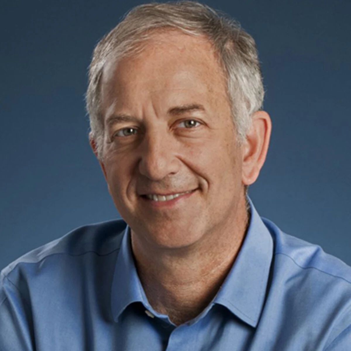 Steven Gallinger合作论文数Department of Surgery, Temerty Faculty of Medicine, University of Toronto44
 Jenny Chang-Claude合作论文数Division of Cancer Epidemiology, German Cancer Research Center44
 Le Marchand Loïc合作论文数University of Hawaiʻi Cancer Center;John A. Burns School of Medicine, University of Hawaiʻi at Mānoa42
 Mark A. Jenkins合作论文数Centre for Epidemiology and Biostatistics, School of Population and Global Health, University of Melbourne41Casey Graham合作论文数41
 Ulrike Peters合作论文数Department of Epidemiology, School of Public Health, University of Washington40
 Castellví-Bel Sergi合作论文数Gastrointestinal & Pancreat Oncol Grp, Inst Invest Biomed August Pi i Sunyer IDIBAPS40
 Christopher Mathew合作论文数King’s Global Health Institute, School of Basic & Medical Biosciences, Faculty of Life Sciences and Medicine, Kings College London;Department of Medical and Molecular Genetics, School of Basic & Medical Biosciences, Faculty of Life Sciences and Medicine, Kings College London;Sydney Brenner Institute for Molecular Bioscience, Faculty of Health Sciences, Witwatersrand University39
 Alicja Wolk合作论文数Institute of Environmental Medicine, Karolinska Institutet39
 Anna H Wu合作论文数Keck School of Medicine, University of Southern California39
 Andrea Gsur合作论文数39Fränzel J B van Duijnhoven合作论文数Wageningen Univ & Res, Div Human Nutr & Hlth, Wageningen, Netherlands38
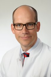 Sebastian Hinz合作论文数Department of Surgery, University of Kiel38Holger Kalthoff合作论文数Kiel University37
 Pavel Vodicka合作论文数37
 Gad Rennert合作论文数Department of Community Medicine and Epidemiology, Carmel Medical Center;Ruth and Bruce Rappaport Faculty of Medicine, Technion - Israel Institute of Technology36
 Thomas Berg合作论文数Division of Hepatology, Department of Medicine II, University of Leipzig Medical Center36
 Li Hsu合作论文数Fred Hutchinson Canc Res Ctr, Div Publ Hlth Sci, 1124 Columbia St, Seattle, WA 98104 USA35
 Peter Nürnberg合作论文数Cologne Center for Genomics, University of Cologne35
 Jane C Figueiredo合作论文数Departamento de Engenharia Florestal;Instituto Superior de Agronomia;Institute da Conserva35
 Bethany Van Guelpen合作论文数Dept Radiat Sci, Umea Univ34
 Heather Hampel合作论文数Department of Medical Oncology & Therapeutics Research, City of Hope;Division of Clinical Cancer Genomics, Beckman Research Institute, City of Hope34
 Robert E. Schoen合作论文数Division of Gastroenterology, Hepatology and Nutrition, Department of Medicine, School of Medicine, University of Pittsburgh34Renate Schmelz合作论文数34Michael Hoffmeister合作论文数33
 Temitope O Keku合作论文数33
 Frank Lammert合作论文数Medizinischen Kliniken, Universitätsklinikums des Saarlandes31
 Peter T. Campbell合作论文数Department of Epidemiology, American Cancer Society31Conghui Qu合作论文数Fred Hutchinson Canc Res Ctr, Div Publ Hlth Sci, 1124 Columbia St, Seattle, WA 98104 USA30
 Elizabeth A. Platz合作论文数Division of Cancer Epidemiology, Department of Epidemiology, Bloomberg School of Public Health, Johns Hopkins University30
 Lori C. Sakoda合作论文数Division of Public Health Sciences, Fred Hutchinson Cancer Research Center;Division of Research, Kaiser Permanente Northern California;Kaiser Permanente Bernard J. Tyson School of Medicine30Yu-Ru Su合作论文数29
 Mingyang Song合作论文数Harvard T.H. Chan School of Public Health, Harvard University28




