This is a comprehensive review on current utilization and challenges of digital pathology adoption in clinical trials and aims to provide a broad view on its impact on pathology review processes in clinical trials. It provides an overview of current pathology review practices in clinical trials and unique advantages digital pathology adoption can offer. The key areas including existing workflows, use case scenarios in different disease areas in clinical trials, including but not limited to patient identification and pre-screening, and regulatory aspects have been described with relevance. In addition, the review delves into the integration of genomics, AI, image analysis, radiology, and advanced computational pathology, to propose measures to enhance clinical trial outcomes. The current regulatory landscape around digital pathology adoption and potential future advancements in this field are also discussed as appropriate.
Context.— Computational pathology combines clinical pathology with computational analysis, aiming to enhance diagnostic capabilities and improve clinical productivity. However, communication barriers between pathologists and developers often hinder the full realization of this potential. Objective.— To propose a standardized framework that improves mutual understanding of clinical objectives and computational methodologies. The goal is to enhance the development and application of computer-aided diagnostic (CAD) tools. Design.— This article suggests pivotal roles for pathologists and computer scientists in the CAD development process. It calls for increased understanding of computational terminologies, processes, and limitations among pathologists. Similarly, it argues that computer scientists should better comprehend the true use cases of the developed algorithms to avoid clinically meaningless metrics. Results.— CAD tools improve pathology practice significantly. Some tools have even received US Food and Drug Administration approval. However, improved understanding of machine learning models among pathologists is essential to prevent misuse and misinterpretation. There is also a need for a more accurate representation of the algorithms’ performance compared to that of pathologists. Conclusions.— A comprehensive understanding of computational and clinical paradigms is crucial for overcoming the translational gap in computational pathology. This mutual comprehension will improve patient care through more accurate and efficient disease diagnosis.
Measuring virus in biofluids is complicated by confounding biomolecules coisolated with viral nucleic acids. To address this, we developed an affinity-based microfluidic device for specific capture of intact severe acute respiratory syndrome coronavirus 2 (SARS-CoV-2). Our approach used an engineered angiotensin-converting enzyme 2 to capture intact virus from plasma and other complex biofluids. Our device leverages a staggered herringbone pattern, nanoparticle surface coating, and processing conditions to achieve detection of as few as 3 viral copies per milliliter. We further validated our microfluidic assay on 103 plasma, 36 saliva, and 29 stool samples collected from unique patients with COVID-19, showing SARS-CoV-2 detection in 72% of plasma samples. Longitudinal monitoring in the plasma revealed our device’s capacity for ultrasensitive detection of active viral infections over time. Our technology can be adapted to target other viruses using relevant cell entry molecules for affinity capture. This versatility underscores the potential for widespread application in viral load monitoring and disease management.
BackgroundGrowing evidence supports the importance of characterizing the organizational patterns of various cellular constituents in the tumor microenvironment in precision oncology. Most existing data on immune cell infiltrates in tumors, which are based on immune cell counts or nearest neighbor-type analyses, have failed to fully capture the cellular organization and heterogeneity.MethodsWe introduce a computational algorithm, termed Tumor-Immune Partitioning and Clustering (TIPC), that jointly measures immune cell partitioning between tumor epithelial and stromal areas and immune cell clustering versus dispersion. As proof-of-principle, we applied TIPC to a prospective cohort incident tumor biobank containing 931 colorectal carcinoma cases. TIPC identified tumor subtypes with unique spatial patterns between tumor cells and T lymphocytes linked to certain molecular pathologic and prognostic features. T lymphocyte identification and phenotyping were achieved using multiplexed (multispectral) immunofluorescence. In a separate hepatocellular carcinoma cohort, we replaced the stromal component with specific immune cell types-CXCR3+CD68+ or CD8+-to profile their spatial relationships with CXCL9+CD68+ cells.ResultsSix unsupervised TIPC subtypes based on T lymphocyte distribution patterns were identified, comprising two cold and four hot subtypes. Three of the four hot subtypes were associated with significantly longer colorectal cancer (CRC)-specific survival compared to a reference cold subtype. Our analysis showed that variations in T-cell densities among the TIPC subtypes did not strictly correlate with prognostic benefits, underscoring the prognostic significance of immune cell spatial patterns. Additionally, TIPC revealed two spatially distinct and cell density-specific subtypes among microsatellite instability-high colorectal cancers, indicating its potential to upgrade tumor subtyping. TIPC was also applied to additional immune cell types, eosinophils and neutrophils, identified using morphology and supervised machine learning; here two tumor subtypes with similarly low densities, namely 'cold, tumor-rich' and 'cold, stroma-rich', exhibited differential prognostic associations. Lastly, we validated our methods and results using The Cancer Genome Atlas colon and rectal adenocarcinoma data (n = 570). Moreover, applying TIPC to hepatocellular carcinoma cases (n = 27) highlighted critical cell interactions like CXCL9-CXCR3 and CXCL9-CD8.ConclusionsUnsupervised discoveries of microgeometric tissue organizational patterns and novel tumor subtypes using the TIPC algorithm can deepen our understanding of the tumor immune microenvironment and likely inform precision cancer immunotherapy.
This study aimed to assess the correlation between RNA sequencing (RNA-seq) and immunohistochemistry (IHC) in detecting key cancer biomarkers across solid tumors, and then, to establish RNA-seq thresholds that accurately reflect clinical IHC classifications. Expression levels of nine biomarkers—ESR1, PGR, AR, MKI67, ERBB2, CD274, CDX2, KRT7, and KRT20—were analyzed in 365 formalin-fixed, paraffin-embedded samples from breast, lung, gastrointestinal, and other solid carcinomas. Correlations between RNA-seq data and IHC scores were determined using Spearman’s correlation coefficients, with RNA-seq cut-offs established to distinguish positive from negative IHC scores. The results revealed strong correlations for most biomarkers, with coefficients ranging from 0.53 to 0.89. RNA-seq thresholds were confirmed across internal and external cohorts, demonstrating high diagnostic accuracy (up to 98%) and precision in identifying biomarker expression levels. The analysis also highlighted the influence of tumor microenvironment and purity, particularly in the moderate correlation of 0.63 observed for PD-L1. Our study demonstrates that RNA-seq can serve as a robust complementary tool to IHC, offering objective and high-throughput biomarker assessment. The RNA-seq thresholds established provide a reliable method for determining biomarker positivity, supporting the integration of RNA-seq in clinical diagnostics to enhance precision, especially where tumor purity and microenvironment factors are significant.
INTRODUCTION:First-line mesothelioma treatment paradigms prioritize histology without integrating molecular features. Findings from other thoracic cancers suggest that tumor immune microenvironment (TME) composition and immunotherapy efficacy are informed by genomic profile. Mesothelioma studies exploring the relationship between molecular alterations, immune infiltrate, and immunotherapy outcomes are needed. METHODS:Exome and transcriptomic sequencing and multiplex immunofluorescence were performed on pleural and peritoneal mesotheliomas annotated for BAP1, CDKN2A, MTAP, and NF2 (merlin) status to infer immune cell abundance and TME composition. Progression-free survival and overall survival on ipilimumab plus nivolumab was retrospectively determined according to molecular profile. RESULTS:Transcriptional analysis segregated 113 mesothelioma specimens (n = 85 epithelioid, n = 28 non-epithelioid) into the following four predefined TME groups: fibrotic (n = 14), immune desert (n = 52), immune-enriched fibrotic (n = 13), and immune-enriched nonfibrotic (n = 34). The composition of the immune infiltrate was similar when tumors with BAP1 alterations were compared with BAP1 wild-type tumors. In contrast, specimens with MTAP or CDKN2A loss had global decrease in immune populations with predominance of the immune desert phenotype. There was nonsignificant increase in T lymphocytes in NF2-altered tumors. Multiplex immunofluorescence similarly demonstrated increased T lymphoid infiltrate in mesotheliomas with merlin loss, including regulatory T cells. On ipilimumab plus nivolumab, patients with BAP1 alterations had improved survival whereas those with NF2 and CDKN2A alterations had shorter survival. CONCLUSIONS:Composition of the immune infiltrate may be distinct for mesotheliomas with loss of 9p21 genes (i.e., MTAP, CDKN2A) and NF2 alterations. Overall immune infiltrate abundance did not align with immunotherapy outcomes. Future immunotherapy biomarker development strategies should consider molecular background and functional characterization of mesothelioma tumor-immune interactions.
CONTEXT.—:Technology companies and research groups are increasingly exploring applications of generative artificial intelligence (GenAI) in pathology and laboratory medicine. Although GenAI holds considerable promise, it also introduces novel risks for patients, communities, professionals, and the scientific process. OBJECTIVE.—:To summarize the current frameworks for the ethical development and management of GenAI within health care settings. DATA SOURCES.—:The analysis draws from scientific journals, organizational websites, and recent guidelines on artificial intelligence ethics and regulation. CONCLUSIONS.—:The literature on the ethical management of artificial intelligence in medicine is extensive but is still in its nascent stages because of the evolving nature of the technology. Effective and ethical integration of GenAI requires robust processes and shared accountability among technology vendors, health care organizations, regulatory bodies, medical professionals, and professional societies. As the technology continues to develop, a multifaceted ecosystem of safety mechanisms and ethical oversight is crucial to maximize benefits and mitigate risks.
Cell division drives somatic evolution but is challenging to quantify. We developed a framework to count cell divisions with DNA replication-related mutations in polyguanine homopolymers. Analyzing 505 samples from 37 patients, we studied the milestones of colorectal cancer evolution. Primary tumors diversify at ~250 divisions from the founder cell, while distant metastasis divergence occurs significantly later, at ~500 divisions. Notably, distant but not lymph node metastases originate from primary tumor regions that have undergone surplus divisions, tying subclonal expansion to metastatic capacity. Then, we analyzed a cohort of 73 multifocal lung cancers and showed that the cell division burden of the tumors’ common ancestor distinguishes independent primary tumors from intrapulmonary metastases and correlates with patient survival. In lung cancer too, metastatic capacity is tied to more extensive proliferation. The cell division history of human cancers is easily accessible using our simple framework and contains valuable biological and clinical information. This work presents a framework for estimating cell division numbers using DNA replication-associated polyguanine tract mutations, with applications for understanding tumor natural histories and origins.
Current breast cancer classification methods, particularly immunohistochemistry and PAM50, face challenges in accurately characterizing the HER2-low subtype, a therapeutically relevant entity with distinct biological features. This notable gap can lead to misclassification, resulting in inappropriate treatment decisions and suboptimal patient outcomes. Leveraging RNA-seq and machine-learning algorithms, we developed the Breast Cancer Classifier (BCC), a unique transcriptomic classifier for more precise breast cancer subtyping, specifically by delineating and incorporating HER2-low as a distinct subtype. BCC also redefined the PAM50 Normal subtype into other subtypes, disputing its classification as a unique molecular group. Our statistical analysis not only confirmed the reproducibility and accuracy of BCC, but also revealed similarities in prognostic characteristics between the HER2-low and Basal subtypes. Addressing this gap in breast cancer classification is clinically significant because it not only improves treatment stratification, but also uncovers novel molecular and immunohistochemical features associated with the HER2-low and HER2-high subtypes, thereby advancing our understanding of breast cancer heterogeneity and providing guidance in precision oncology.
Digital innovation in precision diagnostics requires addressing complex challenges, such as implementation, adoption, equity, and sustainability. This study introduces a co-creation framework that leverages the pre-competitive space to drive collaborative innovation in personalized diagnostics. Over 5 years, a multidisciplinary community of stakeholders from computational pathology, oncology, genetics, digital medicine, and industry engaged in design-thinking workshops to identify unmet medical needs and co-develop solutions. These efforts led to 15 pilot projects, with 7 successfully implemented, including an automated lab system enhancing workflow efficiency. The co-creation approach fostered strategic alignment, community building, and integration of diverse perspectives, resulting in tangible outputs (datasets, publications, and resources) and intangible benefits (networking, market insight). This framework demonstrates how collaborative ecosystems accelerate diagnostic innovations and offer a scalable model for advancing personalized healthcare. Co-creation addresses interdisciplinary silos, promotes patient-centered solutions, and adapts to evolving regulatory landscapes, making it a catalyst for impactful healthcare transformation.
Whether metastasis in humans can be accomplished by most primary tumor cells or requires the evolution of a specialized trait remains an open question. To evaluate whether metastases are founded by non-random subsets of primary tumor lineages requires extensive, difficult-to-implement sampling. We have realized an unusually dense multi-region sampling scheme in a cohort of 26 colorectal cancer patients with peritoneal metastases, reconstructing the evolutionary history of on average 28.8 tissue samples per patient with a microsatellite-based fingerprinting assay. To assess metastatic randomness, we evaluate inter- and intra-metastatic heterogeneity relative to the primary tumor and find that peritoneal metastases are more heterogeneous than liver metastases but less diverse than locoregional metastases. Metachronous peritoneal metastases exposed to systemic chemotherapy show significantly higher inter-lesion diversity than synchronous, untreated metastases. Projection of peritoneal metastasis origins onto a spatial map of the primary tumor reveals that they often originate at the deep-invading edge, in contrast to liver and lymph node metastases which exhibit no such preference. Furthermore, peritoneal metastases typically do not share a common subclonal origin with distant metastases in more remote organs. Synthesizing these insights into an evolutionary portrait of peritoneal metastases, we conclude that the peritoneal-metastatic process imposes milder selective pressures onto disseminating cancer cells than the liver-metastatic process. Peritoneal metastases’ unique evolutionary features have potential implications for staging and treatment. Emma CE Wassenaar, Alexander N. Gorelick, Wei-Ting Hung, David M. Cheek, Emre Kucukkose, I-Hsiu Lee, Martin Blohmer, Sebastian Degner, Peter Giunta, Rene MJ Wiezer, Mihaela G. Raicu, Inge Ubink, Sjoerd J. Klaasen, Nico Lansu, Emma V. Watson, Ryan B. Corcoran, Genevieve Boland, Gad Getz, Geert JPL Kops, Dejan Juric, Jochen K. Lennerz, Djamila Boerma, Onno Kranenburg, Kamila Naxerova. Access and selection shapes peritoneal metastasis evolution in colorectal cancer [abstract]. In: Proceedings of the American Association for Cancer Research Annual Meeting 2025; Part 1 (Regular Abstracts); 2025 Apr 25-30; Chicago, IL. Philadelphia (PA): AACR; Cancer Res 2025;85(8_Suppl_1):Abstract nr 3889.
Although the development of multiple primary tumors in smokers with lung cancer can be attributed to carcinogen-induced field cancerization, the occurrence of multiple tumors at presentation in individuals with EGFR-mutant lung cancer who lack known environmental exposures remains unexplained. In the present study, we identified ten patients with early stage, resectable, non-small cell lung cancer who presented with multiple, anatomically distinct, EGFR-mutant tumors. We analyzed the phylogenetic relationships among multiple tumors from each patient using whole-exome sequencing (WES) and hypermutable poly(guanine) (poly(G)) repeat genotyping as orthogonal methods for lineage tracing. In four patients, developmental mosaicism, assessed by WES and poly(G) lineage tracing, indicates a common non-germline cell of origin. In two other patients, we identified germline EGFR variants, which confer moderately enhanced signaling when modeled in vitro. Thus, in addition to germline variants, developmental mosaicism defines a distinct mechanism of genetic predisposition to multiple EGFR-mutant primary tumors, with implications for their etiology and clinical management.
BACKGROUND:Viral infections can cause acute respiratory distress syndrome (ARDS), systemic inflammation, and secondary cardiovascular complications. Lung macrophage subsets change during ARDS, but the role of heart macrophages in cardiac injury during viral ARDS remains unknown. Here we investigate how immune signals typical for viral ARDS affect cardiac macrophage subsets, cardiovascular health, and systemic inflammation.METHODS:We assessed cardiac macrophage subsets using immunofluorescence histology of autopsy specimens from 21 patients with COVID-19 with SARS-CoV-2-associated ARDS and 33 patients who died from other causes. In mice, we compared cardiac immune cell dynamics after SARS-CoV-2 infection with ARDS induced by intratracheal instillation of Toll-like receptor ligands and an ACE2 (angiotensin-converting enzyme 2) inhibitor.RESULTS:In humans, SARS-CoV-2 increased total cardiac macrophage counts and led to a higher proportion of CCR2+ (C-C chemokine receptor type 2 positive) macrophages. In mice, SARS-CoV-2 and virus-free lung injury triggered profound remodeling of cardiac resident macrophages, recapitulating the clinical expansion of CCR2+ macrophages. Treating mice exposed to virus-like ARDS with a tumor necrosis factor alpha-neutralizing antibody reduced cardiac monocytes and inflammatory MHCIIlo CCR2+ macrophages while also preserving cardiac function. Virus-like ARDS elevated mortality in mice with pre-existing heart failure.CONCLUSIONS:Our data suggest that viral ARDS promotes cardiac inflammation by expanding the CCR2+ macrophage subset, and the associated cardiac phenotypes in mice can be elicited by activating the host immune system even without viral presence in the heart.
Recent advances in the field of immuno-oncology have brought transformative changes in the management of cancer patients. The immune profile of tumours has been found to have key value in predicting disease prognosis and treatment response in various cancers. Multiplex immunohistochemistry and immunofluorescence have emerged as potent tools for the simultaneous detection of multiple protein biomarkers in a single tissue section, thereby expanding opportunities for molecular and immune profiling while preserving tissue samples. By establishing the phenotype of individual tumour cells when distributed within a mixed cell population, the identification of clinically relevant biomarkers with high-throughput multiplex immunophenotyping of tumour samples has great potential to guide appropriate treatment choices. Moreover, the emergence of novel multi-marker imaging approaches can now provide unprecedented insights into the tumour microenvironment, including the potential interplay between various cell types. However, there are significant challenges to widespread integration of these technologies in daily research and clinical practice. This review addresses the challenges and potential solutions within a structured framework of action from a regulatory and clinical trial perspective. New developments within the field of immunophenotyping using multiplexed tissue imaging platforms and associated digital pathology are also described, with a specific focus on translational implications across different subtypes of cancer. © 2024 The Authors. The Journal of Pathology published by John Wiley & Sons Ltd on behalf of The Pathological Society of Great Britain and Ireland.
Objective With the increasing energy surrounding the development of artificial intelligence and machine learning (AI/ML) models, the use of the same external validation dataset by various developers allows for a direct comparison of model performance. Through our High Throughput Truthing project, we are creating a validation dataset for AI/ML models trained in the assessment of stromal tumor-infiltrating lymphocytes (sTILs) in triple negative breast cancer (TNBC). Materials and methods We obtained clinical metadata for hematoxylin and eosin-stained glass slides and corresponding scanned whole slide images (WSIs) of TNBC core biopsies from two US academic medical centers. We selected regions of interest (ROIs) from the WSIs to target regions with various tissue morphologies and sTILs densities. Given the selected ROIs, we implemented a hierarchical rank-sort method for case prioritization. Results We received 122 glass slides and clinical metadata on 105 unique patients with TNBC. All received cases were female, and the mean age was 63.44 years. 60% of all cases were White patients, and 38.1% were Black or African American. After case prioritization, the skewness of the sTILs density distribution improved from 0.60 to 0.46 with a corresponding increase in the entropy of the sTILs density bins from 1.20 to 1.24. We retained cases with less prevalent metadata elements. Conclusion This method allows us to prioritize underrepresented subgroups based on important clinical factors. In this manuscript, we discuss how we sourced the clinical metadata, selected ROIs, and developed our approach to prioritizing cases for inclusion in our pivotal study.
Abstract PIK3CA is among the most frequently mutated kinases in multiple types of cancer, including in 40% of hormone receptor-positive, HER2-negative breast cancers. The PI3Kα-selective orthosteric inhibitor alpelisib is the only drug targeting PIK3CA currently approved in this patient population, in combination with fulvestrant. As commonly observed with other targeted therapies, resistance often develops during treatment with PI3Kα-selective inhibitors. In one of the largest patient cohorts to date, including serial liquid biopsies and rapid autopsies in 39 patients, our group has observed genomic alterations within the PI3K/AKT pathway as one of the major mechanisms of resistance to these agents. Among them, we have identified for the first-time secondary mutations in PIK3CA (W780R and Q859K) that decrease the affinity of the PI3Kα-selective inhibitors alpelisib and inavolisib, leading to acquired resistance. Multiple small molecule inhibitors designed to suppress signaling through the PI3K/AKT axis are currently in pre-clinical development. These include AKT inhibitors, isoform-selective as well as pan-PI3K inhibitors, and a novel subclass of allosteric PI3Kα-selective inhibitors designed to preferentially inhibit both kinase (H1047R) and helical (E545K) domain mutant PI3Kα activity. We have introduced all the emergent alterations observed in our patient cohort into breast cancer models and screen how they modify the effect of a structurally diverse array of PI3K/AKT inhibitors. While some acquired mutations compromise the effect of specific drugs, we have determined that the W780R mutation could drive universal resistance to ATP-competitive PI3K inhibitors that bind the catalytic pocket. Importantly, vertical pathway inhibition or allosteric inhibition with pan-mutant-selective PI3Kα inhibitors such as RLY-2608 or STX-478 could overcome resistance induced by all emerging secondary PIK3CA mutations.Dose-limiting toxicity in the form of hyperglycemia, rash or gastrointestinal issues are common in patients treated with orthosteric PI3Kα inhibitors. While mutant selective PI3Kα inhibitors induce less adverse effects in patients, they have a decreased potency targeting wildtype PIK3CA. We have identified wildtype PIK3CA-mediated feedback pathway reactivation as a potential mechanism of resistance to this type of inhibitors. Our work suggests that combining orthosteric and allosteric PI3Kα inhibitors as well as downstream pathway deactivation with AKT inhibitors could result in an increased therapeutic index and delay the emergence of resistance in patients. This work provides an insightful characterization of the current knowledge on clinical acquired resistance to PI3Kα inhibitors and proposes a detailed strategy to overcome resistance mediated by most PI3K/AKT alterations described to date. Citation Format: Ferran Fece de la Cruz, Andreas Varkaris, Elizabeth E. Martin, Bryanna L. Norden, Nicholas Chevalier, Allison M. Kehlmann, Ignaty Leshchiner, Haley Barnes, Sara Ehnstrom, Parasvi Patel, Janice S. Kim, Haley Ellis, Ioannis Sanidas, Kayao T. Lau, Aditya Bardia, Laura M. Spring, Steven J. Isakoff, Jochen K. Lennerz, Gad Getz, Ryan B. Corcoran, Dejan Juric. Strategies to overcome resistance to PI3Kalpha-selective inhibitors mediated by acquired alterations in the PI3K/AKT pathway [abstract]. In: Proceedings of the American Association for Cancer Research Annual Meeting 2024; Part 2 (Late-Breaking, Clinical Trial, and Invited Abstracts); 2024 Apr 5-10; San Diego, CA. Philadelphia (PA): AACR; Cancer Res 2024;84(7_Suppl):Abstract nr LB449.
MYB has been shown to play a central role in oncogenesis in a majority of adenoid cystic carcinomas (ACC). Testing for MYB expression via immunohistochemistry (IHC) or testing for the MYB gene fusion by next-generation sequencing (NGS) have become useful tools for the diagnosis of ACC. In addition, detection of MYB expression may have implications for patient management. A cohort of 35 ACC cases was identified from the archival pathology files of the Massachusetts General Hospital. Cases were tested for MYB expression using a panel of 4 different commercially available MYB antibodies and scored using a modified Allred system. RNA-based NGS for MYB gene fusion detection was also performed. Among 4 different MYB antibodies, the sensitivity for MYB detection ranged from 26 to 97
Cancer remains a significant global health challenge due to its high morbidity and mortality rates. Early detection is essential for improving patient outcomes, yet current diagnostic methods lack the sensitivity and specificity needed for identifying early-stage cancers. Here, we explore the potential of multi-omics approaches, which integrate genomic, transcriptomic, proteomic, and metabolomic data, to enhance early cancer detection. We highlight the challenges and benefits of data integration from these diverse sources and discuss successful examples of multi-omics applications in other fields. By leveraging these advanced technologies, multi-omics can significantly improve the sensitivity and specificity of early cancer diagnostics, leading to better patient outcomes and more personalized cancer care. We underscore the transformative potential of multi-omics approaches in revolutionizing early cancer detection and the need for continued research and clinical integration.
 Anthony Iafrate合作论文数Department of Pathology, Massachusetts General Hospital Cancer Center;Harvard Medical School86
 Theodore Hong合作论文数Tucker Gosnell Center for Gastrointestinal Cancers, Massachusetts General Hospital;Harvard Medical School43
 Ibiayi Dagogo-Jack合作论文数Massachusetts General Hospital Cancer Center and Department of Medicine, Massachusetts General Hospital40
 Dora Dias-Santagata合作论文数Massachusetts General Hospital;Harvard Medical School, Harvard University38
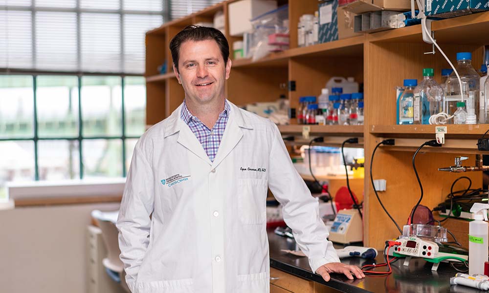 Ryan B. Corcoran合作论文数Department of Medicine, Harvard Medical School35
 Valentina Nardi合作论文数Harvard Medical School and Division of Pediatric Hematology/Oncology, Dana Farber Cancer Institute34Gainor Justin F合作论文数Massachusetts Gen Hosp, Boston, MA 02114 USA32
 Long Phi Le合作论文数29
 Mari Mino-Kenudson合作论文数Harvard Medical School;Massachusetts General Hospital27
 Ritterhouse Lauren L合作论文数Dept Pathol, Univ Chicago27
 Alice Shaw合作论文数Harvard Medical School;Department of Medical Oncology, Dana-Farber Cancer Institute25
 Lipika Goyal合作论文数Division of Oncology, Department of Medicine, School of Medicine, Stanford University;Stanford Cancer Institute25
 Julia Thierauf合作论文数Klinik für Hals-, Nasen-, Ohrenheilkunde, Universitätsklinikum Ulm24
 Eliezer Van Allen合作论文数Division of Population Sciences, Dana-Farber Cancer Institute;Harvard Medical School23Kamran Sophia C合作论文数23
 Dejan Juric合作论文数MiNDNET;Frankfurt School of Finance & Management;JK GmbH23
 Henning Willers合作论文数Department of Radiation Oncology, Massachusetts General Hospital/Radiation Oncology, Harvard Medical School23
 Subba Digumarthy合作论文数Department of Radiology, Massachusetts General Hospital;Harvard Medical School23
 Jessica Lin合作论文数Department of Microbiology and Immunology, School of Medicine, The University of North Carolina at Chapel Hil23
 Marble Hetal D合作论文数22
 Peter Möller合作论文数Universitätsklinikum Ulm;Comprehensive Cancer Center Ulm21
 Chin-Lee Wu合作论文数Pathology Department, Massachusetts General Hospital;Harvard Medical School21
 Gad A. Getz合作论文数Harvard Medical School;Department of Pathology, Massachusetts General Hospital;Broad Institute of Harvard and MIT20
 Vikram Deshpande合作论文数Department of Pathology, Beth Israel Deaconess Medical Center, Harvard Medical School20Albrecht Stenzinger合作论文数Institute of Pathology, University Hospital20
 Jason Efstathiou合作论文数Mass General Research Institute;Harvard Medical School20
 David Miyamoto合作论文数Massachusetts General Hospital Cancer Center, Harvard Medical School19
 Elai Davicioni合作论文数Veracyte, Inc.19
 Kent W. Mouw合作论文数Harvard Medical School;Bladder Cancer Center, Dana-Farber Cancer Institute;Brigham And Women's Hospital19
 Stephanie Ellen Weissinger合作论文数19Drumm Michael合作论文数Harvard University19
 William U. Shipley合作论文数Dana-Farber/Harvard Cancer Center;Department of Radiation Oncology, Massachusetts General Hospital;Harvard Medical School19
 Ewan A. Gibb合作论文数Department of Microbiology, University of Manitoba19Yukako Otani合作论文数18Keisuke Otani合作论文数18Shulin Wu合作论文数18Wszolek Matthew F合作论文数18
 Spring Laura合作论文数Massachusetts General Hospital;Harvard Medical School18Richard Lee合作论文数Department of Medical Physics, British Columbia Cancer Agency18
 Adam S Feldman合作论文数Boston18Philip J. Saylor合作论文数18
 Aditya Bardia合作论文数Dana-Farber/Harvard Cancer Center17
 Andrew X. Zhu (朱秀轩)合作论文数Jiahui International Cancer Center, Jiahui International Hospital (Shanghai)17Yuzhen Zhou合作论文数16Alexander Farahani合作论文数Massachusetts General Hospital16
 Jonathan Nowak合作论文数Harvard Medical School, Harvard University;Hale Family Center For Pancreatic Cancer Research, Dana-Farber Cancer Institute;Department Of Pathology, Brigham And Women’s Hospital;Department Of Pathology, Dana-Farber Cancer Institute16
 Jeffrey W Clark合作论文数Harvard Cancer Center, Harvard Medical School;Dana-Farber/Partners Cancer Care, Massachusetts General Hospital;Cancer Center Protocol Office, Massachusetts General Hospital16
 Jeffrey A. Meyerhardt合作论文数Colon and Rectal Cancer Center, Dana-Farber Cancer Institute;Harvard Medical School16
 Charles S. Fuchs合作论文数Genentech and Roche;EvolveImmune Therapeutics16




