Metastatic disease remains the primary cause of mortality in cancer patients. Yet the number of available in vitro models to study metastasis is limited by challenges in the recapitulation of the metastatic microenvironment in vitro, and by difficulties in maintaining colonized-tissue specificity in the expansion and maintenance of metastatic cells. Here, we show that decellularized scaffolds that retain tissue-specific extracellular-matrix components and bound signalling molecules enable, when seeded with colorectal cancer cells, the spontaneous formation of three-dimensional cell colonies that histologically, molecularly and phenotypically resemble in vivo metastases. Lung and liver metastases obtained by culturing colorectal cancer cells on, respectively, lung and liver decellularized scaffolds retained their tissue-specific tropism when injected in mice. We also found that the engineered metastases contained signet ring cells, which has not previously been observed ex vivo. A culture system with tissue-specific decellularized scaffolds represents a simple and powerful approach for the study of organ-specific cancer metastases.
514 Background: Stereotactic body radiation therapy (SBRT) is increasingly used to treat hepatocellular carcinoma (HCC), but its safety in the treatment of multiple synchronous or recurrent lesions is underreported. We aim to better characterize SBRT-related hepatic toxicity in this population. Methods: We conducted a retrospective analysis of patients with primary HCC who underwent SBRT for 2 or more synchronous or recurrent liver lesions. We collected patient characteristics and dosimetric data (mean liver dose, cumulative effective volume [Veff], cumulative volume of liver receiving 15Gy [V15Gy], and cumulative planning target volume [PTV]) along with liver-related toxicity (measured by albumin-bilirubin [ALBI] and Child-Pugh [CP] scores). We employed a linear mixed-effects model to assess the effect of multi-target SBRT on changes in ALBI. Results: There were 25 patients and 56 lesions with median follow-up of 29 months. Eleven patients had synchronous lesions and 14 had recurrent lesions treated with separate SBRT courses. Eight local failures occurred at a median of 8 months (range: 4 – 25 months) after SBRT. Among those receiving multiple SBRT courses, there were 7 lesions with overlap of V15Gy (median V15Gy overlap: 35mL, range: 0.5 – 388mL). There was no association between cumulative Veff, V15Gy, or PTV and change in ALBI. Four of 25 patients an increase of Child-Pugh (CP) score by ≥ 2 points, within 3 to 6 months after SBRT. Neither increase in CP nor ALBI were associated with cumulative Veff, V15Gy, and PTV. Comparing the groups that received SBRT in a single course versus multiple courses revealed no statistically significant differences in liver toxicity. Conclusions: LiverSBRT for multiple lesions in a single or in separate courses results in worsening of CP scores or ALBI in a small percentage of patients, suggesting a low risk of RILD. Prospective studies with a larger cohort are needed to better characterize safety in this population.
This issue of the Red Journal presents novel combinatorial approaches (eg, advanced imaging with sophisticated planning and delivery tools). We herein take this opportunity to again highlight (1) our tendency toward an overreliance on imaging and (2) the utility of continuing to extend beyond imaging to incorporate anatomic and physiological concepts into the treatment planning process. 1 Marks LB Tepper JE Wisest is he who knows what he does not know. Int J Radiation Oncol Biol Phys. 2018; 102: 687-690 Abstract Full Text Full Text PDF PubMed Scopus (2) Google Scholar ,2 Marks LB "Error bars" in medical imaging: Stealth and treacherous. Radiology. 2015; 277: 318-328 Crossref PubMed Scopus (3) Google Scholar
Background:We aim to better characterize stereotactic body radiation therapy (SBRT)-related hepatic biochemical toxicity in patients with multiple intrahepatic lesions from hepatocellular carcinoma (HCC).Methods:We conducted a retrospective analysis of patients with HCC who underwent SBRT for 2 or more synchronous or metachronous liver lesions. We collected patient characteristics and dosimetric data (mean liver dose [MLD], cumulative effective volume [Veff], cumulative volume of liver receiving 15 Gy [V15Gy], and cumulative planning target volume [PTV]) along with liver-related toxicity (measured by albumin-bilirubin [ALBI] and Child-Pugh [CP] scores). A linear mixed-effects model was used to assess the effect of multi-target SBRT on changes in ALBI.Results:There were 25 patients and 56 lesions with median follow-up of 29 months. Eleven patients had synchronous lesions, and 14 had recurrent lesions treated with separate SBRT courses. Among those receiving multiple SBRT courses, there were 7 lesions with overlap of V15Gy (median V15Gy overlap: 35 mL, range: 0.5-388 mL). There was no association between cumulative MLD, Veff, V15Gy, or PTV and change in ALBI. Four of 25 patients experienced non-classic radiation-induced liver disease (RILD), due to an increase of CP score by ≥2 points 3 to 6 months after SBRT. Sixteen of 25 patients experienced an increase in ALBI grade by 1 or more points 3 to 6 months after SBRT. Comparing the groups that received SBRT in a single course versus multiple courses revealed no statistically significant differences in liver toxicity.Conclusion:Liver SBRT for multiple lesions in a single or in separate courses is feasible and with acceptable risk of hepatotoxicity. Prospective studies with a larger cohort are needed to better characterize safety in this population.
Abstract OBJECTIVE: The toxicities associated with stereotactic radiosurgery (SRS) are important factors when considering treatment options and supportive management for patients with brain metastases. We assessed the association between brain metastasis location and rates of toxicity after SRS. METHODS: We conducted a retrospective single-institution review of 170 patients treated with SRS for brain metastases from 2008–2016 with median follow-up of 8.6 months. Typical SRS doses were 18-20Gy in 1 fraction (lesions < 2cm), 18-21Gy in 3 fractions (lesions 2-3cm), and 25-30Gy in 5 fractions (lesions >3cm). Toxicity measures evaluated included radiation necrosis, seizure, and dexamethasone requirement. RESULTS: A total of 221 lesions were treated among frontal (29%), cerebellar (23%), parietal (16%), temporal (15%), occipital (14%), and other (brainstem, thalamus, basal ganglia) (4%) regions. The rate of SRS-related radionecrosis was 4% for all patients and significantly correlated with metastasis volume (increasing from 1% to 7% for lesions ≤1cm3 to >3cm3) and prior whole brain radiotherapy (WBRT) but not with metastasis location or prior resection on multi-variable analysis (P< 0.05). Post-SRS seizure occurred in 9% of all patients but was significantly higher for primary motor cortex and sensory cortex lesions, associated with 52% and 33% seizure rates, respectively (P< 0.05). Of patients who initially presented with seizure and were on anti-epileptic medication during SRS, 53% had no further seizures, while 47% did have post-SRS seizures, nearly all with motor cortex lesions. Only 5% of patients had new-onset seizure after SRS, related to lesion hemorrhage or motor cortex location. Dexamethasone use >3 months post-SRS was higher for motor strip lesions. CONCLUSION: Brain metastasis location in the primary motor cortex was associated with higher rates of post-SRS seizure, including new-onset seizures and breakthrough seizures on anti-epileptic medication during SRS. Rates of radionecrosis were associated with lesion volume and prior WBRT but not with metastasis location.
485 Background: External beam radiotherapy (EBRT) is a highly effective treatment in select patients with hepatocellular carcinoma (HCC). However, there are many types of EBRT described in the literature with no formal definition of what constitutes “ablative.” Thus, we convened a group of international experts to provide consensus on the parameters that define ablative EBRT in HCC. Methods: A Steering Committee was convened to generate a series of Key Criteria (KC) that could be used to define ablative EBRT for HCC. KC were based on factors related to dose, fractionation, radiobiology, target identification, and delivery technique. An international panel of experts participated in a modified Delphi (mDelphi) process to independently answer questions for each KC. Respondents were given 30 days to respond in round 1 of the mDelphi and 14 days to respond in round 2. A threshold of ≥ 70% was used to define consensus for answers to each KC. Results: Invitations were sent to 40 individuals and 35 (88%) returned responses. In the first round of the mDelphi, 3 of 7 KC reached consensus. In the second round, 100% of participants returned responses and consensus was reached in 3 of the remaining 4 KC. Based on this mDelphi analysis, there was expert agreement that ablative EBRT for HCC should be defined as 1) a BED 10 ≥ 80 Gy, 2) daily imaging and multi-phasic contrast used for target delineation should be used, 3) treatment breaks (e.g., for adaptive EBRT) are allowed, but the total treatment time should be ≤ 6 weeks, 4) the equivalent dose when treating with protons should use a conversion factor of 1.1, and 5) there is no single conversion factor for carbon ions. In one KC, which questioned the expert’s opinion on the α/β ratio of HCC, consensus was not achieved. Conclusions: Using an mDelphi method assessing expert opinion, we provide the first consensus definition of ablative EBRT for HCC. Empiric data are required to define the α/β of HCC.
Aim: Hepatocellular carcinoma (HCC) localized to the liver has various treatment options, including external beam radiotherapy (EBRT). Despite many prospective and retrospective reports showing excellent local control (LC) and favorable toxicity, EBRT has not been widely adopted in the first-line setting and this may be due to a perceived lack of evidence. This study aims to share a decade of experience with Stereotactic Body Radiotherapy (SBRT) for HCC, leveraging a homogenous treatment technique. Methods: This retrospective study at a single institution included patients with HCC treated with SBRT, with a standardized treatment protocol. Freedom from local progression (FFLP), overall survival (OS), and rates of hepatotoxicity post-treatment using child-pugh (CP) and albumin-bilirubin (ALBI) scores were analyzed. A mixed-effects multivariable analysis (MVA) was also performed to assess various factors’ impact on outcomes. Results: A total of 138 lesions in 106 patients were treated between 2009 and 2020. FFLP was 91% at one year and 86% at three years. OS was 80% and 46% at 1 and 3 years, respectively. Baseline liver function was a significant predictor of FFLP and OS on MVA. CP scores and ALBI grades were stable 3 months after treatment in ≥ 70% of patients. Conclusion: SBRT provides excellent LC and low toxicity for HCC patients. While long-term survival remains challenging, treatment decisions should consider overall clinical status. Multidisciplinary review and forthcoming prospective trial results will further clarify radiotherapy’s role in HCC management.
PURPOSE The optimal neoadjuvant treatment for resectable carcinoma of the thoracic esophagus (TE) or gastroesophageal junction (GEJ) remains a matter of debate. We performed an individual participant data (IPD) network meta-analysis (NMA) of randomized controlled trials (RCTs) to study the effect of chemotherapy or chemoradiotherapy, with a focus on tumor location and histology subgroups. PATIENTS AND METHODS All, published or unpublished, RCTs closed to accrual before December 31, 2015 and having compared at least two of the following strategies were eligible: upfront surgery (S), chemotherapy followed by surgery (CS), and chemoradiotherapy followed by surgery (CRS). All analyses were conducted on IPD obtained from investigators. The primary end point was overall survival (OS). The IPD-NMA was analyzed by a one-step mixed-effect Cox model adjusted for age, sex, tumor location, and histology. The NMA was registered in PROSPERO (CRD42018107158). RESULTS IPD were obtained for 26 of 35 RCTs (4,985 of 5,807 patients) corresponding to 12 comparisons for CS-S, 12 for CRS-S, and four for CRS-CS. CS and CRS led to increased OS when compared with S with hazard ratio (HR) = 0.86 (0.75 to 0.99), P = .03 and HR = 0.77 (0.68 to 0.87), P < .001 respectively. The NMA comparison of CRS versus CS for OS gave a HR of 0.90 (0.74 to 1.09), P = .27 (consistency P = .26, heterogeneity P = .0038). For CS versus S, a larger effect on OS was observed for GEJ versus TE tumors ( P = .036). For the CRS versus S and CRS versus CS, a larger effect on OS was observed for women ( P = .003, .012, respectively). CONCLUSION Neoadjuvant chemotherapy and chemoradiotherapy were consistently better than S alone across histology, but with some variation in the magnitude of treatment effect by sex for CRS and tumor location for CS. A strong OS difference between CS and CRS was not identified.
We thank Dr. Khosla and colleagues for the reply1 to our manuscript2 and would like to address their comments. First, the authors highlight the potential for differences in the stereotactic body radiation therapy (SBRT) technique among the included centers. We acknowledge site-level variability but would like to point out that we limited the study to specific tertiary centers highly experienced in SBRT. Technical details from each center are included in the supplementary materials. We considered the inclusion of several centers with different techniques as a strength since it improves generalizability, as variability can exist in the SBRT technique. Internationally, there are variable definitions for SBRT itself and no agreement on what defines ablative dose radiotherapy. There is a need for consensus definitions on ablative dose radiotherapy and more research on optimal radiotherapy treatment techniques. Second, these authors point out that the increased risk of Child-Pugh score worsening ≥ 2 points among SBRT treatments compared with thermal ablation was statistically significant at 3 months but not 6 months. They suggest that the differences could have been attributable to the larger median lesion size for SBRT (2.4 cm) compared with ablation (2.1 cm), but differences remained statistically significant after adjustment for tumor size. Furthermore, the lower proportion of SBRT-treated patients with Child-Pugh increase at 6 months (11.1%) compared with 3 months (13.0%) could be in part attributable to the 13 SBRT-treated patients who experienced Child-Pugh increase at 3 months and died before the 6-month mark. While we were unable to ascertain the cause of death, it is possible that deaths related to liver toxicity in the SBRT group could have contributed to the lower proportion experiencing Child-Pugh increase at 6 months. Lastly, Khosla et al raise concerns of residual confounding, including tumor biology and medical comorbidities. We agree with this point. Each institution clearly had biases in making their treatment decisions, and this certainly could impact outcomes. Ultimately, randomized controlled trials would be beneficial to assess the comparative effectiveness and toxicity of these treatments. There are challenges in completing such trials given that thermal ablation has been proposed as the ablative “treatment of choice” by many, including the recent American Association for the Study of Liver Diseases guidelines.3 Trials may be particularly challenging in populations with decompensated liver disease or comorbidities. Multicenter observational studies such as our study, while subject to biases, provide important information supporting the use of both thermal ablation and SBRT while we await prospective comparative effectiveness data.
Background & Aims: Early-stage HCC can be treated with thermal ablation or stereotactic body radiation therapy (SBRT). We retrospectively compared local progression, mortality, and toxicity among patients with HCC treated with ablation or SBRT in a multicenter, US cohort. Approach & Results: We included adult patients with treatment-naïve HCC lesions without vascular invasion treated with thermal ablation or SBRT per individual physician or institutional preference from January 2012 to December 2018. Outcomes included local progression after a 3-month landmark period assessed at the lesion level and overall survival at the patient level. Inverse probability of treatment weighting was used to account for imbalances in treatment groups. The Cox proportional hazard modeling was used to compare progression and overall survival, and logistic regression was used for toxicity. There were 642 patients with 786 lesions (median size: 2.1 cm) treated with ablation or SBRT. In adjusted analyses, SBRT was associated with a reduced risk of local progression compared to ablation (aHR 0.30, 95% CI: 0.15–0.60). However, SBRT-treated patients had an increased risk of liver dysfunction at 3 months (absolute difference 5.5%, aOR 2.31, 95% CI: 1.13–4.73) and death (aHR 2.04, 95% CI: 1.44–2.88, p < 0.0001). Conclusions: In this multicenter study of patients with HCC, SBRT was associated with a lower risk of local progression compared to thermal ablation but higher all-cause mortality. Survival differences may be attributable to residual confounding, patient selection, or downstream treatments. These retrospective real-world data help guide treatment decisions while demonstrating the need for a prospective clinical trial.
Evaluation of skin toxicity following drug with radiation (XRT) treatment. (A) Representative images and (B) quantification of skin toxicities observed in animals 49 days following treatment with PBS (Control), CPT, or CRLX101 with radiation.
William Blackstock was an individual on whom one could always rely to get things done when they needed to be done, and who did them with a laugh and a smile. He was an individual who was liked by all and who excelled in getting the most out of others—be they residents, junior faculty, senior faculty, or scientific collaborators.William was born in 1963 in Eden, North Carolina. He had the advantage of having parents who were teachers in the public school system, and that association seemed to carry with him throughout his life as he became a teacher as well as a physician and clinical researcher. He went to the public schools in Eden, received a scholarship to attend Wake Forest University (Winston-Salem, NC), and then went on to medical school at East Carolina University School of Medicine (Greenville, NC).I had the pleasure of first meeting William in the late 1980s, when he did a radiation oncology student rotation at the University of North Carolina (UNC; Chapel Hill, NC). His enthusiasm and intelligence were apparent, and we were pleased to be able to offer him a residency position at UNC. As his residency progressed, it was clear that William had skills that would be a hallmark of his career. More than almost any resident I have seen, he sought out research opportunities, both within and importantly outside the department, trying to find those opportunities that would have a meaningful impact on clinical care. He talked to anyone who could provide ideas or insights, and then collaborated with those people. This is a signature trait through his career: his ability to learn from and collaborate with a wide variety of individuals. When he recognized opportunities, he collaborated with others and pursued these ideas to completion.His initial work focused on gemcitabine radiation sensitization for pancreatic cancer, and he developed a new approach based on the nonlinear pharmacokinetics of gemcitabine with an alternative dose delivery scheme. This approach was then tested in both the single-institution study and then through the cooperative group system. These interests in radiation sensitization based on science stayed with him throughout his career.After his residency at UNC, he stayed on at UNC as faculty for a short while and then took a faculty position at Wake Forest University School of Medicine (Winston-Salem, NC), where he rose through the ranks to become Professor and then Chair of the Department of Radiation Oncology. His clinical focus was on the treatment of gastrointestinal and thoracic malignancies, with substantive research efforts both at a local and a national level. He participated and led multiple local and national clinical trials in these areas. His position allowed him to combine his interests in clinical care, scientific research, and teaching, as well as to pursue his deep interest in cancer disparities and the social determinants of health.His primary concern was always his patients. He related to his patients and spent the time and effort that was needed to develop special rapport with them.The quality of his work naturally was noticed by others in both the clinical cancer and cancer research communities. He has been a member of the NCI clinical oncology study section and the NCI Board of Scientific Counselors (intramural). He has been co-chair of the NCI Thoracic Malignancy Steering Committee and has served on multiple committees of the American Society of Clinical Oncology and the American Association for Cancer Research. He was, as well, a member of a large number of special panels and symposia.One example of how he worked relates to events surrounding the time when the Wake Forest Comprehensive Cancer Center was having some issues, and they naturally went to William to get the situation resolved. He became the Acting Director of the Cancer Center, a position he did not wish to have. He took on a very difficult situation, and he was able to get the problems resolved before the leadership (to his delight) was transferred to others. Amazingly, he took on this role while he was dealing with multiple treatments for metastatic and progressive prostate cancer. He knew that he would die of this disease, but he was loyal to the institution and to his colleagues at Wake Forest. Being who he was, he could do nothing less. Despite many years of treatment for his prostate cancer, he told very few people of his illness. He was active in both clinical and research activities until very shortly before his death and he was still planning what his next steps would be.William spent virtually his entire academic life in North Carolina, but his impact has been much wider. He leaves behind his two daughters, Jessica Blackstock (Logan) and Ansley Blackstock (Jiri), and granddaughter Bryn Blackstock, all of Winston-Salem. He will be missed by his family, and his many friends and colleagues in the state and far beyond. As his mentor, and especially as his colleague, I will personally miss him very much.
There is a linear relationship between the number of HT-29-Luc2 cells and the luminescence intensity.
Table S1. Clinical and demographic characteristics of recruited cancer patients; Table S2. Demographic characteristics of recruited healthy donors; Figure S1. Reproducibility confirmation of CapioCyte-D by testing duplicated blood samples from three cancer patients (A-C) using separately prepared capture surfaces (slides 1 and 2); Figure S2. A comparison of the captured epithelial cell number from two healthy donor groups with different age ranges (under 35 years vs. above 50 years).
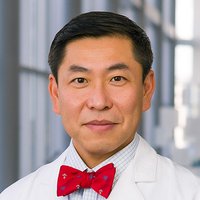 Andrew Wang合作论文数Department of Radiation Oncology, University of Texas Southwestern Medical Center;Medical School, University of Texas Southwestern Medical Center;Archimmune Therapeutics Inc.;Capio Biosciences28
 Donna Niedzwiecki合作论文数Department of Biostatistics and Bioinformatics, School of Medicine, Duke University;Duke Cancer Institute, Duke University;Toray Industries, Inc.20
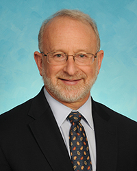 Richard M Goldberg合作论文数Department of Medicine, School of Medicine, West Virginia University;Cancer Institute, West Virginia University18
 Benjamin Calvo合作论文数Department of Surgical Oncology, Roswell Park Comprehensive Cancer Center18
 Christopher G. Willett合作论文数Department of Radiation Oncology, Duke University School of Medicine;Duke Cancer Institute, Duke University18
 Wang Kyle合作论文数16
 Leonard L. Gunderson合作论文数Department of Radiation Oncology Mayo Clinic College of Medicine15A. William Blackstock合作论文数15
 Al B Benson合作论文数Division of Hematology and Oncology, Department of Medicine, Feinberg School of Medicine, Northwestern University;Robert H. Lurie Comprehensive Cancer Center, Feinberg School of Medicine, Northwestern University13
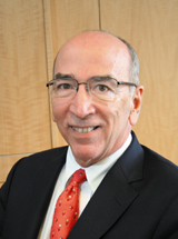 Daniel G. Haller合作论文数Department of Medicine, Perelman School of Medicine, University of Pennsylvania13
 Bert H. O’Neil合作论文数12Rj Mayer合作论文数11Robert J. Mayer合作论文数10
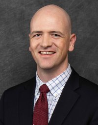 Andrew Moon合作论文数10William F. Sindelar合作论文数10
 Michael O. Meyers合作论文数Division of Surgical Oncology, Department of Surgery, School of Medicine, The University of North Carolina at Chapel Hill10
 Hanna K. Sanoff合作论文数Division of Oncology, Department of Medicine, School of Medicine, The University of North Carolina at Chapel Hill;North Carolina Basnight Cancer Hospital, Lineberger Comprehensive Cancer Center, School of Medicine, The University of North Carolina at Chapel Hill10Joseph M. Caster合作论文数10
 William U. Shipley合作论文数Dana-Farber/Harvard Cancer Center;Department of Radiation Oncology, Massachusetts General Hospital;Harvard Medical School9
 Charles S. Fuchs合作论文数Genentech and Roche;EvolveImmune Therapeutics9
 Allison M. Deal合作论文数Lineberger Comprehensive Cancer Center, School of Medicine, University of North Carolina at Chapel Hill9
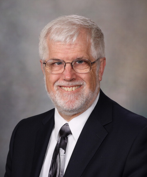 James Martenson合作论文数Department of Radiation Oncology, Mayo Clinic9
 Herman D. Suit合作论文数Department of Radiation Oncology, Massachusetts General Hospital;Harvard Medical School, Harvard University8
 Hong-Jin Kim合作论文数Department of HBP Surgery and Liver Transplantation, Yeungnam University Hospital8
 Timothy Kinsella合作论文数Department of Radiation Oncology, School of Medicine, Case Western Reserve University;Department of Radiation Oncology, Warren Alpert Medical School, Brown University;Department of Radiation Oncology, School of Medicine, Tufts University8Julian G. Rosenman合作论文数7
 Neal Meropol合作论文数Flatiron Health;School of Medicine, Case Western Reserve University7Garmey Edward G合作论文数7Ted K. Yanagihara合作论文数7
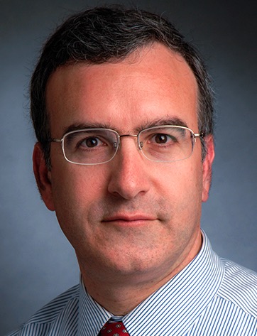 Harvey J. Mamon合作论文数Brigham and Women's Hospital;Harvard Medical School;Division of Gastrointestinal Radiation Oncology, Dana-Farber Cancer Institute7Xi Tian合作论文数6D R Hollis合作论文数6
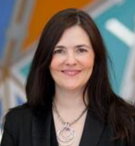 Nancy Baxter合作论文数School of Population and Global Health, University of Melbourne6
 William K Funkhouser合作论文数6John S. Macdonald合作论文数6
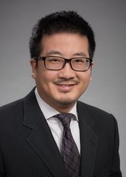 Smith Apisarnthanarax合作论文数Experimental Radiation Oncology, M.D. Anderson Cancer Center6Donna Hollis合作论文数6Scott L. Sailer合作论文数6
 William C. Wood合作论文数Department of Surgery, Emory University School of Medicine6
 Kyle C Roche合作论文数5Bert H. O'Neil合作论文数5Minh Nguyen合作论文数5
 Bhisham Chera合作论文数Department of Radiation Oncology, University of North Carolina at Chapel Hill;Institute for Healthcare Quality Improvement, School of Medicine, University of North Carolina at Chapel Hill5
 Michael J O'Connell合作论文数5Harris Stephen L合作论文数5
 Autumn J Mcree合作论文数5
 Xianming Tan合作论文数Department of Biostatistics, Gillings School of Global Public Health, The University of North Carolina at Chapel Hill5
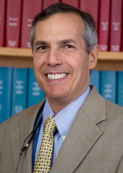 Lawrence B. Marks合作论文数Department of Radiation Oncology, UNC School of Medicine5




