Allogeneic T cell therapies are a highly desirable option to circumvent the cost and complexity of using autologous T cells to treat diseases. Allogeneic CD8+ T cells can be made from pluripotent stem cells (PSCs), but deriving CD4+ T cells from PSCs has remained a significant challenge. Using feeder- and serum-free conditions, we found that CD4+ vs. CD8+ T cell commitment from PSCs can be controlled by fine-tuning the dynamics of Notch and T cell receptor (TCR) signaling delivered to CD4+CD8+ double-positive T cells. Notch signaling negatively impacts CD4+ T cell commitment, and its timed removal allows generation of clonally diverse and expandable CD4+ T cells from PSCs. The resulting CD4+ T cells respond to cytokine-mediated polarization by differentiating into Th1, Th2, or Th17 cells, recapitulating canonical helper cell function. These findings represent a significant step toward using PSC-derived CD4+ T cells as a low-cost, off-the-shelf cell therapy.
T cells develop from hematopoietic progenitors in the thymus and protect against pathogens and cancer. However, the emergence of human T cell-competent blood progenitors and their subsequent specification to the T lineage have been challenging to capture in real time. Here, we leveraged a pluripotent stem cell differentiation system to understand the transcriptional dynamics and cell fate restriction events that underlie this critical developmental process. Time-resolved single-cell RNA sequencing revealed that downregulation of the multipotent hematopoietic program, upregulation of >90 lineage-associated transcription factors, and cell-cycle exit all occur within a highly coordinated developmental window. Gene-regulatory network inference uncovered a role for YBX1 in T lineage specification. We mapped the differentiation cell fate hierarchy using transcribed lineage barcoding and discovered that mast and myeloid potential bifurcate from each other early in hematopoiesis, upstream of T lineage restriction. Our systems-level analyses provide a quantitative, time-resolved model of human T cell fate specification. A record of this paper’s transparent peer review process is included in the supplemental information.
SummaryWithin the thymus, regulation of the cellular cross-talk directing T cell development is dependent on spatial interactions within specialized niches. To create a holistic, spatially defined map of tissue niches guiding postnatal T cell development we employed the multidimensional imaging platform CO-detection by indEXing (CODEX), as well as CITE-seq and ATAC-seq. We generated age-matched 4–5-month-old postnatal thymus datasets for male and female donors, and identify significant sex differences in both T cell and thymus biology. We demonstrate a crucial role for JAG ligands in directing thymic-like dendritic cell development, reveal important functions of a novel population of ECM-fibroblasts, and characterize the medullary niches surrounding Hassall’s corpuscles. Together, these data represent a unique age-matched spatial multiomic resource to investigate how sex-based differences in thymus regulation and T cell development arise, and provide an essential resource to understand the mechanisms underlying immune function and dysfunction in males and females.
T cells develop from multi-potent hematopoietic progenitors in the thymus and provide adaptive protection against pathogens and cancer. However, the emergence of human T cell-competent blood progenitors, and their subsequent specification to the T lineage, has been challenging to capture in real time. Here, we leveraged a pluripotent stem cell differentiation system to understand the transcriptional dynamics and cell fate restriction events that underlie this critical developmental process. Time-resolved single cell RNA sequencing revealed that cell-cycle exit, downregulation of the multipotent hematopoietic program, and upregulation of >90 lineage-associated transcription factors all occur within a highly co-ordinated and narrow developmental window. Computational gene-regulatory network inference elucidated the transcriptional logic of T lineage specification, uncovering an important role for YBX1. We mapped the differentiation cell fate hierarchy using transcribed lineage barcoding and mathematical trajectory inference and discovered that mast and myeloid potential bifurcate from each other early in haematopoiesis, upstream of T lineage restriction. Collectively, our analyses provide a quantitative, time-resolved model of human T cell specification with relevance for regenerative medicine and developmental immunology.
The generation of T-cells from stem cells in vitro could provide an alternative source of cells for immunotherapies. T-cell development from hematopoietic stem and progenitor cells (HSPCs) is tightly regulated through Notch pathway activation by Delta-like (DL) ligands 1 and 4. Other molecules, such as stem cell factor (SCF) and interleukin (IL)-7, play a supportive role in regulating the survival, differentiation, and proliferation of developing T-cells. Numerous other signaling molecules influence T-lineage development in vivo, but little work has been done to understand and optimize their use for T-cell production. Using a defined engineered thymic niche system, we undertook a multi-stage statistical learning-based optimization campaign and identified IL-3 and tumor necrosis factor α (TNFα) as a stage- and dose-specific enhancers of cell proliferation and T-lineage differentiation. We used this information to construct an efficient three-stage process for generating conventional TCRαβ+CD8+ T-cells expressing a diverse TCR repertoire from blood stem cells. Our work provides new insight into T-cell development and a robust system for generating T-cells to enable clinical therapies for treating cancer and immune disorders.
T cells show tremendous efficacy as cellular therapeutics. However, obtaining primary T cells from human donors is expensive and variable. Pluripotent stem cells (PSCs) have the potential to provide a renewable source of T cells, but differentiating PSCs into hematopoietic progenitors with T cell potential remains an important challenge. Here, we report an efficient serum- and feeder-free system for differentiating human PSCs into hematopoietic progenitors and T cells. This fully defined approach allowed us to study the impact of individual proteins on blood emergence and differentiation. Providing DLL4 and VCAM1 during the endothelial-to-hematopoietic transition enhanced downstream progenitor T cell output by ~80-fold. These two proteins synergized to activate notch signaling in nascent hematopoietic stem and progenitor cells, and VCAM1 additionally promoted an inflammatory transcriptional program. We also established optimized medium formulations that enabled efficient and chemically defined maturation of functional CD8αβ + , CD4 − , CD3 + , TCRαβ + T cells with a diverse TCR repertoire.
T cells are key mediators of the adaptive immune response and show tremendous efficacy as cellular therapeutics. However, obtaining primary T cells from human donors is expensive and variable. Pluripotent stem cells (PSCs) have the potential to serve as a consistent and renewable source of T cells, but differentiating PSCs into hematopoietic progenitors with T cell potential remains a significant challenge. Here, we developed an efficient serum- and feeder-free protocol for differentiating human PSCs into hematopoietic progenitors and T cells. This defined method allowed us to study the impact of individual recombinant proteins on blood emergence and lineage potential. We demonstrate that the presence of DLL4 and VCAM1 during the endothelial-to-hematopoietic transition (EHT) enhances downstream progenitor T cell output by >80-fold. Using single cell transcriptomics, we showed that these two proteins synergise to drive strong notch signalling in nascent hematopoietic stem and progenitor cells and that VCAM1 additionally drives a pro-inflammatory transcriptional program. Finally, we applied this differentiation method to study the impact of cytokine concentration dynamics on T cell maturation. We established optimised media formulations that enabled efficient and chemically defined differentiation of CD8αβ+, CD4-, CD3+, TCRαβ+ T cells from PSCs.
T-cell development from hematopoietic stem and progenitor cells (HSPCs) is tightly regulated through Notch pathway activation by the Notch ligands Delta-like (DL) 1 and 4 and Jagged-2. Other molecules, such as stem cell factor (SCF), FMS-like tyrosine kinase 3 ligand (Flt3L) and interleukin (IL)-7, play a supportive role in regulating the survival, differentiation, and proliferation of developing progenitor (pro)T-cells. Numerous other signaling molecules are known to instruct T-lineage development in vivo , but little work has been done to optimize their use for T-cell production in vitro . Using a defined T-lineage differentiation assay consisting of plates coated with the Notch ligand DL4 and adhesion molecule VCAM-1, we performed a cytokine screen that identified IL-3 and tumor necrosis factor α (TNFα) as enhancers of proT-cell differentiation and expansion. Mechanistically, we found that TNFα induced T-lineage differentiation through the positive regulation of T-lineage genes GATA3, TCF7 , and BCL11b . TNFα also synergized with IL-3 to induce proliferation by upregulating the expression of the IL-3 receptor on CD34+ HSPCs, yielding 753.2 (532.4-1026.9; 5-95 percentile)-fold expansion of total cells after 14 days compared to 8.9 (4.3-21.5)-fold expansion in conditions without IL-3 and TNFα. We then optimized cytokine concentrations for T-cell maturation. Focusing on T-cell maturation, we used quantitative models to optimize dynamically changing cytokine requirements and used these to construct a three-stage assay for generating CD3+CD4+CD8+ and CD3+CD4−CD8+ T-cells. Our work provides new insight into T-cell development and a robust in vitro assay for generating T-cells to enable clinical therapies for treating cancer and immune disorders. ### Competing Interest Statement Intellectual property related to this work is being evaluated under an option to license. P.W.Z. is a scientific founder and consultant of Notch Therapeutics, a biotechnology company developing stem cell-derived T-cell immunotherapies.
We hypothesized that generating spinal motor neurons (sMNs) from human induced pluripotent stem cell (hiPSC)-derived neural aggregates (NAs) using a chemically -defined differentiation protocol would be more effective inside of 3D fibrin hydrogels compared to 2D poly-L-ornithine(PLO)/laminin-coated tissue culture plastic surfaces. We performed targeted RNA-Seq using next generation sequencing to determine the substrate -specific differences in gene expression that regulate cell phenotype. Cells cultured on both substrates expressed sMN genes CHAT and MNX1, though persistent WNT signaling contributed to a higher expression of genes associated with interneurons in NAs cultured in 3D fibrin scaffolds. Cells in fibrin also expressed lower levels of astrocyte progenitor genes and higher levels of the neuronal specific gene TUBB3, suggesting a purer population of neurons compared to 2D cultures.Statement of SignificanceFibrin scaffolds can support the neuronal differentiation of pluripotent stem cells. This study provides insight into how fibrin hydrogels affect neuronal induction by analyzing of the signaling pathways activated during the differentiation process. These insights can then be used to tailor the properties of these hydrogels to optimize the generation of sMNs for regenerative medicine applications. Crown Copyright (c) 2017 Published by Elsevier Ltd on behalf of Acta Materialia Inc. All rights reserved.
This paper describes a fully defined, nonxenogeneic in vitro niche for the differentiation of haematopoietic stem and progenitor cells to progenitor T cells in mouse and human.
Event Abstract Back to Event Genipin crosslinked fibrin as a potential bioscaffold for delivering spinal motor neurons derived from human induced pluripotent stem cells Meghan Robinson1, Andrew Agbay2, Stephanie M. Willerth2, 3 and John M. Edgar1 1 University of Victoria, Department of Biomedical Engineering, Canada 2 University of Victoria, Division of Medical Sciences, Canada 3 University of Victoria, Department of Mechanical Engineering, Canada Introduction: Human induced pluripotent stem cells (hiPSCs) can generate any type of cell in the body, making them attractive for cell therapy applications. hiPSCs can be directed towards a neural lineage for use in regenerative therapy through delivery of physical and chemical cues[1],[2]. Personalized neural tissue derived from hiPSCs can survive and differentiate into relevant neural phenotypes to replace damaged central nervous system (CNS) tissues and promote functional recovery[4]. The physical microenvironment of regenerating tissues has a significant influence on the behavior of differentiating hiPSCs, therefore such engineered tissues require a 3D biomaterial scaffold with physical properties closely resembling those of the native extracellular matrix found in the CNS[2],[3]. Fibrin, a natural biomaterial involved in blood clotting, has proven to be a suitable scaffold for promoting the generation and viability of neurons from hiPSCs[2],[4]. However, the fast degradation rate of fibrin does not provide an adequate time window to accommodate the regeneration of neural tissue by hiPSCs. Degradation rates of fibrin scaffolds can be decreased by increasing the amount of crosslinking through the addition of a crosslinking agent. Genipin, a plant-derived agent that promotes crosslink formation in fibrin scaffolds, possesses neuritogenic, neurotrophic and neuroprotective properties[5]-[9]. As the addition of genipin alters the physical characteristics of the scaffold, it may also influence the behaviour of the differentiating cells[11]-[13]. This study determines the concentrations of fibrin and genipin that provide the optimal physical properties for the generation of spinal motor neurons from iPSCs. Materials and Methods: Human iPSCs were cultured in AggreWell™ 800 plates using neural induction medium (NIM) for 5 days to form neural aggregates. These were then seeded in 3D fibrin-genipin scaffolds of genipin concentrations 1 mM, 2.5 mM, 5.0 mM, or 10 mM, with NIM + 1 µM retinoic acid + 1 µM purmorphamine, and cultured for an additional 21 days. Cell viability, adhesion and phenotype were assessed using flow cytometry or immunocytochemistry on days 14 and 21. The elastic modulus and degree of crosslinking for each scaffold was characterized by rheometry and scanning electron microscope. Results and Discussion: Human iPSCs formed aggregates 24 hours after being placed into the AggreWell™ 800 plates. When seeded into fibrin-genipin scaffolds, neural aggregates adhered and extended neurites. LIVE-DEAD staining showed that neurons were viable, while neural identity was confirmed by immunocytochemistry. The formation of crosslinks due to the addition of genipin to fibrin scaffolds was found to provide superior mechanical stability for the induction of spinal motor neurons from hiPSCs without negatively impacting their differentiation. Figure 1. Spinal motor neurons induced from hiPSCs in 3-D fibrin-genipin bioscaffolds, immunostained for A) TUJ1, a neuronal marker, and B) HB9, a marker specific to spinal motor neuron precursors. Conclusion: The degradation rates of fibrin scaffolds can be tailored to accomodate the induction of spinal motor neurons from hiPSCs by the addition of the crosslinking agent genipin, while maintaining the necessary physical properties to promote differentiation. The authors would like to thank the Natural Sciences and Engineering Research Council of Canada (NSERC).References:[1] Takahashi, K. et al. Induction of Pluripotent Stem Cells from Adult Human Fibroblasts by Defined Factors. Cell (2007). doi:10.1016/j.cell.2007.11.019[2] Montgomery, A., Wong, A., Gabers, N. & Willerth, S. M. Engineering personalized neural tissue by combining induced pluripotent stem cells with fibrin scaffolds. Biomater Sci 3, 401–13 (2015).[3] Engler, A., Sen, S., Sweeney, L. & Discher, D. Matrix Elasticity Directs Stem Cell Lineage Specification. Cell 126, (2005).[4] Lu, P., Woodruff, G., Wang, Y., Graham, L., Hunt, M., Wu, D., ... & Tuszynski, M. H. (2014). Long-distance axonal growth from human induced pluripotent stem cells after spinal cord injury. Neuron, 83(4), 789-796.[5] Yamazaki, M., & Chiba, K. (2005). Neurotrophic effects of genipin on Neuro2a cells. Journal of health science, 51(6), 687-692.[6] Yamazaki, M., Chiba, K., & Satoh, K. (2008). Neuro2a cell death induced by 6- hydroxydopamine is attenuated by genipin. Journal of health science, 54(6), 638- 644.[7] Nam, K. N., Choi, Y. S., Jung, H. J., Park, G. H., Park, J. M., Moon, S. K., ... & Lee, E. H. (2010). Genipin inhibits the inflammatory response of rat brain microglial cells. International immunopharmacology, 10(4), 493-499.[8] Yamazaki, M., Chiba, K., & Yoshikawa, C. (2009). Genipin suppresses A23187- induced cytotoxicity in neuro2a cells. Biological and Pharmaceutical Bulletin, 32(6), 1043-1046.[9] Yamazaki, M., Chiba, K., Mohri, T., & Hatanaka, H. (2004). Cyclic GMP- dependent neurite outgrowth by genipin and nerve growth factor in PC12h cells.European journal of pharmacology, 488(1), 35-43.[10] Schek, R. M., Michalek, A. J., & Iatridis, J. C. (2011). Genipin-crosslinked fibrin hydrogels as a potential adhesive to augment intervertebral disc annulus repair. European cells & materials, 21, 373.[11] Gamboa‐Martínez, T. C., Luque‐Guillén, V., González‐García, C., Gómez Ribelles, J. L., & Gallego‐Ferrer, G. (2015). Crosslinked fibrin gels for tissue engineering: Two approaches to improve their properties. Journal of Biomedical Materials Research Part A, 103(2), 614-621.[12] Liu, B. S., Yao, C. H., Hsu, S. H., Yeh, T. S., Chen, Y. S., & Kao, S. T. (2004). A novel use of genipin-fixed gelatin as extracellular matrix for peripheral nerve regeneration. Journal of biomaterials applications, 19(1), 21-34.[13] McKay, C. A., Pomrenke, R. D., McLane, J. S., Schaub, N. J., DeSimone, E. K., Ligon, L. A., & Gilbert, R. J. (2014). An Injectable, Calcium Responsive Composite Hydrogel for the Treatment of Acute Spinal Cord Injury. ACS applied materials & interfaces, 6(3), 1424-1438. Keywords: Regenerative Medicine, Tissue Engineering, stem cell, 3D scaffold Conference: 10th World Biomaterials Congress, Montréal, Canada, 17 May - 22 May, 2016. Presentation Type: Poster Topic: Biomaterials for cellular programming Citation: Robinson M, Agbay A, Willerth SM and Edgar JM (2016). Genipin crosslinked fibrin as a potential bioscaffold for delivering spinal motor neurons derived from human induced pluripotent stem cells. Front. Bioeng. Biotechnol. Conference Abstract: 10th World Biomaterials Congress. doi: 10.3389/conf.FBIOE.2016.01.01685 Copyright: The abstracts in this collection have not been subject to any Frontiers peer review or checks, and are not endorsed by Frontiers. They are made available through the Frontiers publishing platform as a service to conference organizers and presenters. The copyright in the individual abstracts is owned by the author of each abstract or his/her employer unless otherwise stated. Each abstract, as well as the collection of abstracts, are published under a Creative Commons CC-BY 4.0 (attribution) licence (https://creativecommons.org/licenses/by/4.0/) and may thus be reproduced, translated, adapted and be the subject of derivative works provided the authors and Frontiers are attributed. For Frontiers’ terms and conditions please see https://www.frontiersin.org/legal/terms-and-conditions. Received: 27 Mar 2016; Published Online: 30 Mar 2016. Login Required This action requires you to be registered with Frontiers and logged in. To register or login click here. Abstract Info Abstract The Authors in Frontiers Meghan Robinson Andrew Agbay Stephanie M Willerth John M Edgar Google Meghan Robinson Andrew Agbay Stephanie M Willerth John M Edgar Google Scholar Meghan Robinson Andrew Agbay Stephanie M Willerth John M Edgar PubMed Meghan Robinson Andrew Agbay Stephanie M Willerth John M Edgar Related Article in Frontiers Google Scholar PubMed Abstract Close Back to top Javascript is disabled. Please enable Javascript in your browser settings in order to see all the content on this page.
Ongoing clinical trials are evaluating the use of stem cells as a way to treat traumatic spinal cord injury (SCI). However, the inhibitory environment present in the injured spinal cord makes it challenging to achieve the survival of these cells along with desired differentiation into the appropriate phenotypes necessary to regain function. Transplanting stem cells along with an instructive biomaterial scaffold can increase cell survival and improve differentiation efficiency. This study reviews the literature discussing different types of instructive biomaterial scaffolds developed for transplanting stem cells into the injured spinal cord. We have chosen to focus specifically on biomaterial scaffolds that direct the differentiation of neural stem cells and pluripotent stem cells since they offer the most promise for producing the cell phenotypes that could restore function after SCI. In terms of biomaterial scaffolds, this article reviews the literature associated with using hydrogels made from natural biomaterials and electrospun scaffolds for differentiating stem cells into neural phenotypes. It then presents new data showing how these different types of scaffolds can be combined for neural tissue engineering applications and provides directions for future studies. (C) 2016 S. Karger AG, Basel
Ongoing clinical trials are evaluating the use of stem cells as a way to treat traumatic spinal cord injury (SCI). However, the inhibitory environment present in the injured spinal cord makes it challenging to achieve the survival of these cells along with desired differentiation into the appropriate phenotypes necessary to regain function. Transplanting stem cells along with an instructive biomaterial scaffold can increase cell survival and improve differentiation efficiency. This study reviews the literature discussing different types of instructive biomaterial scaffolds developed for transplanting stem cells into the injured spinal cord. We have chosen to focus specifically on biomaterial scaffolds that direct the differentiation of neural stem cells and pluripotent stem cells since they offer the most promise for producing the cell phenotypes that could restore function after SCI. In terms of biomaterial scaffolds, this article reviews the literature associated with using hydrogels made from natural biomaterials and electrospun scaffolds for differentiating stem cells into neural phenotypes. It then presents new data showing how these different types of scaffolds can be combined for neural tissue engineering applications and provides directions for future studies.
Event Abstract Back to Event Differential gene expression and phenotype during motor neuron differentiation of human induced pluripotent stem cells in 3D fibrin scaffolds John M. Edgar1, Meghan Robinson1, Andrew Agbay2 and Stephanie M. Willerth2, 3 1 University of Victoria, Department of Biomedical Engineering, Canada 2 University of Victoria, Division of Medical Sciences, Canada 3 University of Victoria, Department of Mechanical Engineering, Canada Introduction: Research in the field of regenerative medicine took a giant leap in 2007 with the discovery of human induced pluripotent stem cells (iPSCs)—adult cells that could be induced to an embryonic stem cell-like state and then differentiated into any other cell-type[1]. This created a need for biomaterial scaffolds that could both mimic the three-dimensional structure of human tissue and provide cues to direct iPSC differentiation into a desired cell-type. Fibrin is a promising biomaterial scaffold for supporting the differentiation of iPSCs into neural cell-types for treating patients with spinal cord injuries[2]. A fibrous hydrogel derived from blood, fibrin has been extensively characterized for its biocompatibility, and was recently found to support embryonic and neural stem cell survival, differentiation, and engraftment in mouse models of spinal cord injury[3]-[5]. The physical properties of scaffolds have been shown to direct differentiation[6]; however, little is known about the mechanisms by which they effect this change. Here, we use flow cytometry, immunocytochemistry (ICC), and next-generation RNA sequencing (RNA-seq) to compare human iPSC differentiation into spinal motor neurons in fibrin scaffolds with traditional two-dimensional culture methods. These experiments seek to define the ways that cell substrates can affect gene expression using standardized exogenous chemical signaling in order to optimize fibrin scaffolds for motor neuron differentiation and tissue formation. Materials and Methods: Human iPSCs were cultured in AggreWell™ 800 plates with neural induction medium (NIM) for 5 days to form neural aggregates. These were then seeded in fibrin or on laminin-coated cell culture plates with NIM + 1 µM retinoic acid + 1 µM purmorphamine and cultured for an additional 15 days. Cell phenotype was assessed on day 0, 5, 12, and 20 using flow cytometry or ICC. RNA was purified from cells at those same intervals and a custom, 200-target RNA-seq library was created for the Illumina MiSeq next-generation sequencer. Results and Discussion: Human iPSCs formed tight aggregates after 5 days in AggreWell™ plates that readily spread and extended neurites when placed in fibrin or on laminin. On laminin, aggregates adhered and spread in two dimensions. In fibrin, the aggregates maintained a spherical morphology while spreading and extending neurites in three dimensions, as is desirable for tissue formation. ICC showed that cell aggregates in fibrin began to express the neuronal marker Tuj1 and were viable (see Figure 1). It was observed that human iPSCs degraded the fibrin faster than murine iPSCs used in previous experiments, possibly due to increased proteolysis. A protease inhibitor such as aprotinin may reduce the rate if degradation in future experiments. Further analysis of RNA-seq data will clarify the stages of neuronal differentiation preceding spinal motor neuron development and how they influence cell patterning and morphology. Figure 1. Cell aggregates seeded in fibrin were A) positive for viability marker and B) express neuronal marker Tuj1. Conclusion: Human iPSCs can be successfully differentiated into spinal motor neurons in fibrin scaffolds with the addition of retinoic acid and purmorphamine. Fibrin provides a suitable substrate for motor neuron differentiation by allowing cell development in an environment that better recapitulates human tissue than traditional cell culture techniques. JME was supported through the Canadian Institute of Health Research (CIHR) Summer Studentship Award - Institute Community Support (#139968); Funding provided by the Canadian Foundation for Innovation (CFI); Funding provided by the British Columbia Knowledge Development Fund (BCKDF)References:[1] Takahashi, K. et al. Induction of Pluripotent Stem Cells from Adult Human Fibroblasts by Defined Factors. Cell (2007). doi:10.1016/j.cell.2007.11.019[2] Montgomery, A., Wong, A., Gabers, N. & Willerth, S. M. Engineering personalized neural tissue by combining induced pluripotent stem cells with fibrin scaffolds. Biomater Sci 3, 401–13 (2015).[3] McCreedy, D. A. et al. Survival, Differentiation, and Migration of High-Purity Mouse Embryonic Stem Cell-derived Progenitor Motor Neurons in Fibrin Scaffolds after Sub-Acute Spinal Cord Injury. Biomater Sci 2, 1672–1682 (2014).[4] Lu, P., Graham, L., Wang, Y., Wu, D. & Tuszynski, M. Promotion of survival and differentiation of neural stem cells with fibrin and growth factor cocktails after severe spinal cord injury. J Vis Exp e50641 (2014). doi:10.3791/50641[5] Sharp, K. G., Yee, K. M. & Steward, O. A re-assessment of long distance growth and connectivity of neural stem cells after severe spinal cord injury. Exp. Neurol. 257, 186–204 (2014).[6] Engler, A., Sen, S., Sweeney, L. & Discher, D. Matrix Elasticity Directs Stem Cell Lineage Specification. Cell 126, (2005). Keywords: Gene Expression, stem cell, 3D scaffold, matrix-cell interaction Conference: 10th World Biomaterials Congress, Montréal, Canada, 17 May - 22 May, 2016. Presentation Type: Poster Topic: Regenerative medicine: biomaterials for control of tissue induction Citation: Edgar JM, Robinson M, Agbay A and Willerth SM (2016). Differential gene expression and phenotype during motor neuron differentiation of human induced pluripotent stem cells in 3D fibrin scaffolds. Front. Bioeng. Biotechnol. Conference Abstract: 10th World Biomaterials Congress. doi: 10.3389/conf.FBIOE.2016.01.01033 Copyright: The abstracts in this collection have not been subject to any Frontiers peer review or checks, and are not endorsed by Frontiers. They are made available through the Frontiers publishing platform as a service to conference organizers and presenters. The copyright in the individual abstracts is owned by the author of each abstract or his/her employer unless otherwise stated. Each abstract, as well as the collection of abstracts, are published under a Creative Commons CC-BY 4.0 (attribution) licence (https://creativecommons.org/licenses/by/4.0/) and may thus be reproduced, translated, adapted and be the subject of derivative works provided the authors and Frontiers are attributed. For Frontiers’ terms and conditions please see https://www.frontiersin.org/legal/terms-and-conditions. Received: 27 Mar 2016; Published Online: 30 Mar 2016. Login Required This action requires you to be registered with Frontiers and logged in. To register or login click here. Abstract Info Abstract The Authors in Frontiers John M Edgar Meghan Robinson Andrew Agbay Stephanie M Willerth Google John M Edgar Meghan Robinson Andrew Agbay Stephanie M Willerth Google Scholar John M Edgar Meghan Robinson Andrew Agbay Stephanie M Willerth PubMed John M Edgar Meghan Robinson Andrew Agbay Stephanie M Willerth Related Article in Frontiers Google Scholar PubMed Abstract Close Back to top Javascript is disabled. Please enable Javascript in your browser settings in order to see all the content on this page.
Pluripotent stem cells (PSCs) can form any specialized cell type found in the body making them an excellent tool for regenerative medicine applications. Directed differentiation of PSCs into specific phenotypes can be accomplished by introducing specific chemical cues such as the small molecule retinoic acid (RA). Expressed in the developing nervous system, RA can induce differentiation of PSCs into neural phenotypes including neurons. In this study, we encapsulated all-trans RA within poly (ɛ-caprolactone) (PCL) microspheres to generate controlled morphogen release over 28 days. RA/PCL microspheres less than ~10 µ m in diameter were readily incorporated within the interstitial sites of human induced pluripotent stem cell (hiPSC) aggregates. After 5 days of culture, the microspheres did not induce cytotoxic effects and the hiPSC aggregates containing microspheres showed a decrease in the pluripotency marker SSEA-4. After 7 days of culture on laminin surfaces, aggregates expressed the neuronal marker TUJ1 and displayed extended neurite outgrowth. This approach provides consistent RA delivery throughout the aggregate and could be an effective strategy for differentiating cells in vivo. Overall, our results demonstrate that it is possible to combine hiPSC aggregates with RA/PCL microspheres for neural tissue engineering applications.
Introduction Protein-based biomaterials can be combined with stem cells to develop therapies that aid in the regeneration of injured spinal cords. These approaches use various protein scaffolds to support the survival and differentiation of implanted stem cellderived populations into functional neurons and glial cells with the aim of overcoming the inhibitory scarring that restricts cell regrowth after spinal cord injury. The following review evaluates two key protein-based biomaterials–fibrin and collagen–in combination with both pluripotent and multipotent stem cells as strategies for spinal cord injury repair. Recent studies on the protein network of decellularised extracellular matrix are also highlighted as an exciting area for future work. Conclusion The combination of protein-based biomaterials with stem cells shows significant promise as a strategy for spinal cord injury repair.
 Peter Zandstra合作论文数School of Biomedical Engineering, The University of British Columbia;Department of Medical Genetics, The University of British Columbia;Michael Smith Laboratories, The University of British Columbia8
 Stephanie Willerth合作论文数Faculty of Medicine, The University of British Columbia6
 Andrew Agbay合作论文数6
 Carla Zimmerman合作论文数5
 Meghan Robinson合作论文数Neuroimaging Research for Veterans Center (NeRVe),, VA Boston Healthcare System;Translational Research Center for TBI and Stress Disorders (TRACTS),, VA Boston Healthcare System;Neuroimaging Research for Veterans Center (NeRVe),, VA Boston Healthcare System5Matthew C. Major合作论文数4
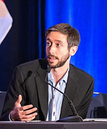 Yale S Michaels合作论文数Univ Oxford, Weatherall Inst Mol Med, Radcliffe Dept Med, Oxford OX3 9DS, England4
 Nima Khadem Mohtaram合作论文数4Charles Lau合作论文数3Elizabeth L. Castle合作论文数3Lauren J. Durland合作论文数3Tara Styan合作论文数3
 David J. H. F. Knapp合作论文数Terry Fox Laboratory, University of British Columbia3Tara R Stach合作论文数2Kevin Salim合作论文数2Tiam Heydari合作论文数Michael Smith Laboratories, University of British Columbia2
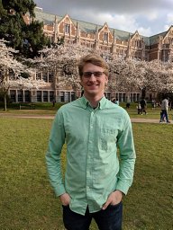 Ross D Jones合作论文数School of Biomedical Engineering, University of British Columbia2
 Laura Greenstreet合作论文数Department of Mathematics, University of British Columbia, 121-1984 Mathematics Road, Vancouver, BC V6T 1Z2, Canada.2
 Geoffrey Schiebinger合作论文数Institute of Applied Mathematics, Faculty of Science, The University of British Columbia2
 Fabio Rossi合作论文数School of Biomedical Engineering, Faculty of Applied Science, The University of British Columbia/School of Biomedical Engineering, Faculty of Medicine, The University of British Columbia2
 Megan Levings合作论文数Department of Surgery, Faculty of Medicine, University of British Columbia;School of Biomedical Engineering, Faculty of Applied Science, University of British Columbia;BC Children's Hospital Research Institute2
 Junghyuk Ko合作论文数2Aden Forrow合作论文数Mathematical Institute, University of Oxford2
 Roser Vento-Tormo合作论文数Wellcome Sanger Institute2Vilarrasa-Blasi Roser合作论文数2Montgomery Amy合作论文数2Ting Yin合作论文数2Jastaranpreet Singh合作论文数1
 Matthew Langley合作论文数1Jana K Gillies合作论文数1Laura N Stankiewicz合作论文数1Mona Siu合作论文数1nicole gabers合作论文数1Emma Bibault合作论文数1
 Andrew Hagner合作论文数1
 Shreya Shukla合作论文数Notch Therapeut1
 Alixandra Wong合作论文数Sch Med, Univ Queensland1
 Grace C Bingham合作论文数University of Virginia1David G Rattray合作论文数1
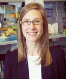 Elizabeth Rideout合作论文数Department of Cellular & Physiological Sciences, Faculty of Medicine, The University of British Columbia1
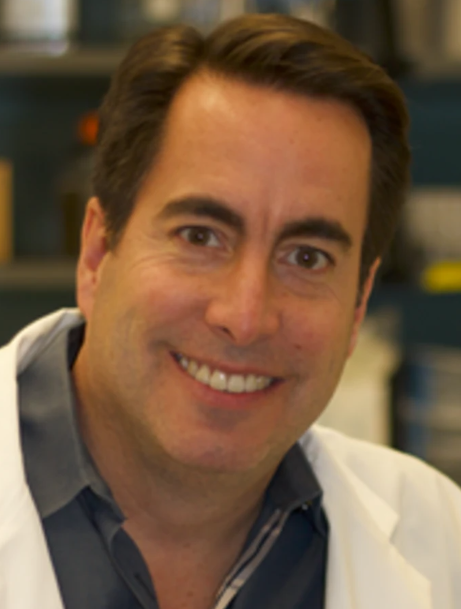 Juan Carlos Zuniga-Pflucker合作论文数Department of Immunology, Temerty Faculty of Medicine, University of Toronto;Sunnybrook Research Institute1
 King Craig合作论文数1
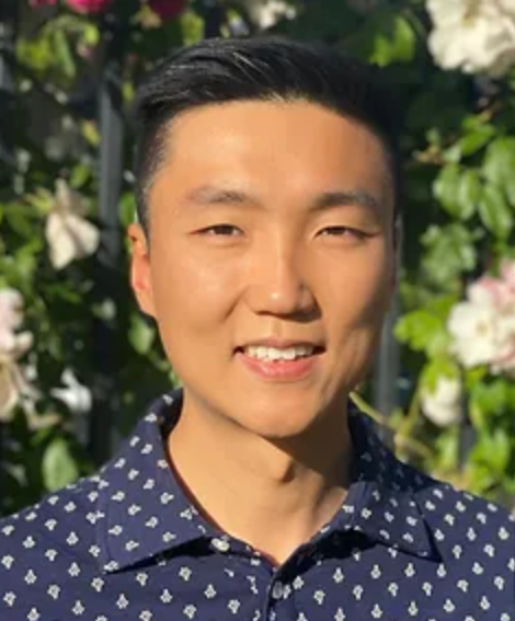 Yu Xin (Will) Wang合作论文数The Will Wang Lab, Sanford Burnham Prebys Medical Discovery Institute1
 Ryan A. Koppes合作论文数Rensselaer Polytechnic Institute1
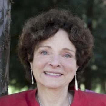 Helen M. Blau合作论文数Department of Psychiatry and Behavioral Sciences, School of Medicine, Stanford University;Baxter Laboratory for Stem Cell Biology, Stanford University1
 Martin B.G. Jun合作论文数Department of Mechanical and Industrial Engineering, University of Illinois at Urbana-Champaign1Mahmood Mohtashami合作论文数1




