Microvascular decompression (MVD) surgery is traditionally performed via a retrosigmoid craniotomy. Recently, neurosurgeons have increasingly adopted smaller approaches; however, the available evidence has not yet been systematically compiled. This is the first systematic review and meta-analysis on the efficacy and safety of ≤ 2-cm-keyhole retrosigmoid MVD. PubMed and Embase databases were searched for studies involving adults with trigeminal neuralgia (TN), hemifacial spasm (HFS), or glossopharyngeal neuralgia (GPN), who underwent first-time retrosigmoid keyhole MVD (diameter ≤ 2 cm). Primary outcomes were symptom relief and complications. Study quality was assessed using the Methodological Index for Non-Randomized Studies (MINORS). Pooled estimates were calculated using a random-effects single-arm meta-analysis. 32 publications met the inclusion criteria. 93.7
BACKGROUND AND OBJECTIVES:In endoscopic endonasal trans-sphenoidal pituitary surgery, the semisitting position (SSP) may reduce intraoperative bleeding compared with the supine position (SP); however, supporting evidence is lacking. The aim of this study was to compare the SSP with the SP and assess its effect on intraoperative bleeding. METHODS:This randomized, controlled, single-blind superiority trial was conducted at a Swiss tertiary hospital from 2021 to 2024. Of 107 patients screened, 64 were randomized and 56 included in the final analysis. In the intervention group, patients were placed in the SSP, with the torso elevated 30° and the legs raised, and compared with those in the SP. All patients were monitored for venous air embolism using a precordial Doppler. The primary outcome was intraoperative blood loss. Secondary outcomes included, among others, the frequency of interruptions for suctioning or hemostatic maneuvers, as well as the incidence of air embolism. RESULTS:Twenty-eight patients were allocated to each group. Most patients had a pituitary neuroendocrine tumor (25 [89.3%] in the SSP group and 27 [96.4%] in the SP group). The mean (SD) intraoperative blood loss was lower in the SSP group than in the SP group (184.7 [130.3] mL vs 277.3 [181.0] mL), corresponding to a mean reduction of 92.6 mL (33.4%) (95% CI, -177.1 to -8.1 mL; P = .03). Similarly, the median (IQR) frequency of intraoperative interruptions was lower in the SSP group than in the SP group (85.5 [62.3-109.0] vs 104.0 [77.5-135.5]), with a rate ratio of 0.80 (95% CI, 0.66-0.98; P = .03). Venous air embolism occurred in 3 patients (10.7%) in the SSP group and none in the SP group (P = .09); all events were hemodynamically insignificant and resolved spontaneously. CONCLUSION:The SSP in endoscopic endonasal pituitary surgery reduces intraoperative bleeding and may improve surgical workflow, without raising major safety concerns.
Abstract Background Accurate catheter positioning is essential for optimal outcomes in ventriculoperitoneal shunt (VPS) surgery, while shorter operative times lower infection risk and costs. Navigated VPS placement, using either intraoperative ultrasound-guided (US-G) or stereotactically guided (ST-G) navigation, enhances catheter accuracy and reduces revision rates. However, high-quality studies comparing the two navigation methods are lacking. We aim to compare surgical intervention time, accuracy, and safety of US-G to ST-G VPS placement. Methods The Navigated VPS (NAVPS) trial was an investigator-initiated, randomized trial conducted from February 2020 to June 2024 in the Neurosurgical Department of the University Hospital of Basel. Consecutive adults undergoing VPS placement were included. Out of 153 screened participants, 134 participants were included. Participants were randomized 1:1 to receive either US-G or ST-G insertion of the ventricular catheter. The primary outcome was surgical intervention time. An intention to treat analysis was performed calculating surgical intervention time differences. Secondary outcomes were accuracy of catheter positioning, number of ventricular puncture attempts, and VPS dysfunction and complication rates. The study follow-up lasted 6 months. Results Of 134 participants, 66 were assigned to US-G and 68 to ST-G. The mean (SD) age was 73 (55.3 to 78) and 66 (54.5 to 73) years for the US-G and ST-G, respectively, and 58 participants (45.7%) were female. The US-G group had significantly shorter surgical intervention times compared to the ST-G group (-11.5 min; 95% CI -18.5 to -4.5; P = 0.002). The number of ventricular puncture attempts was significantly higher in the US-G group, while accuracy of catheter placement, and VPS dysfunction and complications rates were comparable in both groups. Conclusions The NAVPS trial shows US-G VPS placement to be more time-efficient, while accuracy of catheter placement and complication rates seem to be comparable to ST-G placement. US-G can be efficiently and safely used in clinical practice. Trial registration clinicalTrials.gov Identifier: NCT04450797, date of registration: 22.06.2020.
OBJECTIVE Mastery of the posterior clinoidectomy technique is of utmost importance for neurosurgeons who specialize in endoscopic endonasal approaches, because the posterior clinoid process (PCP) is commonly involved in chondroid tumor resection. Three main techniques for posterior clinoidectomy have been developed: intradural, extradural, and transcavernous interdural. The authors introduce here a novel technical variant in which the transcavernous approach is extended to the dorsal clinoidal space after transection of the caroticoclinoid ligament, and they elaborate on its clinical application on the basis of anatomical dissections and radiological studies. METHODS The authors reviewed CT angiography images and 3D reconstruction of the PCP in 50 adults to analyze the height and presence of ossified ligament attachments. In addition, endoscopic endonasal posterior clinoidectomy was performed in 20 lightly embalmed postmortem human heads. Three techniques, including extradural, transcavernous, and extended transcavernous posterior clinoidectomy, were performed sequentially, and anatomical landmarks and areas exposed with each technique were investigated and compared. RESULTS Using radiological studies, the authors categorized the PCPs as 1 of 2 types: 1) normal, defined as less than or equal to 8 mm high with no ossified ligament attachments; or 2) complex, defined as greater than 8 mm high with or without an ossified ligament attachment. Compared with extradural (exposed PCP height 4.7 ± 0.5 mm) and transcavernous (exposed PCP height 7.3 ± 0.8 mm) posterior clinoidectomies, the extended transcavernous posterior clinoidectomy provided the maximally exposed PCP height (9.6 ± 0.4 mm; p < 0.0001). CONCLUSIONS This report details the extended transcavernous posterior clinoidectomy as a novel technical variant for achieving maximal exposure of the PCP in endoscopic endonasal surgery. In addition, the positive results establish the importance of preoperative skull base imaging for surgical planning.
BACKGROUND AND OBJECTIVES:The development of sonolucent cranial implants has led to the possibility of sonographic visualization of intracranial structures. The use of ultrasound instead of computed tomography imaging for ventricular monitoring during external ventricular drain (EVD) weaning has not yet been described. This study aims to describe the feasibility of bedside sonographic ventricular monitoring for EVD weaning after aneurysmal subarachnoid hemorrhage and to report our preliminary clinical experience. METHODS:A 24-mm sonolucent cranial implant was incorporated in the bone flap after clipping of a ruptured aneurysm. Baseline sonographic measurements of ventricular size were obtained with the EVD set at 10 cm. After raising the EVD to 15 cm for 24 hours, a second sonographic assessment was performed. If ventricular size, intracranial pressure and the examination remained stable, the EVD was clamped. A third sonographic assessment was conducted 24 hours after clamping. If ventricular size, intracranial pressure, and the examination remained stable, the EVD was removed. A fourth sonographic assessment was performed 24 hours after removal, with 1 final assessment before discharge of the patient. RESULTS:A total of 6 patients underwent the placement of an EVD, surgical clipping, and sonographic weaning of the EVD. Adequate sonographic visualization of the ventricles for EVD weaning was obtained in all 6 patients. Sonographic increase in ventricular size, accompanied by clinical deterioration, was observed in 3 patients, leading to the decision to place a ventriculoperitoneal shunt. There were no instances of missed ventricular enlargement nor postoperative infection or need for revision within the mean follow-up period of 6 weeks. CONCLUSION:Bedside sonographic ventricular monitoring through a sonolucent cranial implant represents a new and valid method for weaning of EVD after aneurysmal subarachnoid hemorrhage. Using ultrasound instead of computed tomography offers several advantages, including enhanced safety by avoiding intrahospital transport, reduced costs, and elimination of radiation exposure.
Superior cerebellar artery (SCA) aneurysms are rare. They represent 1%-2% of all intracranial aneurysms and are usually treated endovascularly.1 However, when the aneurysm configuration is not optimal for endovascular treatment, microsurgical clipping remains a valid alternative treatment option.2,3 We present the case of a 66-year-old male patient, with an incidental 7-mm wide-neck SCA aneurysm, with the parent vessel coming directly out of the aneurysm neck (Video 1). The different possible endovascular treatment options were reviewed and deemed not optimal for this specific aneurysm, potentially resulting either in incomplete aneurysm occlusion or vessel occlusion. Microsurgical clipping was considered the most suitable treatment option for this specific aneurysm. Surgery was facilitated by the high-riding localization of the aneurysm, waiving the need for a posterior clinoidectomy to obtain proximal control at the basilar artery. An orbitozygomatic approach was chosen to obtain an optimal caudocranial working angle and reduce the need for brain retraction. This video highlights the thought process in choosing the optimal treatment for this rare type of cerebral aneurysm, and the important surgical steps to safely approach and clip an SCA aneurysm.
IntroductionWhile endovascular treatment has gained popularity for its minimally invasive approach, microsurgical clipping for unruptured intracranial aneurysms (UIAs) has also evolved using surgical adjuncts such as virtual reality (VR) and keyhole techniques to enhance patient outcomes. Immersive 3D VR images allow for the creation of an accurate 3D anatomical model, making it a valuable tool for surgical planning. This study investigated the impact of VR-based surgical planning on approach type and craniotomy size.MethodsThis retrospective cohort study included all patients who underwent elective microsurgical clipping for UIAs from 1 January 2009 to 31 December 2024 at the University Hospital of Basel, Switzerland, and was approved by the local ethics board. Demographic, surgical, and outcome parameters were collected. SpectoMedical®, developed at the University of Basel, was the VR platform used. The primary outcome was craniotomy size (cm2) measured using the anterior-posterior method. Descriptive and comparative statistics were conducted. To assess the factors influencing the craniotomy size, we calculated a multivariable linear regression model.ResultsWe included a total of 163 aneurysms in 159 patients with a mean age of 58.52 (±10.23), and 114 (69.9%) were female. VR-based surgical planning resulted in a significantly smaller craniotomy size [no VR vs. VR, 20.31 (±19.21) cm2 vs. 13.22 (±7.85) cm2, p = 0.007] and shorter hospital stay [no VR vs. VR, 10.04 (±5.58) vs. 7.89 (±2.79) days, p = 0.031]. Operative time was shorter in the VR group but lacked statistical significance [no VR vs. VR, 226.41 (±86.18) min vs. 207.93 (±54.92) min, p = 0.160]. The multivariable regression model showed that the use of VR-based surgical planning reduced the craniotomy size by 6.2 cm2.ConclusionVR-based surgical planning was associated with significantly smaller craniotomy sizes and shorter hospital stays. It results in an intraoperative déjà-vu effect for the surgeon, which supports its use as a valuable adjunct in preoperative planning.
OBJECTIVE The modern management of patients with Koos grade IV vestibular schwannomas (VSs) aims at functional preservation and long-term tumor control. Gross-total resection (GTR) leads to optimal tumor control but frequently also results in permanent facial nerve (FN) palsy. Subtotal resection (STR) or near-total resection (NTR) followed by a waitand-scan protocol and second-line radiation therapy (RT) in case of progressive residuals yields excellent tumor control rates with less permanent morbidity. METHODS The authors present the results of their prospective cohort of Koos grade IV VS patients who underwent less-than-total resection followed by a wait-and-scan protocol between January 2009 and December 2019 and discuss the latest evidence on this controversial subject. The cohort was followed up with annual clinical and volumetric outcome analyses after standardized MRI. RESULTS Forty-eight patients were included in the analysis. The mean extent of resection was 87% (median 91%, range 45%-100%), best fitting into the definition of STR rather than NTR. In 2 cases, the proximal portion of the FN at the brainstem could not be reliably identified and monitored during the initial operation, and a second-stage resection was necessary. At 4.4 years after surgery, 81% (39/48) of the tumor residuals regressed or were stable in size. The percentage of regressive tumor residuals increased over time. Nineteen percent (9/48) of the tumor residuals displayed volumetric progression within a mean time of 35 months (median 36 months, range 14-72 months), resulting in a Kaplan-Meier estimate for progression-free survival of 79% after 4 years; higher postoperative volume showed a linear correlation with higher volumetric progression (factor 1.96, 95% CI 1.67-2.30; p < 0.001). Thirty-four of the 48 (71%) patients continue to undergo a wait-and-scan protocol. Second-line RT was performed in 14 patients (29%) within a mean time of 25 months (median 23 months, range 5-54 months), 12 (86%) of whom responded with post-RT pseudoprogression, resulting in an overall tumor control rate of 96%. At the 4.4-year follow-up from the initial resection, 92% of the patients had a good facial outcome (House-Brackmann [HB] grade I or II), 6% had a fair facial outcome (HB grade III), and 2% had a poor facial outcome (HB grades IV-VI). So far, there has been no need for salvage surgery after RT. CONCLUSIONS STR followed by observation and second-line RT in cases of progression leads to good facial outcome and an excellent tumor control rate in the longer term.
OBJECTIVE Despite its potential advantages, robotic surgery has yet to be applied to skull base procedures. Complex anatomy and restricted access have limited the development of robotic skull base surgery. The authors' aim was to conduct a feasibility study of robotic surgery for posterior fossa skull base lesions. METHODS Six silicone-injected postmortem human heads were prepared for the robotic surgery. Because there was no drilling tool with the robot, specimens were dissected in advance using an endoscope and microscope. The following approaches were investigated: 1) supracerebellar-infratentorial; 2) retromastoid; and 3) posterior occipitocervical junction surgeries. For each approach specific anatomical landmarks were identified, and the surgical freedom (vertical distance angle between the tools) was measured. RESULTS In the case of the supracerebellar-infratentorial approach, the authors used 3 burrs with 1.5 cm of diameter: 1 paramedian and 2 laterally. The view of the pineal region was visualized, and sufficient surgical freedom of both tools was secured. The median vertical distance was 1.2 cm (range 1.1-1.8 cm), and the median angle between the tools was 105 degrees (range 92 degrees-110 degrees). On the other hand, in the retromastoid approach, with a single burr 2.5 cm in diameter, the root exit zone of the facial nerve was barely visible, and a space for tools to access was not secured. The median vertical distance was 0.8 cm (range 0.6-1.0 cm), and the median angle between the tools was 10 degrees (range 6 degrees-12 degrees). In the case of the posterior occipitocervical junction approach, the authors used the 3 tubular retractors, 1 in the middle and 2 laterally. Even though the space was narrow, the medulla and adjacent nerves could be identified, and a moderate level of surgical freedom could be obtained for tool mobilization. The median vertical distance was 1.6 cm (range 1.2-2.5 cm), and the median angle between the tools was 90 degrees (range 88 degrees-95 degrees). CONCLUSIONS Although robotic surgery has yet to be applied to neurosurgery, it is expected to be helpful in posterior fossa skull base surgery if appropriate tools can be developed.
OBJECTIVE:Recurrent cerebrospinal fluid (CSF) rhinorrhea caused by sequential, anatomically separated skull base defects is rarely reported in the literature. Neither management nor etiology has been sufficiently investigated. We present an illustrative case and a systematic review of the literature regarding etiology, diagnostics, and management of this rare phenomenon. METHODS:A systematic literature search looking for articles reporting sequential CSF leaks with multiple skull base defects was performed. Data from included articles were descriptively reported, and the quality of the included studies was assessed with Grading of Recommendations Assessment, Development and Evaluation. RESULTS:A 71-year-old woman with posttraumatic CSF rhinorrhea and left-sided CSF otorrhea due to a left-sided horizontal fracture of the petrous bone presented at our institution. After initial surgical repair and a 10-week symptom-free interval, CSF rhinorrhea recurred. Imaging revealed a preexisting contralateral meningoencephalocele of the lateral sphenoid recess causing recurrent CSF rhinorrhea most likely after initial traumatic laceration. The defect was successfully treated. A literature search identified 366 reports, 6 of which were included in the systematic review with a total of 10 cases. Quality was deemed good in 8 of 10 cases. The most common location for primary and sequential CSF leaks was along the sphenoid bone (4/10 and 5/10 patients, respectively). All publications except one reported the presence of a meningo (encephalo)cele as cause of the sequential CSF leak. CONCLUSIONS:Occurrence of recurrent CSF rhinorrhea due to an anatomically separated sequential skull base lesion remains a rare phenomenon. Reassessment of imaging studies and a structured diagnostic workup to detect sequential CSF leaks independent of the primary lesion should is recommended.
OBJECTIVE:The mainstay of treatment for skull base chordoma (SBC) is maximal safe resection followed by radiotherapy. However, even after gross-total resection (GTR), the recurrence rate is high due to microscopic disease in the resection margins. Therefore, supramarginal resection (SMR) could be beneficial, as has been shown for sacral chordoma. The paradigm of postoperative radiation therapy for every patient has also begun to change, as molecular profiling has shown variability in the risk of recurrence. The aim of this study was to present the concept of SMR applied to SBC, along with an individualized decision for postoperative radiation therapy. METHODS:This is a retrospective analysis of all SBCs operated on by the senior author between 2018 and 2023. SMR was defined as negative histological margins of bone and/or dura mater, along with evidence of bone resection beyond the tumor margins in the craniocaudal and lateral planes on postoperative imaging. Tumors were classified into 3 molecular recurrence risk groups (group A, low risk; group B, intermediate risk; and group C, high risk). Postoperative radiation therapy was indicated in group C tumors, in group B chordomas without SMR, or in cases of patient preference. RESULTS:Twenty-two cases of SBC fulfilled the inclusion criteria. SMR was achieved in 12 (55%) cases, with a mean (range) amount of bone resection beyond the tumor margins of 10 (2-20) mm (+40%) in the craniocaudal axis and 6 (1-15) mm (+31%) in the lateral plane. GTR and near-total resection were each achieved in 5 (23%) cases. Three (19%) tumors were classified as group A, 12 (75%) as group B, and 1 (6%) as group C. Although nonsignificant due to the small sample size, the trends showed that patients in the SMR group had smaller tumor volumes (13.9 vs 19.6 cm3, p = 0.35), fewer previous treatments (33% vs 60% of patients, p = 0.39), and less use of postoperative radiotherapy (25% vs 60%, p = 0.19) compared to patients in the non-SMR group. There were no significant differences in postoperative CSF leak (0% vs 10%, p = 0.45), persistent cranial nerve palsy (8% vs 20%, p = 0.57), and tumor recurrence (8% vs 10%, p = 0.99; mean follow-up 15 months) rates between the SMR and non-SMR groups. CONCLUSIONS:In select cases, SMR of SBC appears to be feasible and safe. Larger cohorts and longer follow-up evaluations are necessary to explore the benefit of SMR and individualized postoperative radiation therapy on progression-free survival.
In neuroendoscopy, an assistant surgeon holds the endoscope while the operating surgeon performs brain surgery using surgical instruments in both hands. The assistant surgeon’s task can be strenuous over time, especially in long surgeries, and unsteadiness or tremor might affect the visualization quality that the operating surgeon depends upon. Existing mechanical and pneumatic arms offer limited flexibility. We propose a robotic assistant as a third hand to the surgeon. It holds the endoscope and can be moved freely or held in place. As a proof of concept, we attached a neuroendoscope to an offthe- shelf robot with a custom handle, including a force/torque (F/T) sensor. We qualitatively identified the requirements for the third hand with a surgeon as a participant. We also quantitatively identified the range of forces applied by the endoscope to act as a retractor on two brain phantoms while visualizing the surgical site. With our proof of concept study, we could show the feasibility of robotic assistance in neuroendoscopy. Based on our observations, we found that an intuitive input device to switch the different robot modes, a second F/T sensor to measure the surgeon’s input and tissue interaction separately, and a differently shaped precision grip handle represent promising improvements to our third hand prototype.
Background/Objectives: Detailed morphometric analysis of an aneurysm and the related vascular bifurcation are critical factors when determining rupture risk and planning treatment for unruptured intracranial aneurysms (UIAs). The standard visualization of digital subtraction angiography (DSA) and its 3D reconstruction on a 2D monitor provide precise measurements but are subject to variability based on the rater. Visualization using virtual (VR) and augmented reality platforms can overcome those limitations. It is, however, unclear whether accurate measurements of the aneurysm and adjacent arterial branches can be obtained on VR models. This study aimed to assess interrater reliability and compare measurements between 3D VR, standard 2D DSA, and 3D DSA reconstructions, evaluating the reliability and accuracy of 3D VR as a measurement tool. Methods: A pool of five neurosurgeons performed three individual analyses on each of the ten UIA cases, measuring them in completely immersed 3D VR and the standard on-screen format (2D DSA and 3D reconstruction). This resulted in three independent measurements per modality for each case. Interrater reliability of measurements and morphology characterization, comparative differences, measurement duration, and VR user experience were assessed. Results: Interrater reliability for 3D VR measurements was significantly higher than for 3D DSA measurements (3D VR mean intraclass correlation coefficient [ICC]: 0.69 ± 0.22 vs. 3D DSA mean ICC: 0.36 ± 0.37, p = 0.042). No significant difference was observed between 3D VR and 2D DSA (3D VR mean ICC: 0.69 ± 0.22 vs. 2D DSA mean ICC: 0.43 ± 0.31, p = 0.12). A linear mixed-effects model showed no effect of 3D VR and 3D DSA (95% CI = −0.26–0.28, p = 0.96) or 3D VR and 2D DSA (95% CI = −0.02–0.53, p = 0.066) on absolute measurements of the aneurysm in the anteroposterior, mediolateral, and craniocaudal dimensions. Conclusions: 3D VR technology allows for reproducible, accurate, and reliable measurements comparable to measurements performed on a 2D screen. It may also potentially improve precision for measurements of non-planar aneurysm dimensions.
OBJECTIVE Tumors located in the retrochiasmatic region with extension to the third ventricle might be difficult to access when the pituitary-chiasmatic corridor is narrow. Similarly, tumor extension into the interpeduncular and retrosellar space poses a major surgical challenge. Pituitary transposition techniques have been developed to gain additional access. However, when preoperative pituitary function is already impaired or the risk of postoperative panhypopituitarism (PH) is considered to be particularly high, removal of the pituitary gland (PG) might be the preferred option to increase the working corridor. The aim of this study was to describe the relevant surgical anatomy, operative steps, and clinical experience with the endoscopic endonasal pituitary sacrifice (EEPS) and transsellar approach. METHODS This study comprised anatomical dissections to highlight the relevant surgical steps and a retrospective case series reporting clinical characteristics, indications, and outcomes of patients who underwent EEPS. The surgical technique is as follows: both lateral opticocarotid recesses are exposed laterally, the limbus sphenoidale superiorly, and the sellar floor inferiorly. After opening the dura, the PG is detached circumferentially and mobilized off the medial walls of the cavernous sinuses. The descending branches of the superior hypophyseal artery are coagulated, and the stalk is transected. After removal of the PG, drilling of the dorsum sellae and bilateral posterior clinoidectomies are performed to gain access to the hypothalamic region, interpeduncular, and prepontine cisterns. RESULTS From 2018 to 2023, 11 patients underwent EEPS. The cohort comprised mostly tuberoinfundibular craniopharyngiomas (n = 8, 73%). Seven (64%) patients had partial or complete anterior PG dysfunction preoperatively, while 4 (36%) had preoperative diabetes insipidus. Because of the specific tumor configuration, the chance of preserving endocrine function was estimated to be very low in patients with intact function. The main reasons for pituitary sacrifice were impaired visibility and surgical accessibility to the retrochiasmatic and retrosellar spaces. Gross-total tumor resection was achieved in 10 (91%) patients and near-total resection in 1 (9%) patient. Two (18%) patients experienced a postoperative CSF leak, requiring surgical revision. CONCLUSIONS When preoperative pituitary function is already impaired or the risk for postoperative PH is considered particularly high, the EEPS and transsellar approach appears to be a feasible surgical option to improve visibility and accessibility to the retrochiasmatic hypothalamic and retrosellar spaces, thus increasing tumor resectability.
Arachnoid cysts of the sellar region are rare.(1-3 )When surgery is needed, it is usually performed through the endoscopic endonasal approach.(4-6 )However, some features of the cyst may suggest a transcranial route as a better alternative. We presented the case of a 28-year-old woman with a sellar and suprasellar arachnoid cyst, which displaced the pituitary gland and the optic chiasm cranially. By going through an endoscopic endonasal approach, the cyst could be easily opened; however, fenestration into the basal cisterns would be fairly limited and therefore increasing the risk of recurrence. Furthermore, pituitary function and cranial nerve integrity would be at risk. By going through a transcranial route, we circumvented these problems and were able to achieve wide fenestration into multiple basal cisterns.In7-9 this video, we discussed how the goal of surgery, the particular characteristics of the lesion, and anatomic features of the patient should dictate the surgical approach. The patient consented to the procedure and to the publication of her images.
BACKGROUND H3 K27-altered diffuse midline gliomas (DMGs) are rare tumors, which are, regardless of their histological appearance, classified as World Health Organization grade 4 tumors. They are characterized by a diffuse growth pattern, midline anatomical location, and poor prognosis. Although DMGs occur predominantly in childhood, these tumors can also be found in young adults. OBSERVATIONS The authors present a case of a 29-year-old patient who was found unconscious with a Glasgow Coma Scale score of 4, along with abnormal extensor movements and bilateral middilated nonreactive pupils. Computed tomography revealed obstructive hydrocephalus due to an acute hemorrhage in a right thalamic lesion. To drain the hydrocephalus and relieve the ongoing central herniation, emergent placement of a right-sided, and later a left-sided, extraventricular drain was performed. Despite the postoperative resolution of hydrocephalus, the patient died shortly after because of the central brain herniation that had occurred. Brain autopsy revealed a H3 K27-altered DMG in the right thalamus. LESSONS Although typically described in the pediatric population and located in the pons, H3 K27-altered DMG should also be considered in young adult patients with midline lesions, particularly if they are located in the thalamus or brainstem. In rare cases, H3 K27-altered DMG may present with an acute tumor-related hemorrhage, leading to a fulminant clinical course.
Thyroid-stimulating hormone–secreting adenomas (TSH-oma) are exceptionally rare. 1 The primary treatment is surgical resection with radiation and pharmacotherapy postoperatively if subtotal resection, especially with cavernous sinus invasion. 2 We present the case of a 29-year-old man with TSH-oma with cavernous sinus medial wall invasion. This is the first documented case with selective resection of the cavernous sinus medial wall to achieve a complete resection and biochemical remission in TSH-oma through endoscopic endonasal approach. The patient had elevated TSH and thyroid hormones with symptoms of weight loss, palpitations, excess sweating, and decreased endurance. MRI revealed a 1.3 × 2.1 × 1.2 cm contrast-enhancing sellar mass with rightward pituitary gland displacement without evidence of cavernous sinus invasion (Knosp 2). The patient consented to procedure/publication. No institutional review board approval needed per institution. We performed standard resection of the firm sellar tumor portion and noted that there was tumor invasion into the left cavernous sinus medial wall dura. The bony opening was expanded to expose the anterior wall of the cavernous sinus, which was opened to identify the cavernous internal carotid artery and the medial wall attachments. The thickened medial wall was completely resected. We achieved a complete tumor resection, and the patient's TSH and thyroid hormone dropped to a desired threshold. 3 Tumor stained for GATA3 and PIT1, characterizing the TSH-oma. 4,5 Understanding cavernous sinus vascular and ligamentous anatomy allows for safe separation of invaded medial wall dura from the cavernous internal carotid artery, 6 allowing for a more complete tumor resection, improving surgical cure rates, and sparing the patient from future radiation and pharmacotherapy.
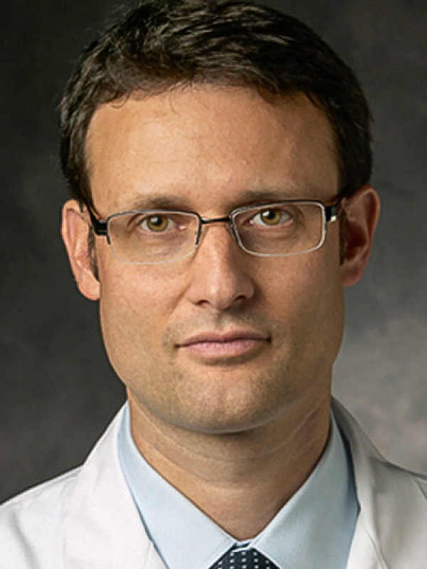 Raphael Guzman合作论文数Department of Neurosurgery, University Hospital Basel;Department of Biomedicine, Medizinische Fakultät, University of Basel25
 Michel Roethlisberger合作论文数18
 Jehuda Soleman合作论文数14
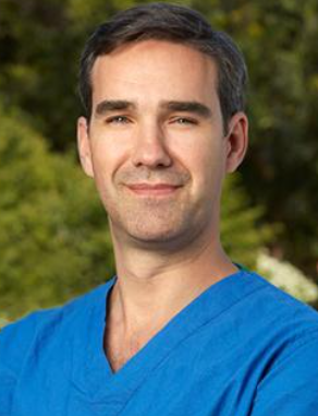 Juan Carlos Fernandez-Miranda合作论文数Department of Neurosurgery, Stanford University School of Medicine;Skull Base and Pituitary Surgery Division, Stanford University School of Medicine11
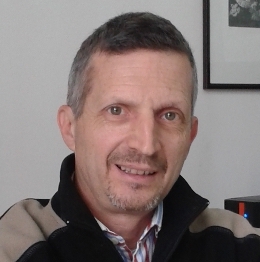 Luigi Mariani合作论文数Istituto Nazionale Tumori9
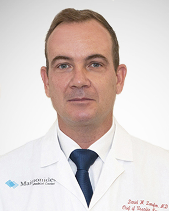 Daniel W. Zumofen合作论文数Maimonides Medical Center9
 Attill Saemann合作论文数8
 Yuanzhi Xu合作论文数7
 Taub Ethan合作论文数7Christine K. Lee合作论文数6
 Alessio Chiappini合作论文数Department of Neurosurgery, University Hospital Basel6
 Felipe Constanzo合作论文数Department of Neurological Surgery, Hospital Clinico Regional de Concepcion6
 Vera Vigo合作论文数6
 Yves Brand合作论文数6Vladimir A. Ljubimov合作论文数5Mariano Rinaldi合作论文数5
 Limin Xiao合作论文数5
 Min Ho Lee合作论文数5
 Marios Psychogios合作论文数Clin Radiol & Nucl Med, Univ Hosp Basel4
 David Bervini合作论文数4
 Howard A. Riina合作论文数Department of Neurology, Grossman School of Medicine, New York University;Department of Radiology, Grossman School of Medicine, New York University;Langone Health Center for Stroke and Neurovascular Diseases, New York University4Enrico Gambatesa合作论文数3Alix Bex合作论文数3Żelechowski Marek合作论文数3
 Florian Ebel合作论文数3Daniel Bodmer合作论文数Clin Otolaryngol Head & Neck Surg, Univ Hosp Basel3
 Erez Nossek合作论文数Department of Neurosurgery, Grossman School of Medicine, New York University3Croci Davide合作论文数3Birgit Westermann合作论文数3
 Christian Zweifel合作论文数3
 Gregor Hutter合作论文数Institute of Neuropathology, University Hospital Zurich3
 Cristina Granziera合作论文数Department of Biomedical Engineering, University of Basel3
 Philippe Claude Cattin合作论文数Department of Biomedical Engineering, University of Basel;Medical Image Analysis Center, Medical Faculty, University of Basel3Tim Hallenberger合作论文数2Elisabeth Pössnecker合作论文数2Jannis Müller合作论文数2
 Johanna Lieb合作论文数2Laila Kulsvehagen合作论文数2Ladina Greuter合作论文数2
 Manina Maja Etter合作论文数2Noëmi Eberhard合作论文数2Maximiliano Alberto Nunez合作论文数2
 Sabrina Hogan合作论文数2
 Tomas A. Martins合作论文数Brain Tumor Immunotherapy Lab, University of Basel2Donato D'Alonzo合作论文数2
 Johannes Goldberg合作论文数2
 Karoline Leuzinger合作论文数2Florian Halbeisen合作论文数Basel Institute for Clinical Epidemiology and Biostatistics, University of Basel2
 Ivan Radovanovic合作论文数Department of Surgery, Temerty Faculty of Medicine, University of Toronto2




