Pulsed field ablation (PFA) has proven to be a safe and effective non-thermal ablation modality for the treatment of atrial fibrillation (AF), but little outcome data beyond 1 year have been reported. Here we present results from the ADVENT-LTO study, which provides extended follow-up of the ADVENT trial, the first randomized trial comparing PFA with conventional thermal ablation. In ADVENT-LTO, 364 patients with paroxysmal AF (183 PFA, 181 thermal; 237 men, 127 women) participated and were followed for 1,332 ± 147 days. For the primary endpoint of 4-year treatment success, PFA demonstrated preserved effectiveness compared to thermal ablation (72.8% PFA, 64.3% thermal; P = 0.12). Moreover, there was a trend favoring PFA as compared to thermal ablation for the prespecified outcome of freedom from hospital-based arrhythmia intervention (85.6% PFA, 78.6% thermal; hazard ratio (HR) = 0.64, 95% confidence interval (CI): 0.38-1.05), including fewer repeat ablations (10.4% PFA, 17.7% thermal; P = 0.04) as well as a trend favoring PFA as compared to thermal ablation for the prespecified outcome of progression to persistent AF (2.6% PFA, 4.6% thermal; HR = 0.55, 95% CI: 0.16-1.88). Taken together, these data demonstrate that the favorable outcomes of PFA are maintained over the course of 4 years. Coupled with the safety advantages of PFA over thermal ablation, these long-term data support widespread adoption of PFA for the treatment of AF. ClinicalTrials.gov registration: NCT06526546 .
BACKGROUND:Pre-participation screening (PPS) in competitive athletes aims to identify cardiovascular diseases associated with sudden cardiac death (SCD). Although the 12‑lead electrocardiogram (ECG) represents the cornerstone of PPS, structural abnormalities may demonstrate limited or incomplete electrical expression, particularly in asymptomatic athletes with physiological remodeling. Artificial intelligence (AI)-enabled ECG models have shown promising performance in hospital-based populations, but their transportability to low-prevalence athlete screening environments remains uncertain. OBJECTIVES:To develop and externally validate a deep learning (DL)-based AI-ECG ensemble model for detecting imaging-confirmed structural heart disease in competitive athletes undergoing PPS. METHODS:A convolutional neural network (CNN) ensemble was trained using hospital-derived ECG images from Beth Israel Deaconess Medical Center (BIDMC, Boston, USA) and externally validated in the Italian Team for Athlete CARDiac evaluation and AI-based Risk prediction (ITACARD-AI) registry. Separate CNNs were developed for valvular heart disease (VHD) and cardiomyopathies (CM) and combined using XGBoost meta-learning. Model performance was assessed using area under the receiver operating characteristic curve (AUROC), subgroup analyses, and threshold-based evaluation. RESULTS:The ITACARD-AI cohort included 1115 competitive athletes (mean age 26 ± 13 years; 70% male), including 48 athletes (4.3%) with VHD and 30 (2.7%) with CM. External validation demonstrated substantial performance degradation compared with hospital-based internal validation. AUROC values decreased to 0.70 (95% CI 0.64-0.75) for VHD and 0.69 (95% CI 0.60-0.78) for CM, indicating only modest discrimination in the screening population. Threshold analyses showed high negative predictive values (∼99%) but persistently low positive predictive values (≤8%), reflecting limited disease enrichment and strong prevalence dependence. CONCLUSIONS:Hospital-trained AI-ECG models demonstrated limited transportability to real-world athlete screening populations. The marked reduction in external performance suggests that low disease prevalence, heterogeneous physiological remodeling, and incomplete ECG expression of structural abnormalities may substantially limit class separability in PPS environments. Although AI-ECG may provide cautious adjunctive support within physician-led workflows, these findings highlight the intrinsic challenges of applying ECG-based AI to low-prevalence sports cardiology screening populations.
BACKGROUND:ECG-based artificial intelligence may enable efficient prediction of incident heart failure (HF) risk to facilitate preventive efforts. Prior models are proprietary, with modest or inconsistent accuracy. We sought to develop and validate a generalizable and publicly available convolutional neural network to predict incident HF using the 12-lead ECG waveform (ECG-to-HF [ECG2HF]). METHODS:We developed ECG2HF in 94 636 patients receiving longitudinal ambulatory care at Massachusetts General Hospital (MGH), and validated it in 3 test sets: MGH, Brigham and Women's Hospital (BWH), and Beth Israel Deaconess Medical Center (BIDMC), among 93 868 individuals aged 30 to 79 years without HF. HF events at 10 years were identified using a validated electronic health record-based natural language processing model. Discrimination was quantified using the area under the receiver operating characteristic curve. We then compared discrimination and net reclassification (at <10%, 10% to 20%, ≥20% 10-year risk categories) using ECG2HF versus the 15-component Pooled Cohorts Equations to Prevent HF score. RESULTS:The test sets comprised MGH (13 954 individuals, 441 events, age 57±13 years, 48% women), BWH (54 396 individuals, 1809 events, age 57±13 years, 55% women), and BIDMC (25 457 individuals, 901 events, age 57±13 years, 53% women). Over 10 years, the cumulative risk of HF was 4.6% (95% CI, 4.1-5.0) in MGH, 5.0% (4.8-5.2) in BWH, and 4.4% (4.1-4.7) in BIDMC. ECG2HF discriminated 10-year incident HF in each test set (area under the receiver operating characteristic curve: MGH 0.86 [0.84-0.87]; BWH 0.85 [0.84-0.86]; BIDMC 0.84 [0.83-0.86]). Compared with the Pooled Cohorts Equations to Prevent HF, ECG2HF provided favorable discrimination (improvement in area under the receiver operating characteristic curve MGH/BWH 0.061 [0.025-0.097]; BIDMC 0.038 [-0.0096 to 0.086]) and net reclassification (NRI MGH/BWH 0.16 [0.077-0.24]; BIDMC 0.23 [0.10-0.35]) of 10-year HF risk. CONCLUSIONS:ECG2HF is a publicly available 12-lead ECG-based artificial intelligence model that discriminates the risk of future HF with favorable and consistent performance across 3 large health care samples from the northeastern United States. ECG2HF may enable efficient prioritization of high-risk individuals for HF-related preventive measures.
BACKGROUND:Pulsed field ablation (PFA) of atrial fibrillation has been rapidly adopted, partly because of safety expectations compared with thermal ablation. Comparative safety data between the 2 modalities remain limited. METHODS:We conducted a prospective registry analyzing consecutive atrial fibrillation ablations at a high-volume US academic center between 2022 and 2026. The primary end point was stroke or transient ischemic attack (TIA) within 30 days, independently adjudicated by blinded neurologists. Secondary end points included death and other procedural complications. Propensity score methods with inverse probability of treatment weighting were used to balance differences in patient characteristics. Differences in the procedural workflow of PFA versus radiofrequency ablation (RFA) were evaluated with exploratory mediation analyses. RESULTS:A total of 4221 ablation procedures (2077 RFA and 2144 PFA: 68.7% Farawave, 23.3% Sphere 9, 6.9% Varipulse, 0.7% PulseSelect) was performed by 12 operators. Patients receiving PFA and RFA had similar baseline characteristics (mean age, 67 years; 31% female; 47% persistent atrial fibrillation; 7% previous stroke/TIA), which were balanced after inverse probability of treatment weighting. Compared with RFA, PFA procedures were shorter (108 versus 144 minutes) and included more frequent posterior wall isolation (57% versus 31%). The 30-day rate of stroke/TIA was significantly higher for PFA cases (10 events; 0.47%) compared with RFA (2 events; 0.10%) in both unweighted and propensity score-weighted analyses (weighted risk difference, 0.36% [95% CI, 0.03%-0.70%]; P=0.03). Stroke/TIA events were evenly distributed throughout the study period with no clustering around the time of PFA adoption or operators (with no operator associated with ≥2 events). Exploratory mediation models were limited by the low event rates and did not identify more extensive ablation as an independent risk factor for stroke. Weighted rates of other procedural complications were low (<1%) and similar between PFA and RFA groups. CONCLUSIONS:In a high-volume, single-center registry with high clinical granularity and systematic patient follow-up, PFA was associated with a significantly higher risk of stroke/TIA at 30 days compared with RFA. These results call for enhanced postmarket surveillance and dedicated prospective evaluation as the PFA procedural volume continues to exponentially increase.
BACKGROUND AND AIMS:Pulsed field ablation (PFA) has emerged as a non-thermal alternative for pulmonary vein isolation (PVI), offering shorter procedural times and a favorable safety profile for atrial fibrillation (AF) ablation. However, large-scale data evaluating its efficacy in patients with persistent AF remain limited. Our objective was to perform a systematic review and meta-analysis evaluating 12-month atrial arrhythmia recurrence following PFA in patients with persistent AF. METHODS:A systematic search of MEDLINE, Embase, Scopus, LILACS, and Cochrane databases was performed. Studies including patients with persistent AF undergoing first-time PVI with PFA and reporting 12-month arrhythmia-free survival were included. When available, outcomes were compared with thermal ablation using time-to-event analyses. Random-effects models were used. Exploratory meta-regression analyses were conducted to assess potential sources of heterogeneity. RESULTS:Among 1699 screened studies, 26 met the inclusion criteria, comprising 3744 patients with persistent AF treated with PFA. The pooled 12-month freedom from atrial arrhythmia recurrence was 72.3% (95% CI, 69.0-75.5), with substantial heterogeneity (I² = 74.4%). Eight studies (31%) included a thermal comparator; pooled time-to-event analysis demonstrated no significant difference between PFA and thermal ablation (HR, 0.91 [95% CI, 0.78-1.07]). Exploratory meta-regression did not identify significant effect modification by baseline characteristics, lesion-set strategy, rhythm monitoring intensity, or year of publication. Ablation-related adverse events occurred in < 1% of cases for both energy sources. CONCLUSION:In patients with persistent AF, PFA is associated with a high arrhythmia-free survival at 12 months and low rates of ablation-related adverse events. Comparative findings versus thermal ablation should be considered exploratory and interpreted cautiously, given the limited and heterogeneous comparator data.
BACKGROUND:Conduction disturbances requiring permanent pacing frequently complicate transcatheter aortic valve replacement (TAVR). The understanding of mechanisms causing conduction block is incomplete. OBJECTIVE:This study aimed to characterize the acute and delayed electrophysiological (EP) effects of TAVR on the atrioventricular (AV) conduction system. METHODS:We conducted a single-center prospective cohort study of 409 patients undergoing TAVR. All patients underwent 12-lead electrocardiography and EP study (EPS) immediately before and after valve implantation, with continuous electrocardiography and EP monitoring during the TAVR. 7 patients with AV block underwent repeat EPS 1-12 days after TAVR. RESULTS:TAVR was associated with significant prolongation of sinus cycle length, atrio-His and His-ventricular (HV) intervals, and Wenckebach cycle length (all P < .0001). Transient AV-nodal conduction block occurred in 8% of patients with intraprocedural AV block and 12% with postprocedural block. Infranodal block occurred in 57 patients, but resolved in 41 by the end of the procedure. Marked HV interval prolongation (>100 ms) without conduction block occurred in 6.4% of patients during TAVR. Intra-His Wenckebach-type block occurred in 9 patients. In 7 patients with follow-up EPS 1-12 days after TAVR, improvement or resolution of AV-nodal and infranodal conduction abnormalities was seen in all. CONCLUSION:Peri-TAVR conduction disturbances may involve both the AV node and His bundle. The usual parameters indicating need for permanent pacing (marked HV prolongation) do not apply to TAVR patients. Distinguishing nodal from infranodal involvement can inform prognosis, anticipated recovery, and pacemaker decision making, particularly given that many conduction abnormalities resolved within 2-4 weeks, supporting a refined post-TAVR risk stratification strategy. CLINICALTRIALS: GOV IDENTIFIER:NCT04982406.
BACKGROUND:Early recurrence of atrial tachyarrhythmias (ERAT) is common after pulmonary vein isolation (PVI) and is traditionally attributed to transient post-procedural inflammation. With the introduction of pulsed field ablation (PFA), the incidence and prognostic significance of ERAT compared with thermal ablation remains unknown. OBJECTIVE:The study aimed to compare the incidence of ERAT during the blanking period in patients undergoing PFA versus radiofrequency (RF) ablation for atrial fibrillation (AF). METHODS:We prospectively enrolled patients undergoing first-time PVI between 2022 and 2025 at our institution. Propensity score matching was performed to address baseline imbalances and group size differences between RF and PFA PVI. The incidence of ERAT and its association with 9-month arrhythmia recurrence, defined as late recurrence of atrial tachyarrhythmias (LRAT), were analyzed. RESULTS:A total of 962 patients were included (420 RF, 542 PFA). Patients with ERAT were more likely to be older, have persistent AF, higher CHA2DS2-VASc scores, larger atria, reduced ejection fraction, and no prior antiarrhythmic drug use. After multivariable adjustment, PFA was independently associated with a lower risk of ERAT compared with RF (hazard ratio [HR] 0.61; 95% confidence interval 0.42-0.88). ERAT was strongly associated with LRAT, with a 3-4-fold higher risk of atrial arrhythmias at 9 months (early ERAT: HR 3.78, P < .001; late ERAT: HR 4.10, P = .001), regardless of ablation modality. CONCLUSION:PFA is associated with a significantly lower risk of ERAT compared with RF ablation. The occurrence of ERAT, irrespective of energy source, predicts a substantially higher risk of LRAT.
Electrocardiography (ECG) is central to cardiovascular care, but conventional AI models are often restricted to common arrhythmias and may generalize poorly across populations or clinically subtle diseases. We developed ECG Contrastive Language-Image Pre-training (ECGCLIP), a signal-language contrastive learning framework that aligns ECG waveforms with expert diagnostic reports. ECGCLIP was pre-trained on 2,837,962 ECG studies from 1,324,856 patients and evaluated on a held-out internal test set plus nine independent external cohorts comprising about 1.5 million ECGs. Evaluation covered 89 downstream tasks, including 45 ECG diagnoses, 39 echocardiographic targets, and 5 rare cardiac diseases, using PRAUC as the primary metric. ECGCLIP consistently improved performance over random initialization and Merl-R18 baselines. On the internal test set, ECGCLIP-R34 achieved strong performance for atrial fibrillation (PRAUC 0.900) and ST-segment elevation myocardial infarction (PRAUC 0.383), with robust generalization across all external cohorts. It also improved low-prevalence and diagnostically elusive diseases, including Ebstein anomaly, constrictive pericarditis, dextrocardia, and cardiac amyloidosis, with internal PRAUC values of 0.253, 0.175, 0.121, and 0.201, respectively. ECGCLIP was data efficient, matching or exceeding full-dataset baseline performance with only 10
INTRODUCTION:Use of high-frequency low-tidal volume (HFLTV) and high-frequency jet ventilation (HFJV) during pulmonary vein isolation (PVI) improves acute procedural success and long-term outcomes compared to conventional ventilation. However, the impact of HFLTV compared to HFJV on acute and long-term outcomes is unknown. METHODS:We prospectively identified 1039 patients who underwent first-time PVI or PVI with posterior wall isolation (PWI) at our institution between 2022 and 2024 with HFLTV or HFJV. Acute procedural and safety outcomes were analyzed. Twelve-month arrhythmia-free survival was evaluated using the Kaplan-Meier and Cox proportional hazards method. RESULTS:After excluding 44 patients who required discontinuation of HFJV, 860 patients receiving HFJV were compared with 179 receiving HFLTV ventilation. Mean age was 65 years, 93% were White, 30% female and 53% had paroxysmal AF. First-pass isolation (FPI) rates were similar between ventilation strategies. HFLTV was associated with a higher rate of intraprocedural hypotension (31% vs 23%, p = 0.02). Complications were generally minor and comparable between groups. Arrhythmia recurrence rates at 12 months were similar (HR: 0.89, p = 0.55). There was a trend toward improved outcomes when FPI was achieved for both pulmonary veins in either group. CONCLUSION:In this observational cohort, no significant differences in acute procedural outcomes, safety outcomes, or long-term arrhythmia recurrence were observed between HFLTV and HFJV. Given the specialized equipment and operator training required for HFJV and its non-negligible rate of discontinuation, HFLTV may offer a modest practical advantage in routine clinical practice.
Contrastive learning is a widely adopted self-supervised pretraining strategy, yet its dependence on cohort composition remains underexplored. We present Contrasting by Augmented Patient Electrocardiograms (CAPE) foundation model and pretrain on four cohorts (n = 5,203,269), from diverse populations across three continents (North America, South America, Asia). We systematically assess how cohort demographics, health status, and population diversity influence the downstream performance for prediction tasks also including two additional cohorts from another continent (Europe). We find that downstream performance depends on the distributional properties of the pretraining cohort, including demographics and health status. Moreover, while pretraining with a multi-centre, demographically diverse cohort improves in-distribution accuracy, it reduces out-of-distribution (OOD) generalisation of our contrastive approach by encoding cohort-specific artifacts. To address this, we propose the In-Distribution Batch (IDB) strategy, which preserves intra-cohort consistency during pretraining, discourages learning of spurious cohort-specific features, and instead promotes clinically meaningful variability within cohorts. This leads to improved out-of-distribution robustness, with gains of 9-40% in downstream label prediction performance. This work provides insights into pretraining strategies for more clinically deployable and generalisable foundation models.
Conduction System Pacing (CSP), particularly Left Bundle Branch Area Pacing (LBBAP), has emerged as a physiological alternative to conventional ventricular pacing. However, optimal lead placement remains challenging. In this study, we investigated whether intraprocedural transesophageal echocardiography (TEE) can facilitate LBBAP implantation and improve procedural efficiency, lead positioning, and electrical outcomes compared with the conventional fluoroscopy-guided approach. In this single-center, retrospective, non-randomized cohort study, we evaluated 405 consecutive patients undergoing LBBAP between January 2018 and March 2025. The study compared a conventional fluoroscopy-only approach in the initial 198 patients and a TEE-guided approach in the subsequent 207 patients. Primary endpoints were categorized as measures of procedural efficiency assessed by total procedure time and fluoroscopy time and electrical performance evaluated by post-procedural QRS duration, R-wave peak time in lead V6, and final lead impedance. The TEE-guided group demonstrated significantly shorter total procedure and fluoroscopy times (65.5 ± 25.2 vs. 89.9 ± 37.9 min; p < 0.001/ 12.8 ± 9.9 vs. 15.8 ± 10.5 min; p < 0.001). Post-procedural QRS duration was narrower in the TEE group (108 ± 16 vs. 116 ± 15 ms; p < 0.001), with a more pronounced reduction in patients with baseline wide QRS (> 140 ms) (ΔQRS: −54 ± 23 vs. −46 ± 21 ms; p < 0.001). Notably, lead impedance was significantly lower with TEE guidance (523.6 ± 115.6 vs. 611.3 ± 155.9 Ω; p < 0.001), while pacing thresholds and R-wave amplitudes were similar. Complication rates were low and comparable. TEE-guided LBBAP implantation is associated with enhanced procedural efficiency and adequate cardiac electrical resynchronization. Incorporation of TEE into CSP implantation protocols warrants further evaluation in randomized trials. Transesophageal echocardiography versus fluoroscopy-guided left bundle branch pacing for pacemaker implant. Representative fluoroscopic and TEE images illustrate left bundle branch pacing with conventional fluoroscopy-only versus adjunctive TEE guidance. TEE allows direct visualization of the interventricular septum and electrode tip, facilitating accurate lead positioning. Quantitative comparisons demonstrate that TEE-guided implantation was associated with significantly shorter fluoroscopy time, comparable or reduced procedure duration, and improved electrical outcomes, including narrower QRS duration and shorter V6 R-wave peak time. RA = right atrium; RV = right ventricle; LA = left atrium; LV = left ventricle; MV = mitral valve; MS = membranous septum; IVS = interventricular septum; TEE = transesophageal echocardiography; LBBP = left bundle branch pacing; RWPT = R-wave peak time
Aims:A significant proportion of type 2 diabetes cases remain undiagnosed despite screening advances, carrying substantial cardiometabolic risk. Artificial intelligence-enhanced electrocardiography (AI-ECG) detects subtle ECG changes in subclinical disease, potentially enabling opportunistic screening. Methods and results:We developed AI-ECG Risk Estimator for Diabetes Mellitus (AIRE-DM), a convolutional neural network with discrete-time survival loss, for diagnosis of prevalent and prediction of incident type 2 diabetes. It was trained on 1 163 401 ECGs from 189 537 individuals from Beth Israel Deaconess Medical Center (BIDMC) and externally validated in UK Biobank (UKB; n = 65 606) and ELSA-Brasil (n = 13 739). AI-ECG Risk Estimator for Diabetes Mellitus demonstrated moderate discrimination for prevalent type 2 diabetes (area under the receiver operating characteristic curve: BIDMC 0.724, UKB 0.733, ELSA-Brasil 0.706) and incident type 2 diabetes (C-index: BIDMC 0.667, UKB 0.688, ELSA-Brasil 0.625). The highest AIRE-DM risk quartile had elevated incident diabetes risk vs. the lowest (hazard ratio: BIDMC 4.75, UKB 7.52, ELSA-Brasil 3.96). AI-ECG Risk Estimator for Diabetes Mellitus was non-inferior to the American Diabetes Association Diabetes Risk Test in BIDMC, with improved predictive accuracy when combined. In normoglycaemic patients, AIRE-DM was superior to glycated haemoglobin (HbA1c) for predicting incident diabetes in BIDMC and non-inferior in ELSA-Brasil. The highest risk quartile reached 5% cumulative type 2 diabetes mellitus incidence 5.4 years (BIDMC) and 4.8 years (ELSA-Brasil) earlier than the lowest risk quartile, after adjusting for HbA1c, age, and sex. Phenome- and genome-wide association studies revealed biologically plausible associations with glucose regulation, cardiac morphology, diastolic dysfunction, arterial stiffness, and lipid metabolism. Conclusion:AI-ECG Risk Estimator for Diabetes Mellitus detects prevalent type 2 diabetes and predicts incident disease, uniquely identifying high-risk individuals within the normoglycaemic range. Combined with clinical scores or biomarkers, it enhances risk stratification, enabling earlier intervention.
Background Low left ventricular ejection fraction (LEF) can progress undiagnosed. Artificial intelligence–based electrocardiogram (ECG-AI) screening may provide a scalable means to detect LEF. Objectives The purpose of this study was to validate a complete ECG-AI software as a medical device for LEF detection. Methods Four geographically diverse sites in the United States identified patients with both ECGs and transthoracic echocardiograms performed within 30 days of each other in clinical practice. Data were electronically extracted to specific guidelines and transmitted to the coordinating center for analysis. Results Records of 16,000 subjects were extracted, resulting in an evaluable set of 13,960 subjects (mean age 66 years; 52% male). The device demonstrated excellent discrimination (AUROC: 0.92 [95% CI: 0.91-0.93]) and was 84.5% (95% CI: 82.2%-86.6%) sensitive and 83.6% (95% CI: 82.9%-84.2%) specific for LEF. The overall prevalence of LEF in the study data set was 7.9%, with LEF among 1.6% of the ECG-AI negative and 30.5% of ECG-AI positive subjects, contributing to positive and negative predictive values of 30.5% (95% CI: 28.8%-32.1%) and 98.4% (95% CI: 98.2%-98.7%), respectively. Conclusions External validation studies such as this one provide a rigorous framework to validate an algorithm’s performance. This study demonstrated the algorithm’s strong diagnostic accuracy over a geographically diverse, independent set of patients. In this generally unselected population, the algorithm produced a test negative result in 78% of the cases, suggesting potential utility as a rule-out strategy to defer echocardiography when other clinical findings are absent.
BACKGROUND:Scalable risk stratification for ischemic stroke remains an unmet need. OBJECTIVES:In this study, the authors sought to assess whether deep learning of 12-lead electrocardiograms (ECGs) can estimate longitudinal ischemic stroke risk and quantify the extent to which risk signals reflect plausible mechanisms (eg, atrial cardiopathy). METHODS:We trained a convolutional neural network to estimate the 10-year risk of incident ischemic stroke with the use of 12-lead ECG among patients receiving longitudinal care at Massachusetts General Hospital (MGH). Neural network-derived stroke probabilities, age, and sex were integrated into a Cox proportional hazards model ("ECG2Stroke"). Within an MGH test set ("MGH Test"), as well as independent samples from Brigham and Women's Hospital (BWH) and Beth Israel Deaconess Medical Center (BIDMC), we assessed model discrimination (area under the curve [AUC]) and calibration (integrated calibration index [ICI]). ECG2Stroke was compared with the revised Framingham Stroke Risk Profile (FSRP). Saliency mapping, associations with clinical factors and structured ECG features, and performance across stroke subtypes were assessed. RESULTS:ECG2Stroke was developed in 101,496 individuals from MGH (age 57 ± 16 years, 48% women), and evaluated in MGH Test (n = 4,771; age 57 ± 16 years, 49% women), BWH (n = 68,884; age 57 ± 16 years, 55% women), and BIDMC (n = 29,882; age 56 ± 17 years, 54% women). At 10 years, there were 346 stroke events in MGH Test, 3,209 in BWH, and 1,236 in BIDMC. ECG2Stroke demonstrated moderate discrimination of incident stroke (10-year AUCs: MGH Test, 0.795; BWH, 0.774; BIDMC, 0.772) and low calibration error (ICIs: MGH Test, 0.030; BWH, 0.005; BIDMC, 0.026). In patients with available data, 10-year AUC for ECG2Stroke was similar to FSRP (MGH/BWH Test: ECG2Stroke, 0.791; FSRP, 0.779; BIDMC: ECG2Stroke, 0.745; FSRP, 0.728). Stratification persisted across subgroups, including patients with and without atrial fibrillation. Saliency maps highlighted the ECG P-wave, and risk estimates correlated with structured P-wave indices. ECG2Stroke was strongly associated with cardioembolic stroke (cause-specific HR: 2.17 per 1-SD of logit-transformed probability; 95% CI: 1.64-2.87) but not noncardioembolic stroke. CONCLUSIONS:ECG-based artificial intelligence (AI) can predict 10-year ischemic stroke with performance similar to a validated clinical score, possibly by encoding markers of abnormal atrial substrate linked to cardioembolism. AI-enabled ECG analysis may enable efficient prioritization for stroke prevention.
Importance:Transcatheter aortic valve replacement (TAVR) to treat aortic stenosis is complicated by heart block requiring permanent pacemaker implantation in at least 10% of cases. Objectives:To better understand mechanisms underlying heart block complicating TAVR and improve prediction of intraprocedural and delayed heart block. Design, Setting, and Participants:This cohort study was conducted at a single academic medical center in Boston, Massachusetts, from May 2021 to January 2024 among all patients undergoing TAVR, except those with preexisting pacemakers. A total of 409 consecutive patients undergoing TAVR were prospectively studied. An electrophysiologic study was performed at the beginning and end of the TAVR procedure. An electrophysiologist monitored the electrocardiogram (ECG) and His bundle recording continuously during the procedure. Patients were followed up for 1 year. Occurrence of high-grade atrioventricular (AV) block was related to ECG and electrophysiological, anatomic, and procedural variables. Data analysis was performed from March 2023 to May 2025. Exposures:An electrophysiologist monitored the ECG and intracardiac electrograms continuously during the valve implant; patients with preexisting right bundle-branch block (RBBB) or periprocedural conduction abnormalities were discharged with an ECG monitor. Main Outcome and Measures:The primary outcome was Mobitz type II or complete heart block. Results:A total of 409 consecutive patients were enrolled, among whom median (IQR) age was 78.5 (73.1-83.5) years and 182 patients (44.5%) were female. Forty patients (9.7%) developed heart block requiring permanent pacemakers: block developed during the TAVR procedure in 15 patients and after TAVR in 25. Block was persistent in all patients developing block during the TAVR but paroxysmal in 20 of 25 patients with post-TAVR block. Block localized to the AV node during TAVR in 6 cases (all resolved) and in 3 patients (7.5%) with delayed block. In the remaining 9 patients that developed intraprocedural block and 22 patients developing postprocedural block, the block was infranodal. Preexisting RBBB was the only ECG or electrophysiological predictor for intraprocedural block, but preexisting RBBB did not predict postprocedural block. The best predictors of delayed heart block were His-ventricular interval of 80 milliseconds or longer at the end of the implant procedure, PR interval longer than 300 milliseconds, and AV Wenckebach cycle length of 500 milliseconds or longer post-TAVR. Conclusions and Relevance:In this cohort study, the characteristics and mechanisms causing AV block during TAVR differed from delayed block. Both AV nodal and infranodal block contributed to heart block accompanying TAVR procedures.
 Daniel B. Kramer合作论文数Beth Israel Deaconess Medical Center, Harvard Medical School, Harvard University57
 Andre d'Avila合作论文数51
 Sau Arunashis合作论文数50
 Alfred E. Buxton合作论文数Beth Israel Deaconess Medical Center, Harvard Medical School, Harvard University44Nicholas S. Peters合作论文数44
 Fu Siong Ng合作论文数Cardiac Function, National Heart and Lung Institute, Faculty of Medicine, Imperial College London43Tung Patricia合作论文数43
 Larisa Tereshchenko合作论文数Department of Medicine, Johns Hopkins University School of Medicine35
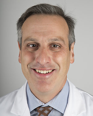 Peter J. Zimetbaum合作论文数Beth Israel Deaconess Medical Center34Andrew H Locke合作论文数33Hans Stabenau合作论文数25
 Mark Josephson合作论文数Harvard Medical School;Beth Israel Deaconess Medical Center24Ewa Sieliwonczyk合作论文数21
 Kabir Muammar M合作论文数Centre for Biomedical Engineering and School of Electrical and Electronic Engineering, The University of Adelaide21Timothy Maher合作论文数20
 Elsayed Z Soliman合作论文数School of Medicine, Wake Forest University;Wake Forest Baptist Health20
 Wendy Post合作论文数Department of Epidemiology, Bloomberg School of Public Health, Johns Hopkins University;School of Medicine, Johns Hopkins University;Ciccarone Center for the Prevention of Cardiovascular Disease, Johns Hopkins University20
 Scott David Solomon合作论文数Heart and Vascular Center, Brigham and Women's Hospital;Clinical Trials Outcomes Center, Brigham and Women's Hospital;Harvard Medical School19
 Libor Pastika合作论文数16
 Kathryn McGurk合作论文数Imperial Coll London, Natl Heart & Lung Inst, London, England16Sotoodehnia Nona合作论文数15
 Tor Biering-Sørensen合作论文数Department of Biomedical Sciences, Faculty of Health and Medical Sciences, Københavns Universitet;Center for Translational Cardiology and Pragmatic Randomized Trials, University of Copenhagen;Cardiovascular Non-Invasive Imaging Research Laboratory Department of Cardiology, Herlev & Gentofte15
 James S. Ware合作论文数Imperial Coll London, Natl Heart & Lung Inst15Charles A. Henrikson合作论文数15
 David S. Siscovick合作论文数Department of Epidemiology, School of Public Health, University of Washington;New York Academy of Medicine13
 Antonio Luiz P. Ribeiro合作论文数Departamento de Clínica Médica, Faculdade de Medicina, Universidade Federal de Minas Gerais13Alexei Shvilkin合作论文数13
 Florian Michaud合作论文数11
 John-Ross Clarke合作论文数10Colleen Sitlani合作论文数Division of General Internal Medicine, School of Medicine, University of Washington10
 Jason Andrew Thomas合作论文数From the School of Psychology, University of Birmingham;P1vital;Department of Psychiatry, University of Oxford;and School of Psychology and Clinical Language Sciences, University of Reading;University of Oxford, University of Reading10
 Danilo P. Mandic合作论文数Communication and Signal Processing Research Group, Department of Electrical and Electronic Engineering, Faculty of Engineering, Imperial College London10
 Mary Lou Biggs合作论文数School of Public Health, University of Washington9
 Antônio H. Ribeiro合作论文数Department of Information Technology, Uppsala University;Einthoven.ai9Alice Haouzi合作论文数8Shu Yang合作论文数7K Patlatzoglou合作论文数7Golriz Sedaghat合作论文数7Robert N D'Angelo合作论文数7
 Kapil kumar合作论文数7
 Robert W. Yeh合作论文数Beth Israel Deaconess Medical Center;Medicine Department, Harvard Medical School;Baim Institute for Clinical Research7
 Declan O'Regan合作论文数MRC Laboratory of Medical Sciences, Imperial College London;Institute of Clinical Sciences, Imperial College London7E Sieliwonczyk合作论文数6Konstantinos Patlatzoglou合作论文数6J. S. Ware合作论文数6
 Nathan Kong合作论文数Dept Internal Med, Univ Chicago6
 Marie-France Poulin合作论文数Laval University;Laval University6
 Roger Laham合作论文数6
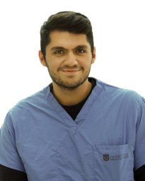 Ghafoori Elyar合作论文数Oregon Health &, The Knight Cardiovascular Institute;Science University;The Knight Cardiovascular Institute, Oregon Health & Science University6




