Purpose To evaluate regeneration after liver venous embolization in healthy and cirrhotic swine and to assess computed tomography (CT) radiomics for predicting these markers. Materials and Methods Twenty-two pigs with healthy (n = 11) or cirrhotic (n = 11) livers underwent portal vein embolization (PVE) with microspheres and coils, n-butyl cyanoacrylate, combined PVE and hepatic vein embolization (PVE + HVE), or sham control. Four weeks after intervention, future liver remnant (FLR) tissue was analyzed for angiogenesis, proliferation, and epithelial integrity using vascular endothelial growth factor (VEGF), angiopoietin-1, Ki-67, and cytokeratin 8/18/19 immunofluorescence and for apoptosis in the embolized lobe using caspase 3. Spearman correlations were computed between biomarker expression and relative FLR hypertrophy. Biomarkers were dichotomized into high versus low groups based on group-specific means. Logistic regression (LR) and random forest (RF) models were trained on baseline CT radiomics using repeated fivefold cross-validation (100 iterations) to predict biomarker expression. Model performance was assessed by mean area under the curve (AUC), with 95% CIs estimated from the 2.5th and 97.5th percentiles across runs. Results All animals completed the study. PVE + HVE resulted in significantly higher expression of VEGF, Ki-67, angiopoietin-1, and caspase 3 than sham controls in both liver conditions (all P < .05). Angiogenesis was greater after PVE + HVE than after PVE in healthy (P < .01) and cirrhotic (P < .001) livers. Ki-67 showed the highest predictive performance (RF AUC, 0.84; LR AUC, 0.81). Conclusions Liver venous embolization induces distinct regenerative responses in healthy and cirrhotic swine. Baseline CT radiomics demonstrated moderate discriminative performance for predicting regenerative biomarkers.
Primary liver cancers, including hepatocellular carcinoma and cholangiocarcinoma, represent a growing global health burden marked by rising incidence and high mortality. Clinical management requires integration of tumour stage, liver function, and patient-related factors to guide treatment decisions, yet current frameworks capture only a fraction of underlying disease complexity. In this setting, artificial intelligence (AI) is emerging as a key enabler of precision medicine in liver oncology. AI encompasses machine learning and deep learning approaches capable of identifying complex, non-linear patterns within large, high-dimensional datasets. These methods are increasingly applied across multimodal data inputs, including electronic health records, radiologic imaging, digital pathology, and omics profiling. In imaging, AI supports surveillance, lesion detection, characterisation, and prediction of recurrence, survival, and treatment response. In computational pathology, models are extending tissue analysis beyond description of morphology and marker expression toward prognostic modelling and inference of tumour biology. Integration of clinical, imaging, and pathological data in multimodal AI frameworks has shown superior performance compared with single-modality models for key outcomes such as recurrence and survival. Despite promising results, clinical translation remains limited by insufficient external and prospective validation, limited interpretability of complex models, and regulatory and workflow integration challenges. Addressing these barriers through federated learning strategies and the development of transparent, explainable systems is essential. If successfully implemented, AI-driven multimodal decision-support systems could substantially refine diagnosis, prognostication, and treatment selection in liver cancer, advancing personalised care in this biologically and clinically complex disease.
Extracellular acidosis is a biologically important feature of the tumor microenvironment in the liver, promoting immune evasion, angiogenesis, and resistance to therapy, and representing a mechanistically important and potentially targetable axis in liver cancer. Imaging extracellular pH (pHe) at high resolution is needed to better understand the immuno-metabolic interplay, especially at the transition regions between the tumor core, tumor margin, and background liver, which is critical for any pharmacological or image-guided intervention. Yet, there is a paucity of imaging techniques capable of providing pHe mapping at high resolution. Here, we demonstrate high-resolution pHe imaging in a mouse Hepa1-6 liver tumor model using 1H Biosensor Imaging of Redundant Deviation in Shifts (BIRDS) with REduced Spherical Encoding with GAussian Weighting (RESEGAW). Eight tumor-bearing C57BL/6J mice were used to demonstrate pHe imaging with RESEGAW using the macrocyclic agent TmDOTP5- at 0.6 mm isotropic resolution on a 9.4 T scanner, which was validated using 31P-MRSI with 3-aminopropylphosphonate (3-APP). pHe imaging with 1H-BIRDS-RESEGAW consistently showed acidic tumor regions (pHe = 6.77 ± 0.14) relative to adjacent normal liver (pHe = 7.14 ± 0.07). Mean pHe values measured by 31P-MRSI with 3-APP and 1H-BIRDS-RESEGAW with TmDOTP5- show no significant differences in tumors (pHe = 6.81 ± 0.13) and normal liver (pHe = 7.14 ± 0.06). Voxelwise comparison after co-registration of 31P-MRSI with 3-APP to 1H-BIRDS-RESEGAW using Bland-Altman analysis demonstrated excellent agreement between the two methods, with minimal mean bias (-0.005 pH units) and variance of less than 0.1 pH units. These results demonstrate the feasibility and quantitative reliability of 1H-BIRDS-RESEGAW for imaging extracellular acidosis in liver tumors at submillimeter resolution, establishing a technical foundation for studying the immuno-metabolic interplay in liver cancer and its response to therapy.
PURPOSE:To evaluate the impact of neoadjuvant systemic PD-1 immune checkpoint inhibition on the local immune response in residual tumors following partial cryoablation in a TIB-75 murine hepatocellular carcinoma (HCC) model. MATERIALS AND METHODS:Forty-eight BALB/c mice (6-12 weeks) were orthotopically implanted with TIB-75 cells to induce a single lesion of HCC. Mice were randomized into 4 treatment groups: (a) control, (b) anti-PD-1, (c) partial cryoablation, and (d) anti-PD-1 and partial cryoablation. Anti-PD-1 was administered on Days 7, 9, and 11 after inoculation, followed by partial cryoablation on Day 13 and tumor harvest on Day 18. The presence of T cell subsets (CD3+, CD4+, and CD8+), tumor-associated macrophages (CD68+ and CD206+), PD-1, and PD-L1 were assessed by histopathological analysis of immunohistochemistry. The percentage of positively stained cells within the tumor was determined using QuPath. RESULTS:Mice treated with anti-PD-1 (n = 12) had greater infiltration of CD3+, CD4+, and CD8+ T cells into residual tumors than control (CD3+: median, 22.4% vs 5.5% [P < .001]; CD4+: median, 19.8% vs 5.1% [P < .001]; CD8+: median, 8.2% vs 3.1% [P = .007]). Partial cryoablation alone (n = 12) increased CD206+ M2-like macrophages (median, 36.6% vs 14.7%; P = .03). Partial cryoablation combined with neoadjuvant anti-PD-1 (n = 12) showed significantly higher infiltration of CD3+ T cells (median, 14.3% vs 4.5%; P = .048) than partial cryoablation alone (n = 12) and significantly lower PD-1 expression than anti-PD-1 alone (median, 2.9% vs 7.3%; P = .004). CONCLUSIONS:In a mouse model of HCC, neoadjuvant PD-1 immune checkpoint inhibition can modulate the immunosuppressive tumor microenvironment observed after cryoablation. This highlights the potential of a combination therapy to treat both early- and advanced-stage HCCs.
Hepatocellular carcinoma (HCC) is biologically heterogeneous, shaped by the interplay between hepatic functional reserve and tumor-related oncologic factors; thus, similar survival outcomes may reflect fundamentally different underlying biological processes. Prognostic modeling in HCC is informed by rich multimodal information from multiparametric MRI and radiology reports from routine clinical practice. Existing prognostic vision-language models (VLMs) learn a single entangled latent representation that blends hepatic and tumor-related factors, limiting both accuracy and biological interpretability. We present BioFact-MoE, a biologically factorized Mixture of Experts (MoE) framework that explicitly decomposes liver and tumor factors via biologically supervised experts within a residual MoE survival architecture. On a HCC cohort of N=588 patients (pretrained on 4,582 3D MRI image-report pairs), BioFact-MoE consistently improves survival prediction over all baselines across time horizons, achieving 12-, 18-, and 24-month AUCs of 75.33
Current hepatocellular carcinoma (HCC) surveillance guidelines rely on manually defined LI-RADS (Liver Imaging Reporting and Data System) features rather than imaging data analysis. This study evaluates the feasibility of machine-learning (ML)-based image analysis frameworks to identify and localize hepatic parenchyma at elevated risk for HCC. In this retrospective study, cirrhotic patients with HCC diagnosis undergoing MRI between 2008 and 2023 were included. The analysis included negative screening MRI preceding a positive screening MRI confirming a LR-5 lesion within 18 months. Volume-of-interest (VOI) annotations of ‘non-malignant’ and ‘malignant’ liver tissue on the screening MRI were manually or automatically placed, mapped from future HCC lesion on positive screening MRI. Radiomics were extracted from these VOIs using PyRadiomics. Logistic regression (LR), random forest (RF), and eXtreme Gradient Boosting (XGB) models were trained and validated across four manual/automatic annotation combinations. Exploratory voxel-level heatmaps were generated to visualize high-risk HCC areas. Model performance was summarized using median values and 95
Manual reporting of 3D MRI studies is time-consuming, yet end-to-end structured report generation for 3D liver MRI remains underexplored due to volumetric complexity and scarce paired data. We propose MRI2Rep, an autoregressive framework for liver MRI report generation. From 3,929 real-world MRI-report pairs acquired over a 10-year single-institution cohort, a Report-to-Label Canonicalization (RLC) module converts free-text reports into structured, closed-vocabulary diagnostic sequences without lesion-level annotations. On a held-out test set, MRI2Rep achieves 76.0
Volumetric assessment of abdominal aortic aneurysms (AAA) offers precise pre- and post-endovascular aortic repair (EVAR) evaluation but is laborious. The primary aim was to train and validate a network facilitating automated segmentation and volume determination of pre- and post-EVAR infrarenal AAAs displayed on computed tomography angiographies (CTA). Secondary aim was evaluation of workflow acceleration. Model was trained on ground truth segmentations. Internal and external validation was performed. AI-generated volumes of total aneurysm, lumen, and thrombus were correlated with ground truth. Model-enabled efficiency gains and semi-automatic AAA segmentations performed by three surgeons were measured. For total aneurysm, mean Dice similarity coefficient was 0.972 ± 0.013 and 0.960 ± 0.035 in internal and external validation. AI-generated thrombus volumes showed a very strong correlation with ground truth in internal (r = 0.996) and external validation (r = 0.940). Mean algorithm-facilitated time savings of 117.1 seconds (56.0
In this study, pre-treatment target lesion vascularisation in either contrast-enhanced (CE) CT or MRI and post-treatment lipiodol deposition in native CT scans were compared in HCC patients who underwent their first cTACE treatment. We analysed the impact of stratification according to cTACE selectivity on these correlations. Seventy-eight HCC patients who underwent their first cTACE procedure were retrospectively included. Pre-treatment tumour vascularisation in arterial contrast phase and post-treatment lipiodol deposition in native CT scans were evaluated using the qEASL (quantitative tumour enhancement) method. Correlations were analysed using scatter plots, the Pearson correlation coefficient (PCC) and linear regression analysis. Subgroup analysis was performed according to lobar, segmental and subsegmental execution of cTACE. Arterial tumour volumes in both baseline CE CT (R2 = 0.83) and CE MR (R2 = 0.82) highly correlated with lipiodol deposition after cTACE. The regression coefficient between lipiodol deposition and enhancing tumour volume was 1.39 for CT and 0.33 for MR respectively, resulting in a ratio of 4.24. After stratification according to selectivity of cTACE, the regression coefficient was 0.94 (R2 = 1) for lobar execution, 1.38 (R2 = 0.96) for segmental execution and 1.88 (R2 = 0.89) for subsegmental execution in the CE CT group. Volumetric lipiodol deposition can be used as a reference to compare different imaging modalities in detecting vital tumour volumes. That approach proved CE MRI to be more sensitive than CE CT. Selectivity of cTACE significantly impacts the respective regression coefficients which allows for an innovative approach to the assessment of technical success after cTACE with a multitude of possible applications. • Lipiodol deposition after cTACE highly correlates with pre-treatment tumour vascularisation and can be used as a reference to compare different imaging modalities in detecting vital tumour volumes. • Lipiodol deposition also correlates with the selectivity of cTACE and can therefore be used to quantify the technical success of the intervention.
BACKGROUND & AIMS:There is growing interest in reducing contrast medium use and the lengthy scan duration in liver imaging. This proof of concept study evaluated the feasibility of deep learning-based generation of synthetic 3D liver contrast-enhanced multiphasic magnetic resonance imaging (MRI) exams, which are similar to ground-truth exams in hepatocellular carcinoma and cirrhosis. METHODS:MRI exams from patients with hepatocellular carcinoma (HCC) or cirrhosis at a single academic center were retrospectively collected. A 3D cycle-consistent generative adversarial network was trained to generate synthetic 3D T1-weighted contrast-enhanced multiphasic liver MRI exams, including arterial, portal venous, delayed, and hepatobiliary phases, using two pre-contrast T1-weighted and T2-weighted input phases. Quantitative performance evaluated similarity, error, and overlap metrics between synthetic and ground-truth exams. For the qualitative multireader study, three blinded radiologists assessed the ground-truth and synthetic MRI exams using a comprehensive questionnaire. Questionnaire tasks 1-5 comprised: visual Turing test (ground-truth vs. synthetic nature), image quality, anatomic accuracy, disease diagnosability, and artifacts. Task 6 comprised Liver Imaging Reporting and Data System features. RESULTS:The study included 3,198 MRI phases from 533 MRI exams from 185 patients with HCC (mean age, 62.1 years ± 9.7 [SD]; 141 men) and 182 patients with cirrhosis (54.4 years ± 10.0; 111 men). Synthetic MRI exams achieved high quantitative and qualitative similarity to ground-truth exams. Quantitative analysis demonstrated high structural similarity index (0.86 ± 0.03), overlap (0.97 ± 0.05), and low symmetric mean absolute percent error (0.63 ± 0.23%). The qualitative multireader study showed no significant difference in tasks 1-5 (p = 0.06-0.50) and high performance metrics in task 6 (accuracy: 0.76-0.86; precision: 0.96-1.00) with moderate to perfect Fleiss's Kappa inter-rater agreement (0.58-1.00, p <0.001). CONCLUSIONS:Deep learning enabled the generation of synthetic 3D liver contrast-enhanced multiphasic MRI exams from pre-contrast sequences, achieving high quantitative and qualitative similarity to ground-truth images. IMPACT AND IMPLICATIONS:This work demonstrates the early feasibility of generating high-quality, 3D contrast-enhanced multiphasic liver MRI exams from pre-contrast sequences, with synthetic exams showing strong agreement with ground-truth across quantitative metrics and key qualitative criteria, including the visual Turing test, image quality, disease diagnosability, anatomic accuracy, artifact severity, and HCC Liver Imaging Reporting and Data System features. Despite the model currently representing a proof of concept based on a moderate single-center dataset, with a need for larger multicenter studies and external validation, the results highlight the potential to transform liver MRI workflows by reducing contrast media costs and potential side effects, significantly shortening acquisition time, especially the prolonged 20-min hepatobiliary phase, and improving accessibility for patients unable to tolerate contrast-enhanced MRI because of renal impairment, contrast agent allergy, or claustrophobia.
Abstract Objective New Zealand White (NZW) rabbits are widely used in interventional radiology research due to their suitability for human-sized treatment and imaging equipment, offering high translational potential. This study aims to define selection criteria for rabbits by correlating body weight (BW) and age with abdominal organ and vessel dimensions measured on cross-sectional imaging. Materials and methods Computed tomography and magnetic resonance imaging scans of 80 male NZW rabbits were analyzed using 3D Slicer to measure abdominal organ volumes and vessel diameters. Additionally, an in-house nnU-Net was built for liver volumetry and validated against manual segmentations. Imaging-based measurements were confirmed by gross anatomy in five animals. Statistics included normality testing and Pearson correlation. Results BW ranged from 2.0 to 4.5 kg (median [IQR]: 3.5 [2.9–3.8]) and age from 10.0 to 24.9 weeks (17.7 [15.0–21.4]); age correlated strongly with BW (p < 0.001). Organ volumes (liver, both kidneys) correlated with BW and age (all p < 0.001), respectively. Additionally, several vessel diameters (left common and internal iliac arteries, inferior vena cava, right common carotid artery) significantly correlated with BW and age, while the celiac trunk (p = 0.010), common hepatic (p = 0.011), and right renal artery (p = 0.031) correlated with BW only. The liver segmentation model achieved a Dice Similarity Coefficient of 0.91. Conclusion BW and age in NZW rabbits correlate with both organ volumes and large vessels relevant to interventional procedures, supporting the use of biometric data as selection criteria to improve standardization, reduce complications, and enhance preclinical research quality. Relevance statement Weight- and age-based selection of NZW rabbits improves anatomical suitability for image-guided interventions, enhancing technical success and reproducibility. It may reduce complications and dropouts, avoiding false attribution of adverse events to the technique rather than biometric unsuitability. Key Points Lack of standardized selection criteria in the VX2 rabbit model increases procedural risks and impairs reproducibility in interventional radiology research. Biometric data correlate with organ and vessel dimensions, enabling estimations of anatomical suitability for image-guided procedures in interventional radiological research. This study establishes anatomical reference data providing a quantitative basis to standardize and refine future VX2 rabbit research. Graphical Abstract
PURPOSE:To evaluate the effectiveness of combined portal vein embolization (PVE) and hepatic vein embolization (HVE) compared with that of PVE alone in cirrhotic and noncirrhotic swine. MATERIALS AND METHODS:Sixteen Yorkshire pigs were included in this study. In the cirrhotic group (n = 8) and noncirrhotic group (n = 8), subjects underwent embolization according to established protocols. Computed tomography (CT) scans were acquired before and at 2- and 4-week intervals following the embolization. Liver volumes were segmented in the portal venous phase. Student t test with a significance level at P < .05 was used. RESULTS:Across all swine, the future liver remnant (FLR) was significantly larger after PVE + HVE than after PVE at 2 weeks (24.12% [95% CI, 15.36%-32.88%] vs 12.75% [95% CI, 7.43%-18.07%]; P = .021) and 4 weeks (23.23% [95% CI, 15.79%-33.47%] vs 15.08% [95% CI, 9.98%-20.87%]; P = .043) after embolization. In the cirrhotic group, the FLR increase was greater following PVE + HVE than after PVE at 2 weeks (20.85% [95% CI, 14.40%-27.30%] vs 8.66% [95% CI, 6.47%-10.86%]; P = .0089) and 4 weeks (19.27% [95% CI, 17.87%-20.67%] vs 13.33% [95% CI, 9.23%-13.33%]; P = .0003) after embolization. CONCLUSIONS:PVE + HVE resulted in greater FLR hypertrophy than PVE alone, indicating that cirrhotic livers may benefit from the addition of HVE.
Background & Aims: Increasing enthusiasm around integrating locoregional therapy with systemic immunotherapy in primary liver cancer underscores the need for non-invasive imaging biomarkers. In this study, we aimed to establish advanced molecular MRI tools for monitoring T-cell responses to cryoablation in murine models, distinguishing between immunologically "hot" and "cold" hepatocellular carcinoma (HCC). Methods: Immunocompetent 7-10-week-old C57BL/6J and BALB/cJ mice (n = 18 each) received carbon tetrachloride for 12 weeks to induce cirrhosis. Intrinsically immunogenic Hepa1-6 ("hot") and non-immunogenic TiB75 ("cold") cells were orthotopically implanted into C57BL/6 or BALB/c mice, respectively, to generate focal HCC lesions. After one week, animals were randomly assigned to (A) partial cryoablation (pCryo) (1.2 mm cryoprobe, -40 degrees C) or (B) no treatment (n = 8 per group and tumor type). Gadolinium 160 (Gd-160)-labeled CD8(+) antibody was administered intravenously either 1 week after tumor induction (control) or 1-week post (pCryo) (treatment). T1-weighted MRI scans were performed using a 9.4 T MRI scanner. Radiological-pathological correlation included imaging mass cytometry and immunohistochemistry. Results: pCryo-treated Hepa1-6 tumors displayed peritumoral ring enhancement on T1-weighted MRI with Gd-160-CD8, correlating with imaging mass cytometry signal patterns. Untreated Hepa1-6 tumors lacked such enhancement. Radiological-pathological correlation confirmed significantly increased tumor-infiltrating CD8(+) T lymphocytes in pCryo Hepa1-6 tumors compared with untreated tumors (p <0.001), and a stronger local response compared with systemic lymph nodes (p = 0.0415). Increased T-lymphocyte infiltration was not observed in TiB75 tumors, as indicated by MRI and histopathology. Conclusion: pCryo induced increased T-cell infiltration in Hepa1-6 tumors compared to TiB75 tumors. T1-weighted MRI, following Gd-160-CD8 antibody administration, reproducibly detected the ablation-induced changes. These findings encourage further investigation of MRI-based molecular imaging biomarkers to assess immune responses to local tumor therapies. (c) 2024 The Authors. Published by Elsevier B.V. on behalf of European Association for the Study of the Liver (EASL). This is an open access article under the CC BY-NC-ND license (http://creativecommons.org/licenses/by-nc-nd/4.0/).
Solid tumors, including hepatocellular carcinoma (HCC), arises most often in cirrhotic livers, where immune exclusion and metabolic reprogramming drive extracellular acidosis (the "Warburg effect") and create an immunosuppressive tumor microenvironment (TME). This study applied a non-invasive MR Spectroscopic Imaging method called Biosensor Imaging of Redundant Deviation in Shifts (BIRDS) to quantify extracellular pH (pHe) dynamics in a mouse model of cirrhosis-associated HCC. Forty-two Mdr2-/- mice received chronic carbon tetrachloride (CCl4), inducing cirrhosis and HCC, confirmed by contrast-enhanced MR and histology. BIRDS revealed significantly lower tumor pHe in untreated tumors (6.78 ± 0.3) compared with liver parenchyma (7.17 ± 0.02). Cryoablation induced tumor pHe normalization (7.08 ± 0.03), coinciding with downregulation of metabolic markers and increased T-cell and macrophage infiltration. These results demonstrate that BIRDS enables non-invasive monitoring of the metabolic and immunologic response to cryoablation in HCC within cirrhotic livers. Cryoablation-induced re-normalization of tumor acidity, coupled with enhanced immune activity, suggests a favorable therapeutic outcome and establishes pHe imaging as a tool for assessing treatment efficacy in acidic TMEs.
"Just Accepted" papers have undergone full peer review and have been accepted for publication in Radiology. This article will undergo copyediting, layout, and proof review before it is published in its final version. Please note that during production of the final copyedited article, errors may be discovered which could affect the content.
Purpose: To develop a machine learning algorithm to improve hepatic resection selection for patients with metastatic colorectal cancer (CRC) by predicting post-portal vein embolization (PVE) outcomes. Materials and Methods: This multicenter retrospective study (2000-2020) included 200 consecutive patients with CRC liver metastases planned for PVE before surgery. Data on radiomic features and laboratory values were collected. Patient-specific eigenvalues for each liver shape were calculated using a statistical shape model approach. After semiautomatic segmentation and review by a board-certified radiologist, the data were split 70%/30% for training and testing. Three machine learning algorithms predicting the total liver volume (TLV) after PVE, sufficient future liver remnant (FLR%), and kinetic growth rate (KGR%) were trained, with performance assessed using accuracy, sensitivity, specificity, area under the curve (AUC), or root mean squared error. Significance between the internal and external test sets was assessed by the Student t-test. One institution was kept separate as an external testing set. Results: A total of 114 (76 men; mean age, 56 years [SD +/- 12]) and 37 (19 men; mean age, 50 years +/- [SD +/- 11]) patients met the inclusion criteria for the internal validation and external validation, respectively. Prediction accuracy and AUC for sufficient FLR% or liver growth potential (KGR%> 0%) were high in the internal testing set-85.81% (SD +/- 1.01) and 0.91 (SD +/- 0.01) or 87.44% (SD +/- 0.10) and 0.66 (SD +/- 0.03), respectively. Similar results occurred in the external testing set-79.66% (SD +/- 0.60) and 0.88 (SD +/- 0.00) or 72.06% (SD +/- 0.30) and 0.69 (SD +/- 0.01), respectively. TLV prediction showed discrepancy rates of 12.56% (SD +/- 4.20%; P = .86) internally and 13.57% (SD +/- 3.76%; P = .91) externally. Conclusions: Machine learning-based models incorporating radiomics and laboratory test results may help predict the FLR%, KGR%, and TLV as metrics for successful PVE.
 MingDe Lin合作论文数86
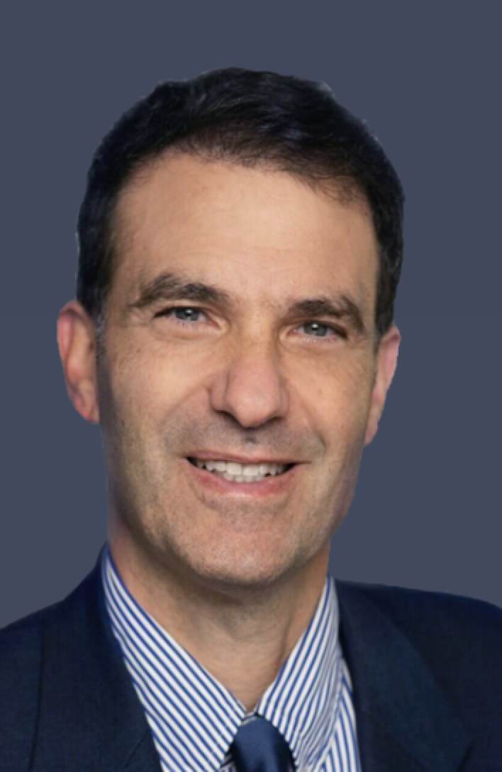 Jeff Geschwind合作论文数Vein Clinics and Oncology Centers, USA Clinics Group;Syntactx;Clinical and Scientific Affairs, Imaging Endpoints, LLC;Cage Pharma;Philips Healthcare86
 Rafael Duran合作论文数56
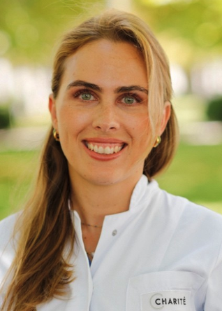 Lynn Jeanette Savic合作论文数Berlin Institute of Health in der Charité;Department of Radiology, Campus Virchow-Klinikum, Charité – Universitätsmedizin Berlin50Ruediger Schernthaner合作论文数37
 James Duncan合作论文数Yale University School of Medicine32
 Bernhard Gebauer合作论文数Klinik für Radiologie, Charité - Universitätsmedizin Berlin31
 Todd Schlachter合作论文数Interventional Radiology, Radiology & Biomedical Imaging, Yale School of Medicine, Yale University30
 Zhijun Wang (王志军)合作论文数Chinese PLA General Hospital19
 Fabian Laage-Gaupp合作论文数Dept Radiol & Biomed Imaging, Yale Univ16
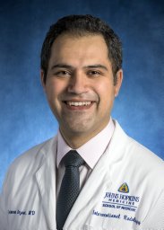 Nezami Nariman合作论文数Drug Appl Res Ctr, Tabriz Univ Med Sci16
 Vania Tacher合作论文数16
 David C. Madoff合作论文数Department of Radiology & Biomedical Imaging, Yale University School of Medicine16Gorodetski Boris合作论文数15
 Junlin Yang合作论文数School of Medicine, Yale University14Annabella Shewarega合作论文数12
 Constantine Frangakis合作论文数Department of Biostatistics, Bloomberg School of Public Health, Johns Hopkins University;Center for Global Health, Bloomberg School of Public Health, Johns Hopkins University12
 Xuchen Zhang合作论文数Pathology Faculty, Yale School of Medicine, Yale University12
 Shanmugasundaram Ganapathy-Kanniappan合作论文数Russell H. Morgan Department of Radiology and Radiological Sciences, Baltimore, MD 21287, USA.12Juvenal Reyes合作论文数12
 Sohn Jae Ho合作论文数Phillips Exeter Academy12Kelvin Hong合作论文数School of Medicine Russell H. Morgan, Johns Hopkins University12Brian S. Letzen合作论文数12Vasily Pekurovsky合作论文数11Jessica G Santana合作论文数11
 Daniel Coman合作论文数Bioimaging Sciences, Radiology & Biomedical Imaging, Yale School of Medicine, Yale University11
 Lawrence H. Staib合作论文数Department of Radiology and Biomedical Imaging, Yale School of Medicine, Yale University;Department of Biomedical Engineering, Yale School of Medicine, Yale University;Department of Electrical Engineering, Yale School of Medicine, Yale University11Ahmet S Kucukkaya合作论文数10
 Moritz Gross合作论文数Charité Center for Diagnostic and Interventional Radiology, Charité-Universitätsmedizin Berlin10
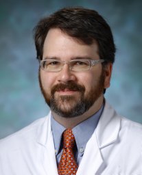 Cornish Toby C合作论文数Dept Pathol, Johns Hopkins Univ10Lin MingDe合作论文数10
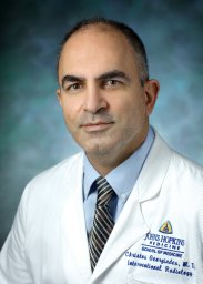 Christos S. Georgiades合作论文数Johns Hopkins Medical Institutions10
 D. S. Fahmeed Hyder合作论文数Department of Biomedical Engineering, Yale University;InnovaCyclics LLC;Magnetic Resonance Research Center, Yale University10
 Bernd Hamm合作论文数Institute of Radiology, Charite - Universitatsmedizin Berlin10
 Alexandra Petukhova合作论文数9
 Nikitha Murali合作论文数Sect Vasc & Intervent Radiol, Yale Sch Med9
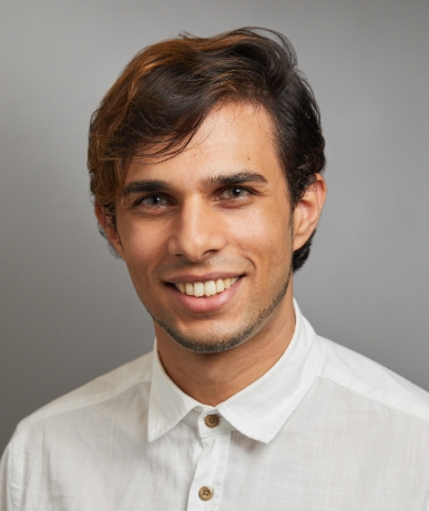 Tal Zeevi合作论文数Yale School of Medicine, Yale University9Milena Miszczuk合作论文数9
 Florian Fleckenstein合作论文数9Sahu Sonia合作论文数9Lee Howard合作论文数9Strazzabosco Mario合作论文数Department of Medicine and surgery, University of Milano-Bicocca9
 Yan Zhao (赵晏)合作论文数Medical College, Xi'an Jiaotong University9Isabel Schobert合作论文数8Susanne Smolka合作论文数Sch Med, Yale Univ8
 Steffen Huber合作论文数Cardiovascular Medicine, Internal Medicine, Yale School of Medicine, Yale University8
 S. Nahum Goldberg合作论文数8
 Sahar Mirpour合作论文数8
 John A. Onofrey合作论文数8




