Peripheral nerve injury (PNI) regeneration involves complex immunoregulatory mechanisms, particularly M2 macrophage‑mediated immunomodulation. Promoting macrophage polarization toward the M2 phenotype represents a potential therapeutic strategy. Exosomes can create favorable microenvironments for tissue regeneration, but their clinical application is often limited by low concentration and purity. Three‑dimensional‑bioprinted exosomes (3D‑Exos) have recently gained attention due to their enhanced concentration and purity. In this study, 3D‑Exos were applied to treat PNI. The regenerative effects of 3D‑Exos were compared with conventionally produced exosomes (2D‑Exos) in vivo. In vitro experiments, RNA sequencing, and miRNA microarray analyses were performed to investigate the underlying mechanisms. The mechanism was further validated in vitro and in vivo. 3D‑Exos significantly enhanced nerve regeneration after PNI and outperformed 2D‑Exos. In vitro studies revealed that 3D‑Exos promoted macrophages polarization toward the M2 macrophages. RNA sequencing indicated that the PI3K/AKT pathway was activated by 3D‑Exos, and miRNA microarray analysis identified miR‑26b‑5p within 3D‑Exos as the key mediator targeting PTEN to activate PI3K/AKT signaling. Subsequently, M2 macrophages facilitated Schwann cell migration, elongation, and myelination, thereby accelerating nerve regeneration. Inhibition of the PI3K/AKT signaling pathway abolished the therapeutic effects of 3D-Exos on nerve regeneration after PNI. This study elucidates a novel mechanism by which 3D‑Exos promote nerve regeneration through reprogramming macrophages polarization via the PI3K/AKT pathway. The work establishes a paradigm‑shifting therapeutic framework that integrates 3D-Exos with immunomodulatory strategies for the treatment of PNI. First demonstration of 3D-Exos as a high-concentration, high-purity therapeutic way to overcome limitations of conventional exosomes applications for PNI. Mechanistic insight into macrophages reprogramming: 3D-Exos drives macrophages polarize into M2 phenotype via PI3K/AKT signaling pathway, fostering an conducive microenvironment critical for SCs myelination, and nerve regeneration. Paradigm-shifting therapeutic framework: Integrates 3D-Exos technology with immunomodulation, establishing a novel crosstalk axis between extracellular vesicles, macrophages, and SCs for functional PNI rehabilitation.
Peripheral nerve injury (PNI) is a intractable disease with poor treatment efficacy, it's suggested that ferroptosis is closely related to the occurrence and development of PNI, but more in-depth studies are needed. Puerarin (Pu), an antioxidant from kudzu root, may mitigate PNI, but its mechanism remains unclear. Here, we investigated Pu's role in PNI based on in vivo and in vitro experiments. We found that Pu could promote nerve regeneration after PNI, then we simulated PNI with the classical oxygen-glucose deprivation (OGD) cell injury model to explore the potential mechanism. The RNA sequencing revealed that Pu attenuated OGD-induced injury via ACSL4-dependent ferroptosis, and the molecular docking showed that Pu competitively binds to the ACSL4 active site of arachidonic acid, and the dynamics simulation suggested that the Leu468 and Thr469 were the main sites that Pu binds to ACSL4. Which were further demonstrated by cellular thermal shift assays, microscale thermophoresis, and pull-down assays. Then, in vivo and in vitro assays showed Pu treatment improved nerve morphology, function, and electrophysiology by inhibiting ACSL4-dependent ferroptosis. What's more, loss-of-function study further validated ACSL4's essential role in Pu's neuroprotective effects. In summary, our study demonstrates the role of Pu in promoting PNI recovery via ACSL4-dependent ferroptosis, thus identifying a promising therapeutic strategy for ferroptosis-related diseases.
Peripheral nerve injury (PNI) disrupts nerve function, there has been increasing focus on the role of macrophages in PNI. This study provides a comprehensive analysis of research trends and key topics related to macrophages in the field of PNI. We conducted a search in the Web of Science Core Collection database for studies published between 2000 and 2023 using "macrophages" and "PNI" as keywords. Bibliometric analysis was performed using visualization tools, including VOSviewer, CiteSpace, and GraphPad Prism 8, and others. As of December 31, 2023, a total of 905 relevant publications were identified. In terms of author contributions, Japanese researchers Kiguchi Norikazu and Kishioka Shiroh shared the highest number of publications, each with 15 articles. In terms of journals, Experimental Neurology published the most articles, while the University of Würzburg in Germany emerged as the leading institution in this field, with the highest publication output. Among the 51 countries involved in this research, the United States ranked first, followed by China. The keyword 'exosomes' first appeared in 2021, making it the most recent keyword. Cluster analysis categorized the keywords into four main groups: the role of macrophages in Wallerian degeneration, the role of macrophages in pathological peripheral neuropathic pain, macrophage-derived products after PNI, and role of macrophages in PNI repair and strategies for modulating their function using biomaterials. Macrophages are increasingly crucial in PNI research, and future studies integrating novel approaches may yield innovative therapies for nerve repair.
BACKGROUND:The optimal management of symptomatic vertebral artery in-stent restenosis or occlusion (SVISRO) after stent-assisted angioplasty (SAA) at the vertebral artery origin remains unclear. This study aimed to investigate the efficacy of vertebral artery reconstruction surgery (VRS) surgery among SVISRO patients. METHODS:A retrospective study was conducted to analyze the clinical data for SVISRO patients admitted to the Third Affiliated Hospital of Sun Yat-sen University between May 2011 and November 2021. The stroke recurrence and stroke-free rates during the follow-up (FU) period and the patients' neurological statuses at the last FU were compared between a VRS group and an intensive medical therapy (IMT) group. RESULTS:Sixty-two SVISRO patients with an average age of 60.1 ± 8.3 years and an average FU duration of 67.6 ± 32.5 months were studied. The VRS group had significantly fewer stroke recurrences than the IMT group did (5.7% vs. 25.9%, p = 0.034). A 154-month stroke-free rate of 73.8% (95% CI, 24.5% to 93.7%) was observed in the VRS group, whereas the IMT group had a stroke-free rate of 33.9% (95% CI, 17.3% to 75.0%). The hazard ratio (log-rank) between the two groups was 0.234 (95% CI, 0.063-0.871; p = 0.048). The modified Rankin scale score for the VRS group was significantly better than that for the IMT group at the final FU (p = 0.032). CONCLUSIONS:In patients with SVISRO secondary to SAA, VRS targeting the extracranial vertebral artery (V1-V2) appears to reduce stroke recurrence, increase the stroke-free rate and improve neurological status.
Endothelial cell glycolysis plays a novel and significant role in Schwann cells and peripheral nerve injury repair, which represents an emerging and important area of research. Glycolysis in endothelial cells is a conserved and tightly regulated biological process that provides essential energy (ATP) and intermediates by ultimately converting glucose into lactate. This metabolic pathway is crucial for maintaining the normal function of endothelial cells. During peripheral nerve injury repair, endothelial cell glycolysis influences the function of Schwann cells and the efficiency of nerve regeneration. Beyond glycolysis, endothelial cells also secrete various factors, including growth factors and extracellular vesicles, which further modulate Schwann cell activity and contribute to the repair process. This review will summarize the role of endothelial cell glycolysis in Schwann cell function and peripheral nerve injury repair, aiming to provide new insights for the development of novel strategies for peripheral nerve injury treatment.
The triglyceride-glucose (TyG) index is related to various cardiovascular diseases, but its relationship with stroke and all-cause mortality (ACM) in individuals with coronary artery disease (CAD) is still not well understood. This research sought to analyze the interaction between the TyG index and the occurrence of stroke and ACM in CAD participants. The dataset was derived from the National Health and Nutrition Examination Survey (NHANES), with 809 CAD patients included from 1999 to 2018. TyG index was determined by ln[fasting triglycerides (mg/dL) × fasting glucose (mg/dL)/2]. Findings showed that heightened TyG index values were markedly associated with a greater risk of stroke; a U-shaped interconnection was detected between the TyG index and stroke risk, with the threshold at 8.14. Individuals with a TyG index exceeding this threshold exhibited a markedly higher rate of stroke occurrence. Additionally, a J-shaped correlation was observed between the TyG index and ACM, with the threshold at 9.25, above which the risk of death increased. These findings indicate that the TyG index could act as a practical indicator for predicting stroke and ACM among CAD patients, particularly when considering threshold values.
This study aimed to develop and validate a nomogram model for predicting cerebral infarction risk after superficial temporal artery–middle cerebral artery (STA-MCA) bypass in patients with intracranial atherosclerotic stenosis (ICAS). Patients with ICAS who received STA-MCA bypass were enrolled in this study. The independent risk factors for post bypass infarction were identified using univariate and multivariate logistic regression analyses. A nomogram model was developed and subsequently evaluated using receiver operating characteristic (ROC) curves, calibration curves, and decision curve analysis (DCA). Eventually, 316 patients with ICAS were included in the study. Diabetes, smoking, and high triglyceride and total cholesterol levels were identified as the independent risk factors, and a nomogram model was developed. The model achieved areas under the curve (AUCs) of 0.88 (95
Over half of patients with chronically ischaemic cerebrovascular disease (CICD) exhibit poor revascularization potential. Tie2-expressing monocytes/macrophages (TEMs) have been reported to promote angiogenesis in tumour tissue; however, whether TEMs promote angiogenesis in chronically ischaemic brain tissue (CIBT) and the regulatory mechanism through which TEMs are recruited to CIBT remain unclear. We first analysed the proportion of TEMs in blood from the internal jugular veins (IJVs) of CICD patients and then isolated TEMs for coculture with human umbilical vein endothelial cells (HUVECs) and for intraventricular injection into nude mice to explore the proangiogenic effects of TEMs in CIBT. Then, molecular biology experiments were performed to verify the upstream regulatory mechanism of the ANGPT2-Tie2 axis, and cell transfection experiments were conducted to confirm the regulatory effects of the detected pathway on Tie2 receptors on the endothelial cell surface. Additionally, a 2-vessel occlusion plus encephalomyosynangiosis rat model was established to confirm the recruitment mechanism of TEMs in CIBT and their ability to improve cerebral blood perfusion (CBP) and cognitive function. The proportion of TEMs from the IJV blood of CICD patients significantly increased, especially in patients who exhibited Matsushima Grade-A revascularization. The viability of HUVECs cocultured with TEMs was significantly increased, and CBP and the expression of CD31 in the CIBT of nude mice treated with TEMs were significantly increased. The above increases were positively correlated with the concentration of TEMs used for coculture and intraventricular injection. Moreover, molecular biology experiments indicated that miR-126-5p can directly bind to the 3'UTR of TRPS1 mRNA and that TRPS1 can directly bind to the promoter of Angpt2. HUVECs transfected with miR-126-5p mimics presented significantly decreased TRPS1 expression, a reduced pTie2/Tie2 ratio, increased ANGPT2 expression, and increased cell viability. Finally, significantly increased TEMs infiltration, downregulated TRPS1 expression, and upregulated ANGPT2, CD31, VEGFA, and IGF1 expression were detected in the CIBT of the rats transfected with the miR-126-5p agomir, accompanied by significant improvements in CBP and cognitive function. TEMs promote angiogenesis in CIBT through a paracrine mechanism, and the recruitment of TEMs to CIBT is regulated by the miR-126-5p/TRPS1/ANGPT2 pathway.
Like the blood-brain barrier and blood-spinal cord barrier, the blood-nerve barrier (BNB) is one of the crucial tissue barriers of the nervous system. It plays a vital role in homeostasis, physiological protection, and pathological reactions. Various factors, such as biological, physical, and chemical factors, can lead to transient or permanent dysfunction of the BNB. With the advancements in biological techniques and the growing peripheral nerve injuries such as trauma and diabetic peripheral neuropathy, the BNB has gained increasing attention. Moreover, the defensive function of the BNB impedes therapeutic deliveries and anesthetic drugs, which compromises the therapeutic experiences and life quality. It should be noted that numerous pathways are involved in the microstructure, function, and opening of the BNB, but the true underlying molecular mechanisms are still under constant exploration and investigation. This review summarizes the microstructure, and signaling pathways of the BNB, and thoroughly discusses the transient or permanent disruptions of the BNB in both physiological and pathological conditions.
Peripheral nerve injury (PNI) can result in severe disabilities, profoundly impacting patients' quality of life and potentially endangering their lives. Therefore, understanding the potential molecular mechanisms that facilitate the regeneration of damaged nerves is crucial. Evidence indicates that Schwann cells (SCs) play a pivotal role in repairing peripheral nerve injuries. Previous studies have shown that RNA, particularly non-coding RNA (ncRNA), plays a crucial role in nerve regeneration, including the proliferation and dedifferentiation of SCs. In this review, the individual roles of ncRNA in SCs and PNI are analyzed. This review not only enhances the understanding of ncRNA's role in nerve injury repair but also provides a significant theoretical foundation and inspiration for the development of new therapeutic strategies.
BACKGROUND AND PURPOSE:The triglyceride-glucose (TyG) index, a novel reliable biomarker for IR that incorporates blood glucose and triglyceride, is linked to intracranial atherosclerotic stenosis (ICAS). In this study, we aimed to further investigate the association between the TyG index and the outcomes of ICAS patients following extracranial-to-intracranial (EC-IC) bypass grafting. METHODS:489 ICAS patients who underwent EC-IC bypass between Jan 2009 and Jan 2022 at our hospital were retrospectively collected. The major adverse cardiac and cerebrovascular events (MACCEs), and anastomotic restenosis, both of which are critical factors leading to poor prognosis of ICAS patients after EC-IC bypass, were mainly recorded and analyzed. Kaplan-Meier survival curve and Log-rank tests were sequentially conducted. Cox regression model was used to investigate the association between the TyG index and MACCEs & anastomotic stenosis. C-statistics, continuous net reclassification improvement (NRI), and integrated discrimination improvement (IDI) evaluated the incremental predictive value of the TyG index. RESULTS:A higher incidence of MACCEs and anastomotic stenosis was found in higher-tertile TyG index group. The TyG index was significantly associated with an increased risk of MACCEs and anastomotic stenosis, independent of confounding factors, with a value of HR (1.30, 95%CI 1.10-1.51, p < 0.001) and (1.27, 95%CI 1.16-1.40, p < 0.001) respectively. The area under the curve (AUC) in the model with the TyG index for predicting the occurrence of MACCEs and anastomotic stenosis were 0.708 (95%CI 0.665-0.748) and 0.731 (95%CI 0.689-0.770) respectively. The addition of the TyG index significantly improved the global performance of the baseline model according to the C-statistics, NRI, and IDI (All p < 0.05). CONCLUSIONS:Higher TyG levels were associated with poorer outcomes in ICAS patients after EC-IC bypass. TyG could be a key factor in managing ICAS risk and standardizing the indications for EC-IC bypass.
Abstract Background Endothelial cell (EC)-driven intraneural revascularization (INRV) and Schwann cells-derived exosomes (SCs-Exos) both play crucial roles in peripheral nerve injury (PNI). However, the interplay between them remains unclear. We aimed to elucidate the effects and underlying mechanisms of SCs-Exos on INRV following PNI. Results We found that GW4869 inhibited INRV, as well as that normoxic SCs-Exos (N-SCs-Exos) exhibited significant pro-INRV effects in vivo and in vitro that were potentiated by hypoxic SCs-Exos (H-SCs-Exos). Upregulation of glycolysis emerged as a pivotal factor for INRV after PNI, as evidenced by the observation that 3PO administration, a glycolytic inhibitor, inhibited the INRV process in vivo and in vitro. H-SCs-Exos more significantly enhanced extracellular acidification rate/oxygen consumption rate ratio, lactate production, and glycolytic gene expression while simultaneously suppressing acetyl-CoA production and pyruvate dehydrogenase E1 subunit alpha (PDH-E1α) expression than N-SCs-Exos both in vivo and in vitro. Furthermore, we determined that H-SCs-Exos were more enriched with miR-21-5p than N-SCs-Exos. Knockdown of miR-21-5p significantly attenuated the pro-glycolysis and pro-INRV effects of H-SCs-Exos. Mechanistically, miR-21-5p orchestrated EC metabolism in favor of glycolysis by targeting von Hippel-Lindau/hypoxia-inducible factor-1α and PDH-E1α, thereby enhancing hypoxia-inducible factor-1α-mediated glycolysis and inhibiting PDH-E1α-mediated oxidative phosphorylation. Conclusion This study unveiled a novel intrinsic mechanism of pro-INRV after PNI, providing a promising therapeutic target for post-injury peripheral nerve regeneration and repair. Graphical Abstract
Objective Despite revascularization has been proven to be the optimal choice of moyamoya disease (MMD), many complications may occur in this context.. This study aimed to analyse the risk factors for new or expanded symptomatic infarctions of patients with MMD after revascularization.Methods Patients with MMD who received revascularization in our department were retrospectively enrolled, new or expanded cerebral infarction within a month after revascularization was the main concern. Basic and clinical information, and intraoperative conditions were evaluated. Univariate and multivariate analyses were performed to evaluate the risk factors. Receiver operating characteristic curve analysis was conducted to evaluate the efficiency of the risk factors.Results Eventually, 108 consecutive patients received 174 surgeries were enrolled, experienced new or expanded infarction occured in 13 (7.47%) surgeries, which showed higher Suzuki stage on the non-operative side, more posterior cerebral artery (PCA) involvement, and more intraoperative hypotension compared to those without infarction(p < .05). The Suzuki stage on the non-operative side had the highest area under the curve (AUC) of 0.737, with a sensitivity of 0.692 and specificity of 0.783. Combination of the three factors showed better efficiency, with an AUC of 0.762, a sensitivity of 0.692, and a specificity of 0.907.Conclusions Revascularization was a safe option for patients with MMD, higher Suzuki stage on the non-operative side, PCA involvement, and intraoperative hypotension might be the risk factors for new or expanded infarction after revascularization in patients with MMD.
Berberine (BBR) has demonstrated potent anti-inflammatory effects by modulating macrophage polarization. Nevertheless, the precise mechanisms through which berberine regulates post-injury inflammation within the peripheral nerve system remain elusive. This study seeks to elucidate the role of BBR and its underlying mechanisms in inflammation following peripheral nerve injury (PNI). Adult male C57BL/6J mice subjected to PNI were administered daily doses of berberine (0, 60, 120, 180, 240 mg/kg) via gavage from day 1 through day 28. Evaluation of the sciatic function index (SFI) and paw withdrawal threshold revealed that BBR dose-dependently enhanced both motor and sensory functions. Immunofluorescent staining for anti-myelin basic protein (anti-MBP) and anti-neurofilament-200 (anti-NF-200), along with histological staining comprising hematoxylin-eosin (HE), luxol fast blue (LFB), and Masson staining, demonstrated that BBR dose-dependently promoted structural regeneration. Molecular analyses including qRT-PCR, Western blotting, enzyme-linked immunosorbent assay (ELISA), and immunofluorescence confirmed that inactivation of the NLRP3 inflammasome by MCC950 shifted macrophages from the pro-inflammatory M1 phenotype to the anti-inflammatory M2 phenotype, while also impeding macrophage infiltration. Furthermore, BBR significantly downregulated the expression of the NLRP3 inflammasome and its associated molecules in macrophages, thereby mitigating NLRP3 inflammasome activationinduced macrophage M1 polarization and inflammation. In summary, BBR's neuroprotective effects were concomitant with the suppression of inflammation after PNI, achieved through the inhibition of NLRP3 inflammasome activation-induced macrophage M1 polarization.
Objective Peripheral nerve injury (PNI) is characterized by high incidence and sequela rate. Recently, there was increasing evidence that has shown ferroptosis may impede functional recovery. Our objective is to explore the novel mechanism that regulates ferroptosis after PNI. Methods LC-MS/MS proteomics was used to explore the possible differential signals, while PCR array was performed to investigate the differential factors. Besides, we also tried to activate or inhibit the key factors and then observe the level of ferroptosis. Regeneration of myelin sheath was finally examined in vivo via transmission electron microscopy. Results Proteomics analysis suggested coagulation signal was activated after sciatic nerve crush injury, in which high expression of F2 (encoding thrombin) and F2r (encoding PAR1) were observed. Both thrombin and PAR1-targeted activator TRAP6 can induce ferroptosis in RSC96 cells, which can be rescued by Vorapaxar (PAR1 targeted inhibitor) in vitro. Further PCR array revealed that activation of PAR1 induced ferroptosis in RSC96 cells by increasing expression of YAP and ACSL4. Immunofluorescence of sciatic nerve confirmed that the expression of YAP and ACSL4 were simultaneously reduced after PAR1 inhibition, which may contribute to myelin regeneration after injury in SD rats. Conclusion Inhibition of PAR1 can relieve ferroptosis after sciatic nerve crush injury in SD rats through Hippo-YAP/ACSL4 pathway, thereby regulating myelin regeneration after injury. In summary, PAR1/Hippo-YAP/ACSL4 pathway may be a promising therapeutic target for promoting functional recovery post-sciatic crush injury.
Backgrounds Persistent trigeminal artery (PTA) is a rare anastomosis between internal carotid artery (ICA) and basilar artery. In rare conditions, the PTA could be combined with others cerebrovascular anomalies, moyamoya disease (MMD) is one of them. Case presentation Here, we reported one rare case of MMD associated with PTA, the patient admitted to our department for severe dizziness and headache, imaging examination suggested MMD combined with right PTA, which arising from the ipsilateral cavernous portion of ICA. The patient received phased bilaterral revascularization with no any complication. In the subsequent follow-up, the patient’s symptoms and intracranial vascular condition gradually improved. Moreover, we conducted a literature review of coexistence of PTA and MMD, the results of a web of science regarding such condition, and a deep discussion providing brief insight into the status of co-occurrence of PTA and MMD, including its manifestation, treatment and outcome. Conclusions The coexistence of PTA and MMD was rarely reported, the pathogenesis of such condition remains unknown. We found that the features of the coexistence of PTA and MMD were diverse, revascularization might be a feasible for such patient.
Background: Computed tomography angiography (CTA) is very popular because it is characterized by rapidity and accessibility. However, CTA is inferior to digital subtraction angiography (DSA) in the diagnosis of intracranial artery stenosis or occlusion. DSA is an invasive examination, so we optimized the quality of cephalic CTA images. Methods: We used 5000 CTA images to train multi-scale residual denoising generative adversarial network (MRDGAN). And then 71 CTA images with intracranial large arterial stenosis were treated by Super-Resolution based on Generative Adversarial Network (SRGAN), Enhanced Super-Resolution based on Generative Adversarial Network (ESRGAN) and post-trained MRDGAN, respectively. Peak signal-to-noise ratio (PSNR) and structural similarity index measurement (SSIM) of the SRGAN, ESRGAN, MRDGAN and original CTA images were measured respectively. The qualities of MRDGAN and original images were visually assessed using a 4-point scale. The diagnostic coherence of digital subtraction angiography (DSA) with MRDGAN and original images was analyzed. Results: The PSNR was significantly higher in the MRDGAN CTA images (35.96 +/- 1.51) than in the original (31.51 +/- 1.43), SRGAN (25.75 +/- 1.18) and ESRGAN (30.36 +/- 1.05) CTA images (all P < 0.001). The SSIM was significantly higher in the MRDGAN CTA images (0.95 +/- 0.02) than in the SRGAN (0.88 +/- 0.03) and ESRGAN (0.90 +/- 0.02) CTA images (all P < 0.01). The visual assessment was significantly higher in the MRDGAN CTA images (3.52 +/- 0.58) than in the original CTA images (2.39 +/- 0.69) (P < 0.05). The diagnostic coherence between MRDGAN and DSA (kappa = 0.89) was superior to that between original images and DSA (kappa = 0.62). Conclusion: Our MRDGAN can effectively optimize original CTA images and improve its clinical diagnostic value for intracranial large artery stenosis.
Vascular reconstruction is indispensable for the regenerative microenvironment after peripheral nerve injury (PNI), while the intrinsic mechanisms remain unclear. Our study found a novel function of Schwann cell facilitating intraneural revascularization and a novel mechanism of miR-21-5p regulating energy metabolism of endothelia cells (ECs) in favor of glycolysis, and determines an important link among exosome, metabolism, angiogenesis, and nerve repair after peripheral nerve injury. Hypoxia-upregulated miR-21-5p in Schwann cells-derived exosomes targets von Hippel-Lindau/Hypoxia-inducible factor-1α (VHL/HIF-1α) pathway and pyruvate dehydrogenase-E1α subunit (PDH-E1α) at once to further skew ECs’ energy metabolism to glycolysis. Interestingly, HIF-1α inactivated PDH-E1α by activating PDK1 to restrain ECs’ oxidative phosphorylation (OXPHOS) finally enhancing glycolysis. Thus, the modulation of ECs’energic metabolism by SCs-Exos-derived miR-21-5p is crucial for intraneural revascularization and nerve regeneration in response to injury.
 Li-Xin Huang合作论文数17Cong Ling (凌聪)合作论文数17Zhimin Wu合作论文数12Baoyu Zhang合作论文数11Hao Li合作论文数7Zhi Liao合作论文数6Sun Tao合作论文数6Zhang-Yu Li合作论文数4
 Chuan Chen (陈川)合作论文数3Junfa Yang合作论文数2Zhangyu Li合作论文数2Jia Qiao合作论文数2Qun Gao合作论文数2Chen Jian-Wen合作论文数2
 Hui Wang (王辉)合作论文数School of Medical Technology, Xinxiang Medical University;Henan Key Laboratory of Immunology and Targeted Drugs2Ya Yang合作论文数1Bufan Xiao合作论文数1Kaddie Kwok Chen合作论文数1Jing Wang合作论文数1Bangjie Chen合作论文数1Zhifeng Wu合作论文数1Feifei Zhang (张菲菲)合作论文数1Cheng Ju合作论文数1Jingtao Zhang合作论文数1Hui Fang (輝芳)合作论文数1Ruihao Zhou合作论文数1JiNian Wang (王继年)合作论文数1LiMing Yang (杨力明)合作论文数1ZhenZhu Dan (单珍珠)合作论文数1Ying Guo (郭英)合作论文数1Guofu Huang (黄国富)合作论文数The First Hospital of Nanchang1Meng Gu合作论文数1
 Xiao-Bin Lv合作论文数1
 Tao Sun (孙涛)合作论文数Institute of Molecular Medicine, School of Biomedical Sciences, School of Medicine, Huaqiao University1
 Xiongwen Pang (庞雄文)合作论文数School of Computer Science, South China Normal University;School of Artificial Intelligence, South China Normal University1
 Qing Wang (王庆)合作论文数School of Chemistry, Nanjing University1
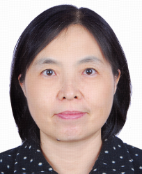 Lei Li (李蕾)合作论文数School of Artificial Intelligence, Beijing University of Posts and Telecommunications1
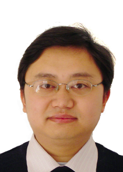 Pengcheng Li (李鹏程)合作论文数School of Biomedical Engineering, Hainan University1Xinyi Wang (汪心怡)合作论文数1
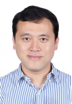 Tao Xu (徐弢)合作论文数Tsinghua University;MEDPRIN;Research Institute of Tsinghua University in Shenzhen1Chaobing Gao (高潮兵)合作论文数1
 Xiaojie Lu (鲁晓杰)合作论文数江苏省无锡市第二人民医院神经外科1




