Background As the indication for immunotherapy is rapidly expanding, it is crucial to accurately identify patients who are likely to respond. Infiltration of B cells into many tumor types correlates with a good response to immune checkpoint inhibitor (ICI) therapy. However, B cells’ roles in the anti-tumor response are far from clear. Methods Based on single-cell transcriptomic data for ICI-treated patients, we identified a B-cell cluster [BIR (ICI-Responsive B) cells] and described the phenotype, cell–cell communication, biological processes, gene signature, and prognosis value of BIR cells through bioinformatic analysis, tissue immunofluorescence, and animal experiments. Surgery samples from 12 non-small cell lung carcinoma (NSCLC) patients with adjuvant checkpoint blockade were evaluated as external validation. Results BIR cells were identified as a subset of CD20+CD22+ADAM28+ B cells with a memory phenotype. Bioinformatic analysis revealed that BIR cells had enhanced cell viability and epigenetic regulation, and that ALOX5AP, MIF, and PTPRC/CD45 expressed by myeloid cells may be critical coordinators of diverse biological processes of BIR cells. Immunofluorescence confirmed the presence of BIR cells in tertiary lymphoid structures (TLSs) in skin SCC, RCC, CRC, and breast cancer. BIR-associated gene signatures correlate with positive outcomes in patients with melanoma, glioblastoma, NSCLC, HNSCC, or RCC treated with ICI therapy, and BIR-cell density predicted NSCLC patients’ response to checkpoint immunotherapy. In line with this, melanoma-bearing mice depleted of BIR cells were resistant to ICIs. Conclusions CD20+CD22+ADAM28+ BIR cells were present in cancer-associated TLS and promoted the response to ICI therapy.
Angiogenesis and neurogenesis play irreplaceable roles in bone repair. Although biomaterial implantation that mimics native skeletal tissue is extensively studied, the nerve-vascular network reconstruction is neglected in the design of biomaterials. Our goal here is to establish a periosteum-simulating bilayer hydrogel and explore the efficiency of bone repair via enhancement of angiogenesis and neurogenesis. In this contribution, we designed a bilayer hydrogel platform incorporated with magnesium-ion-modified black phosphorus (BP) nanosheets for promoting neuro-vascularized bone regeneration. Specifically, we incorporated magnesium-ion-modified black phosphorus (BP@Mg) nanosheets into gelatin methacryloyl (GelMA) hydrogel to prepare the upper hydrogel, whereas the bottom hydrogel was designed as a double-network hydrogel system, consisting of two interpenetrating polymer networks composed of GelMA, PEGDA, and β-TCP nanocrystals. The magnesium ion modification process was developed to enhance BP nanosheet stability and provide a sustained release platform for bioactive ions. Our results demonstrated that the upper layer of hydrogel provided a bionic periosteal structure, which significantly facilitated angiogenesis via induction of endothelial cell migration and presented multiple advantages for the upregulation of nerve-related protein expression in neural stem cells (NSCs). Moreover, the bottom layer of the hydrogel significantly promoted bone marrow mesenchymal stem cells (BMSCs) activity and osteogenic differentiation. We next employed the bilayer hydrogel structure to correct rat skull defects. Based on our radiological and histological examinations, the bilayer hydrogel scaffolds markedly enhanced early vascularization and neurogenesis, which prompted eventual bone regeneration and remodeling. Our current strategy paves way for designing nerve-vascular network biomaterials for bone regeneration.
The molecular pathology of multi-organ injuries in COVID-19 patients remains unclear, preventing effective therapeutics development. Here, we report a proteomic analysis of 144 autopsy samples from seven organs in 19 COVID-19 patients. We quantified 11,394 proteins in these samples, in which 5,336 were perturbed in the COVID-19 patients compared to controls. Our data showed that cathepsin L1, rather than ACE2, was significantly upregulated in the lung from the COVID-19 patients. Systemic hyperinflammation and dysregulation of glucose and fatty acid metabolism were detected in multiple organs. We also observed dysregulation of key factors involved in hypoxia, angiogenesis, blood coagulation, and fibrosis in multiple organs from the COVID-19 patients. Evidence for testicular injuries includes reduced Leydig cells, suppressed cholesterol biosynthesis, and sperm mobility. In summary, this study depicts a multi-organ proteomic landscape of COVID-19 autopsies that furthers our understanding of the biological basis of COVID-19 pathology.
OBJECTIVES:To evaluate the occurrence, abundance, distribution, nature and clinical significance of multinucleated giant cell (MGC) in esophageal cancer.MATERIALS AND METHODS:MGCs were examined with conventional pathology, immunohistochemistry and immunofluorescence in 107 esophageal cancer tissues. The findings were correlated to pathological diagnosis and clinical behavior of the cancers.RESULTS:MGCs were identified in 31.7% (34/107) of the cases. MGCs were positive for CD11c, CD11b, CD32, CD16, HLA-DR and MMP9, and negative for CD163, CD206 and CD64 giving a molecular profile of proinflammatory M1 but not immunosuppressive M2. MGCs were significantly related to decreased lymph node metastasis (p = 0.011), low pTNM stage (p = 0.044), favorable survival (p = 0.04), squamous cell cancer type rather than other histopathological subtypes (p = 0.020) and associated to better differentiation (p = 0.063).CONCLUSIONS:MGCs belong to M1 macrophage and perform phagocytosis and scavenging of cancer cells that would benefit patients' survival and could serve as a prognostic marker.
Background Recent impressive advances in cancer immunotherapy have been largely derived from cellular immunity. The role of humoral immunity in carcinogenesis has been less understood. Based on our previous observations we hypothesize that an immunoglobulin subtype IgG4 plays an essential role in cancer immune evasion. Methods The distribution, abundance, actions, properties and possible mechanisms of IgG4 were investigated with human cancer samples and animal tumor models with an extensive array of techniques both in vitro and in vivo. Results In a cohort of patients with esophageal cancer we found that IgG4-containing B lymphocytes and IgG4 concentration were significantly increased in cancer tissue and IgG4 concentrations increased in serum of patients with cancer. Both were positively related to increased cancer malignancy and poor prognoses, that is, more IgG4 appeared to associate with more aggressive cancer growth. We further found that IgG4, regardless of its antigen specificity, inhibited the classic immune reactions of antibody-dependent cell-mediated cytotoxicity, antibody-dependent cellular phagocytosis and complement-dependent cytotoxicity against cancer cells in vitro, and these effects were obtained through its Fc fragment reacting to the Fc fragments of cancer-specific IgG1 that has been bound to cancer antigens. We also found that IgG4 competed with IgG1 in reacting to Fc receptors of immune effector cells. Therefore, locally increased IgG4 in cancer microenvironment should inhibit antibody-mediated anticancer responses and help cancer to evade local immune attack and indirectly promote cancer growth. This hypothesis was verified in three different immune potent mouse models. We found that local application of IgG4 significantly accelerated growth of inoculated breast and colorectal cancers and carcinogen-induced skin papilloma. We also tested the antibody drug for cancer immunotherapy nivolumab, which was IgG4 in nature with a stabilizing S228P mutation, and found that it significantly promoted cancer growth in mice. This may provide an explanation to the newly appeared hyperprogressive disease sometimes associated with cancer immunotherapy. Conclusion There appears to be a previously unrecognized immune evasion mechanism with IgG4 playing an essential role in cancer microenvironment with implications in cancer diagnosis and immunotherapy.
Objective: To investigate the application of post-mortem tissue sampling under ultrasonography guidance in the autopsy of COVID-19 cases Methods: Ultrasound-guided post-mortem tissue sampling of heart, lungs, liver, kidneys, and spleen were performed in 24 confirmed COVID-19 cases in Union Hospital, Tongji Medical College, Huazhong University of Science and Technology from Feb 20 to Mar 28, 2020 Seventeen males and seven females aged 39-91(66 6±10 6) years old were enrolled The total time required for each post-mortem sampling was recorded, and the size of the samples collected from each organ was measured The success rate of ultrasound-guided post-mortem tissue sampling for each organ was calculated Results: Ultrasound images could clearly show the needle path and enabled accurate placement of the needle within the target organs, including heart, lung, liver, kidney, and spleen The total time required for sampling was about 32-54 (39 8±5 7)min The lengths of heart, lung, liver, kidney, and spleen tissues collected by ultrasound-guided sampling were 10(8, 14)mm, 13(12, 15)mm, 14(13, 15)mm, 13(11, 15)mm, 14(13, 15)mm, respectively The success rates of heart, lung, liver, kidney, and spleen tissue sampling under ultrasound guidance were 87 5% (21/24), 91 7%(44/48), 100%(24/24), 89 6%(43/48) and 83 3%(20/24), respectively Conclusions: Post-mortem sampling under ultrasonography guidance may be a rapid and reliable method for collecting of heart, lung, liver, kidney, and spleen tissues in the autopsy of COVID-19 cases © 2020 Chinese Medical Association
Background: Coronavirus disease 2019 (COVID-19) has become a public health emergency of international concern. About 5·0% of infected patients had severe lung injury or developed multiorgan dysfunction. Secondary infection and sepsis were common complications in critically ill patients, but the underlying pathogen was not clear. We used culture-independent methods to explore the microbiota characteristic of lung tissue from 20 deceased patients infected with COVID-19. Methods: Lung specimens were obtained by minimally invasive autopsy (MIA) from 20 deceased patients with COVID-19. The demographic data, clinical information, laboratory values, comorbidities, and treatments were all collected. The samples underwent pathological evaluation and were analyzed by sequencing the 16S rRNA gene hypervariable region 3-4 (V3-4) and internal transcribed spacer (ITS) DNA regions using barcoded primers for microbial community varieties.Findings: The mean age of the 20 subjects was 67·35 (SD 12·86) year-old. Among them 14 (70%) were men, and 15 (75%) had a comorbidity and the median duration from presentation to death was 33·5 (IQR 27·75–38·25) days. Sepsis was developed in 18 (90%) of 20 patients. The main morphological finding was diffuse alveolar damage (DAD). In 8 of 20 cases, bacteria or fungi were present under the microscope. Using culture-independent techniques, we found that the predominate taxon of the bacterial was Acinetobacter spp.. Meanwhile, gut-specific bacteria (Enterobacteriaceae spp.) were detected commonly in the lung tissues. The fungal population was typically dominated by Cutaneotrichosporon (Cryptococcus) and Issatchenkia at genus level. Furthermore, most fatal COVID-19 patients have a both bacterial and fungal co-infections in lung tissues.Interpretation: Our findings highlight that fatal COVID-19 is associated with complex mixed infections in lung tissue. It is important to serially monitor the microbiota in the lower respiratory tract and peripheral blood, so as to provide the accurate etiological evidence for timely personalized treatment.Funding Statement: The Key Special Project of Ministry of Science and Technology, China (No.2020YFC 0845700); the National Natural Science Foundation of China (No. 81773022); the Fundamental Research Funds for the Central Universities (No. 2020 kfyXG YJ101). Declaration of Interests: The authors declare no competing interests.Ethics Approval Statement: The study was approved by health commission of Hubei province and the ethics committee of Union Hospital, Tongji Medical College, Huazhong University of Science and Technology (protocol code:2020-0043-1).
Diabetic foot ulcer (DFU) is a major complication of diabetes in the elderly population. The aim of this study was to investigate the potential mechanism of DFU at the molecular level and explore a feasible therapy for it. Using data from the Gene Expression Omnibus (GEO) database, we found that phosphatase and tensin homolog (PTEN) is differentially expressed between diabetic patients and those without diabetes. We also found that PTEN expression is regulated by glucose stimulation. In addition, decreased function of human umbilical vein endothelial cells (HUVECs) was found to be associated with reduction of PTEN. We identified microRNA-152-3p (miR-152-3p) to be a putative upstream negative regulator of PTEN, and in vivo and in vitro results indicated that miR-152-3p antagonist could restore HUVEC function and accelerate wound repair. Thus, miR-152-3p-induced downregulation of PTEN appears responsible for the delayed wound healing in DFU, and miR-152-3p inhibition may effectively accelerate wound repair, thereby providing a potential target for DFU therapy.
BACKGROUND:Coronavirus disease 2019 (COVID-19), caused by severe acute respiratory syndrome coronavirus 2 (SARS-CoV-2), involves multiple organs. Testicular involvement is largely unknown. OBJECTIVE:To determine the pathological changes and whether SARS-CoV-2 can be detected in the testes of deceased COVID-19 patients. DESIGN, SETTING, AND PARTICIPANTS:Postmortem examination of the testes from 12 COVID-19 patients was performed using light and electron microscopy, and immunohistochemistry for lymphocytic and histiocytic markers. Reverse transcription-polymerase chain reaction (RT-PCR) was used to detect the virus in testicular tissue. OUTCOME MEASUREMENTS AND STATISTICAL ANALYSIS:Seminiferous tubular injury was assessed as none, mild, moderate, or severe according to the extent of tubular damage. Leydig cells in the interstitium were counted in ten 400× microscopy fields. RESULTS AND LIMITATIONS:Microscopically, Sertoli cells showed swelling, vacuolation and cytoplasmic rarefaction, detachment from tubular basement membranes, and loss and sloughing into lumens of the intratubular cell mass. Two, five, and four of 11 cases showed mild, moderate, and severe injury, respectively. The mean number of Leydig cells in COVID-19 testes was significantly lower than in the control group (2.2 vs 7.8, p < 0.001). In the interstitium there was edema and mild inflammatory infiltrates composed of T lymphocytes and histiocytes. Transmission EM did not identify viral particles in three cases. RT-PCR detected the virus in one of 12 cases. CONCLUSIONS:Testes from COVID-19 patients exhibited significant seminiferous tubular injury, reduced Leydig cells, and mild lymphocytic inflammation. We found no evidence of SARS-CoV-2 virus in the testes in the majority (90%) of the cases by RT-PCR, and in none by electron microscopy. These findings can provide evidence-based guidance for sperm donation and inform management strategies to mitigate the risk of testicular injury during the COVID-19 disease course. PATIENT SUMMARY:We examined the testes of deceased COVID-19 patients. We found significant damage to the testicular parenchyma. However, virus was not detected in testes in the majority of cases.
AIMS:Coronavirus disease 2019 (COVID-19), caused by severe acute respiratory syndrome coronavirus-2 (SARS-CoV-2), infection has been deemed as a global pandemic by the World Health Organisation. While diffuse alveolar damage (DAD) is recognised to be the primary manifestation of COVID-19 pneumonia, there has been little emphasis on the progression to the fibrosing phase of DAD. This topic is of great interest, due to growing concerns regarding the potential long-term complications in prolonged survivors. METHODS AND RESULTS:Here we report a detailed histopathological study of 30 autopsy cases with COVID-19 virus infection, based on minimally invasive autopsies performed between February and March, 2020. The mean age was 69 years, with 20 (67%) males and 10 (33%) females and frequent (70.0%) underlying comorbidities. The duration of illness ranged from 16 to 82 (median = 42) days. Histologically, the most common manifestation was diffuse alveolar damage (DAD) in 28 (93.3%) cases which showed predominantly acute (32%), organising (25%) and/or fibrosing (43%) patterns. Patients with fibrosing DAD were one decade younger (P = 0.034) and they had a longer duration of illness (P = 0.033), hospitalisation (P = 0.037) and mechanical ventilation (P = 0.014) compared to those with acute DAD. Patients with organising DAD had a longer duration of illness (P = 0.032) and hospitalisation (P = 0.023) compared to those with acute DAD. CONCLUSIONS:COVID-19 pneumonia patients who develop DAD can progress to the fibrosing pattern. While we observed fibrosing DAD in fatal cases, whether or not surviving patients are at risk for developing pulmonary fibrosis and the frequency of this complication will require further clinical and radiological follow-up studies.
Objective: To investigate the method and application of ultrasound-guided post-mortem lung tissue sampling in cases of COVID-19 Methods: Ultrasound-guided post-mortem lung tissue sampling was performed with 14G biopsy needles in 17 confirmed COVID-19 cases in Union Hospital, Tongji Medical College, Huazhong University of Science and Technology from Feb to Mar 2020 ①Ten conventional puncture points were designed according to the anatomy of human lung lobes and the projections on the body surface The 3rd and 6th intercostal spaces in the left midclavicular line were the puncture points for the tissues of the medial areas of the upper and lower lobes of the left lung, and the 3rd, 4th, and 6th intercostal spaces in the right midclavicular line were the puncture points of the lung tissues taken from the medial areas of the upper, middle, and lower lobes of the right lung The 4th and 7th intercostal spaces in the left midaxillary line were the puncture points of the lung tissues from the upper and lower lateral lobes, and the 4th, 5th, and 7th intercostal spaces in the right midaxillary line were the puncture points of the lung tissues from the lateral areas of the upper, middle and lower lobes of the right lung ②According to the sonographic findings of the lung tissues at each puncture point, it was divided into three types: air-containing lung, consolidated lung and compressed lung And the corresponding sampling methods were designed to complete multiple points of the medial and lateral areas of the left and right lung lobes ③The time required for each lung sampling was recorded, and the sizes of the specimens were measured The success rates of all puncture points were compared and the success rates of various type lung tissues were compared Results: ①In 17 COVID-19 death cases, 174 puncture points were selected for bilateral lung tissues, and 288 lung tissue specimens were obtained by puncture ②There were differences in the success rates of the puncture points, and the success rate of the 6th intercostal puncture points on the left and right midclavicular lines were lower than those of the other puncture points(P<0 05) ③There were differences in the success rate of corpse lung sampling with different types of ultrasonographic performance The success rates of gas-containing lung sampling was 74 4%, which was lower than that of consolidation lung (86 8%)and compressed lung(84 1%)(P<0 05) ④The length of the compressed lung specimen was 11 4(10 6, 12 3)mm, which was lower than that of gas-containing lung and consolidation lung[14 6(12 5, 15 2)mm, 13 5(12 5, 15 0)mm] (P<0 05) Conclusions: This study introduces an ultrasound-guided minimally invasive puncture method for cadaver lung tissue sampling, and itmay be a reliable method for collecting lung tissues in COVID-19 cases © 2020 Chinese Medical Association
新型冠状病毒肺炎死亡患者的传统尸体解剖工作对场地、人员和环境有严格的要求。我们在抗疫期间开展了超声引导下的新型冠状病毒肺炎死亡病例的经皮穿刺尸检工作,摸索和总结了工作流程和注意事项。我们的实践证明,穿刺尸检对操作场地要求不高,通过对操作流程进行优化,可将人员感染风险及环境污染风险降到最低。病变组织能够及时被取出和固定,最大限度保存了细胞成分和病毒核酸的真实状态。穿刺操作技术相对容易掌握,同时患者及家属思想上相对容易理解和接受。在不具备生物安全保护3级尸体解剖实验室的条件下,穿刺尸检能够获取重要的病变信息,在新型冠状病毒肺炎疫情防控中发挥了重要的作用。同时,穿刺尸检在病理科日常工作中也值得推广。
We read with great interest the article by Yanan Chu and colleagues, accepted for publication in the Journal of Infection.1Chu Y. Li T. Fang Q. Wang X Clinical features of critically ill patients with confirmed COVID-19.J. Infect. Jul 2020; 81 (PubMed PMID: 32360498): 147-178Abstract Full Text Full Text PDF PubMed Scopus (8) Google Scholar Secondary infection and sepsis are common complications in critically ill patients with COVID-19,2Zhou F. Yu T. Du R. Fan G. Liu Y. Liu Z. et al.Clinical course and risk factors for mortality of adult inpatients with COVID-19 in Wuhan.China: Retrosp Cohort Study. Lancet. 2020 Mar 11; (PubMed PMID: 32171076)Google Scholar,3Chen T. Wu D. Chen H. Yan W. Yang D. Chen G. et al.Clinical characteristics of 113 deceased patients with coronavirus disease 2019: retrospective study.Bmj. 2020 Mar 26; 368 (PubMed PMID: 32217556): m1091Crossref PubMed Scopus (2736) Google Scholar but the underlying pathogen is not clear. We investigated the microbiota characteristics of lung tissue from 20 deceased COVID-19 patients. All cases met the COVID-19 clinical diagnostic criteria provided by the National Health Commission of China, and died at Union Hospital, Tongji Medical College, Huazhong University of Science and Technology, Wuhan, between February 1, 2020, and March 31, 2020. The median age was 66 years (interquartile range, IQR, 60.75–77.0 years), 16 (80%) were older than 60 years and 14 (70.0%) were male. One patient had a history of exposure to the Huanan seafood market. On admission, most patients had fever (15 [75.0%]) and cough (15[75.0%]). Half of the patients had dyspnoea (10 [50.0%]) and fatigue or myalgia (10 [50.0%]). One-fifth of the patients had diarrhoea (4 [20%]). Other symptoms included chest pain, vomiting and headache. All 20 (100.0%) patients had findings of bilateral infiltrates on radiographic imaging. Fifteen (75.0%) patients had comorbidities, including cardiovascular disease (10 [50%]), hypertension (9 [45%]), malignancy (7 [35.0%]), diabetes (2 [10.0%]), chronic kidney disease (2 [10.0%]), and chronic lung disease (1 [5%]). The median time from symptom onset to hospital admission was 10 days (IQR, 6.75–14 days). The median time from hospital admission to death was 23 days (IQR, 20–30 days). Measures of vital signs were recorded on the day of hospital admission for all patients. The patients often developed tachypnoea, and the median respiratory rate was 25 (IQR, 20–30 breaths per minute). The patients had a median percutaneous oxygen saturation of 92% (IQR, 86.8%−97.3%)) on admission. The most common complications were respiratory failure (20 [100%]) and sepsis (18 [90%]), followed by liver dysfunction (17 [85%]), acute respiratory distress syndrome (14 [70%]), acute kidney injury (14 [70%]) and acute cardiac injury (14 [60%]). All patients received antibacterial therapy (20 [100%]) and antiviral therapy (20 [100%]). Most patients received glucocorticoid therapy (18 [90%]), and 12 patients received antifungal therapy (60%). In terms of ventilation modes, 4 patients (20%) received noninvasive ventilation, and invasive mechanical ventilation was required in 16 patients (80%). A total of 5 patients (25%) received continuous kidney replacement therapy (Table 1). In agreement with previous studies, our study confirmed that older men (>65 years) and those with comorbidities (especially malignancy) were more vulnerable to SARS-CoV-2 infection. Notably, sepsis was the most frequently observed complication. Immediately after death, postmortem needle core biopsies were performed on bilateral lungs in the negative-pressure isolation ward. The procedures were guided with ultrasound, and specimens were collected aseptically. Tissue cores were fixed in 10% neutral formalin immediately after being removed from the body, fixed for over 24 h, dehydrated in a graded series of ethanol, cleared in xylene, and then embedded in paraffin. A QIAamp DNA Mini Kit (QIAGEN, Hilden, Germany) was used to extract the total DNA from every FFPE tissue. The V3-V4 regions of the 16S rRNA samples were amplified by PCR, and the specific primers were 338F: 5-ACTCCTACGGGAGGCAGCA-3 and 806R: 5-GGACTACHVGGGT WTCTAAT-3. The ITS1 genes were amplified using barcoded ITS 5F: 5-GGAAGTAAAAGTCGTAACAAGG-3 and ITS1R: 5-GCTGCGTTCTTCATCGATGC-3. The quantified amplicons were pooled in equal amounts, and pair-end 2 × 300 bp sequencing was performed using the Illumina MiSeq platform with a MiSeq Reagent Kit v3. The sequencing data were analysed using QIIME V1.8.0 package. The bacterial distribution was characterized by the relative abundance of operational taxonomy units (OTUs). The most prevalent genera were Acinetobacter (80.70% of the total sequences), Chryseobacterium (2.68%), Burkholderia (2.00%), Brevundimonas (1.18%), Sphingobium (0.93%), and Enterobacteriaceae (0.68%), together comprising 92.32% of the total sequences and regularly detected in all subjects. Mycobacterium (3.59%) and Prevotella (0.56%) were detected mainly in patients 19 and 20(Fig. 1A). The fungal community in the lung microbiome of each patient was analysed by ITS gene sequencing. The most common genus was Cutaneotrichosporon (Cryptococcus, 28.14%), followed by Issatchenkia (8.22%), Wallemia (4.77%), Cladosporium (4.67%), Alternaria (4.46%), Dipodascus (4.01%), Mortierella (3.22%), Aspergillus (2.72%), Naganishia (2.53%), Diutina (2.15%), and Candida (1.42%). Each patient's fungal infection status is shown in a heat map (Fig. 1B). More remarkably, the vast majority of patients had mixed bacterial and fungal infections.Table 1Demographic Characteristics of fatal Patients With COVID-19.Characteristicsn = 20, n (%)Median (IQR) age, years66 (60.75–77) <40 years1 (5) 40–60 years3 (15) ≥60 years16 (80)Sex Female6 (30) Male14 (70)Huanan Seafood Market exposure1 (5)Initial common symptoms Fever15 (75) Cough15 (75) dyspnea10 (50) Chest pain1 (5) Headache1 (5) Vomiting1 (5) Diarrhoea4 (20) Fatigue or myalgia10 (50)Chest imaging, infiltratea Unilateral0 (0) Bilateral20 (100)Comorbidities Hypertension9 (45) Diabetes2 (10) Cardiovascular disease10 (50) Chronic lung diseases1 (5) Chronic kidney disease2 (10) Malignancy7 (35)Median (IQR) time from onset of symptom to hospital admission, days10 (6.75–14)Median (IQR) time from hospital admission to death, days23 (20–30)Vital signs on admissionDisorders of consciousness0 (0)Median (IQR) arterial pressure, mm Hg137 (120–150)Median (IQR) heart rate, beat per minute91 (79–103)Median (IQR) respiratory rate, breaths per minute25 (20–30)Median (IQR) percutaneous oxygen saturation,92% (86.8%−97.3%)Complications Sepsis18 (90) Respiratory failure20 (100) Acute respiratory distress syndrome14 (70) Liver dysfunction17 (85) Acute kidney injury14 (70) Acute cardiac injury12 (60)Treatment Antibacterial therapy20 (100) Antiviral therapy20 (100) Antifungal therapy12 (60) Glucocorticoids therapy18 (90) Intravenous immunoglobulin therapy11 (55) Interferon inhalation3 (15)Oxygen treatment High flow nasal cannula1 (5) Noninvasive ventilation3 (15) Invasive mechanical ventilation16 (80) Continuous renal replacement therapy5 (25)Extracorporeal membrane oxygenation0 (0) Open table in a new tab Although originally believed to be sterile, the lung exhibits a microbiota that varies in both physiological and pathological conditions.4Beck J.M. Young V.B. Huffnagle G.B The microbiome of the lung.Transl Res: J Lab Clin Med. Oct 2012; 160 (PubMed PMID: 22683412. Pubmed Central PMCID: 3440512): 258-266Scopus (277) Google Scholar Many authors agree that healthy lung tissue has a low density of microbial populations, represented mainly by genera such as Prevotella, Veillonella, Streptococcus and Tropheryma.5Man W.H. de Steenhuijsen Piters W.A. Bogaert D The microbiota of the respiratory tract: gatekeeper to respiratory health.Nat Rev Microbiol. May 2017; 15 (PubMed PMID: 28316330. Pubmed Central PMCID: 7097736): 259-270Crossref PubMed Scopus (703) Google Scholar In our study, disorder of the lung microbiome was characterized by enrichment with the OTU of the Acinetobacter spp., which is usually comprised of Acinetobacter calcoaceticus, Acinetobacter baumannii, Acinetobacter nosocomialis, and Acinetobacter pittii, with Acinetobacter baumannii (AB) being the most clinically important species responsible for the highest incidence of multidrug resistance and mortality.6Doi Y. Murray G.L. Peleg A.Y Acinetobacter baumannii: evolution of antimicrobial resistance-treatment options.Semin Respir Crit Care Med. Feb 2015; 36 (PubMed PMID: 25643273. Pubmed Central PMCID: 4465586): 85-98Crossref PubMed Scopus (203) Google Scholar It should be noted that Enterobacteriaceae spp., a kind of abundant taxonomic group in the human gut microbiome, was common in the lung tissues of fatal COVID-19 patients. The Enterobacteriaceae family of bacteria (a kind of taxon comprising many potentially pathogenic bacteria including Klebsiella, Escherichia coli, Proteus, Enterobacter, etc.) might release a large amount of endotoxin in the intestinal lumen, which would inhibit protein synthesis in intestinal epithelial cells.7Zeng Y. Chen S. Fu Y. Wu W. Chen T. Chen J. et al.Gut microbiota dysbiosis in patients with hepatitis B virus-induced chronic liver disease covering chronic hepatitis, liver cirrhosis and hepatocellular carcinoma.J. Viral Hepat. Feb 2020; 27 (PubMed PMID: 31600845): 143-155Crossref PubMed Scopus (85) Google Scholar Significantly, we observed that the fungal communities in lung specimens were usually dominated by Cryptococcus spp. Cryptococcus infections have high morbidity and mortality rates worldwide, particularly in the context of immune suppression and central nervous system involvement.8Levitz S.M. Dupont M.P. Smail E.H Direct activity of human T lymphocytes and natural killer cells against Cryptococcus neoformans.Infect. Immun. Jan 1994; 62 (PubMed PMID: 8262627. Pubmed Central PMCID: 186086): 194-202Crossref PubMed Google Scholar Moreover, we successfully detected Issatchenkia spp., Cladosporium spp., Cladosporium spp., Alternaria spp., Aspergillus spp., and Candida spp., all of which are important species for opportunistic invasive mycosis in immunocompromised patients. In general, persistent lymphocytic depletion, mechanical ventilation, corticosteroid therapy and prolonged hospital stays may lead to the development of opportunistic infections in fatal COVID-19. What is more important is that fatal COVID-19 is associated with complex mixed bacteria and fungal infections in the lungs. Therefore, it is urgent to serially monitor the microbiota in the lower respiratory tract for timely personalized treatment. All authors declare no competing interests. The study was approved by the health commission of Hubei Province and the ethics committee of Union Hospital, Tongji Medical College, Huazhong University of Science and Technology (approval number: 2020–0043–1). We thank all patients and their families involved in the study.This study was financially supported by grant from the Key Special Project of Ministry of Science and Technology, China (No.2020YFC 0845700); the National Natural Science Foundation of China (No. 81773022); the Fundamental Research Funds for the Central Universities (No. 2020 kfyXG YJ101).
 Xiu Nie (聂秀)合作论文数Union Hospital, Tongji Medical College, Huazhong University of Science and Technology7
 Sihua Wang (王思桦)合作论文数Union Hospital, Tongji Medical College, Huazhong University of Science and Technology7
 Luo Dan-Ju合作论文数5
 Bo Huang (黄波)合作论文数Institute of Basic Medical Sciences, Chinese Academy of Medical Sciences5Yajun Chen合作论文数Huazhong Univ Sci & Technol, Union Hosp, Tongji Med Coll, Dept Pathol, 1277 Jiefang Ave, Wuhan 430022, Hubei, Peoples R China4Qin Cao合作论文数4
 Jun Fan合作论文数4Junhua Wu合作论文数4Xiaona Chang合作论文数3Shuo Chen合作论文数3Cheng Yu (余铖)合作论文数3Yi Zheng (郑毅)合作论文数3
 Mingxing Xie (谢明星)合作论文数Union Hospital, Tongji Medical College, Huazhong University of Science and Technology3Zhenkao Wang (王振考)合作论文数2Ziqi Zhu合作论文数2Changchun Ma合作论文数2Yan Quan合作论文数2Danqing Zhang合作论文数2Weifeng Zhang合作论文数2
 Li Rui-Ting合作论文数2HuaXiong Pan (潘华雄)合作论文数2Zhu Xiaoqing合作论文数2Qu Yanzhen合作论文数2Chuangzhen Chen合作论文数2
 Li Yan (严丽)合作论文数Tongji Hospital, Tongji Medical College, Huazhong University of Science and Technology2
 Li Xiang (项莉)合作论文数School of Medicine and Health Management, Tongji Medical College, Huazhong University of Science & Technology2
 Hua Su (苏华)合作论文数Union Hospital, Tongji Medical College, Huazhong University of Science and Technology2
 Yan Xu (徐岩)合作论文数Union Hospital, Tongji Medical College, Huazhong University of Science and Technology2
 Qing Lv (吕清)合作论文数Union Hospital, Tongji Medical College, Huazhong University of Science and Technology2Lin Ma (马琳)合作论文数2
 Jiang Gu (顾江)合作论文数Medical College, Shantou University2
 Li Zhang (张丽)合作论文数Union Hospital, Tongji Medical College, Huazhong University of Science and Technology2
 Yali Yang (杨亚利)合作论文数Union Hospital Tongji Medical College, Huazhong University of Science and Technology2
 You Shang (尚游)合作论文数Union Hospital, Tongji Medical College, Huazhong University of Science and Technology;Wuhan Jinyintan Hospital;School of Clinical Medicine, University of Science and Technology of China2Liu Ouyang (欧阳柳)合作论文数2
 Yiqun Geng合作论文数Shantou University, Shantou University2
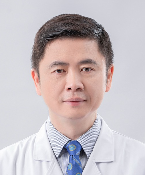 Deyu Duan (段德宇)合作论文数Union Hospital, Tongji Medical College, Huazhong University of Science and Technology2
 Jing Wang (王静)合作论文数Wuhan Union Hospital of China2Shaobin Chen合作论文数Huazhong University of Science and Technology, Huazhong Univ. of Sci. and Technol.2Chao Xu合作论文数1Dashi Zhao合作论文数1Zhuoshuo Xu合作论文数1Zuoqing Su合作论文数1Mengge Lyu合作论文数1Qiongyi Huang合作论文数1Weigang Ge合作论文数1Chanyuan Zhao合作论文数1




