The pathogenesis of Lewy body diseases (LBDs), including Parkinson’s disease (PD), involves α-synuclein (α-Syn) aggregation that originates in peripheral organs and spreads to the brain. PD incidence is increased in individuals with chronic renal failure, but the underlying mechanisms remain unknown. Here we observed α-Syn deposits in the kidneys of patients with LBDs and in the kidney and central nervous system of individuals with end-stage renal disease without documented LBDs. In male mice, we found that the kidney removes α-Syn from the blood, which is reduced in renal failure, causing α-Syn deposition in the kidney and subsequent spread into the brain. Intrarenal injection of α-Syn fibrils induces the propagation of α-Syn pathology from the kidney to the brain, which is blocked by renal denervation. Deletion of α-Syn in blood cells alleviates pathology in α-Syn A53T transgenic mice. Thus, the kidney may act as an initiation site for pathogenic α-Syn spread, and compromised renal function may contribute to the onset of LBDs. Yuan et al. find that the kidney can serve as a site of initiation for the spread of pathological α-synuclein to the brain, contributing to the development of Parkinson’s disease (PD) and providing a mechanistic link between PD and renal dysfunction.
Chronic kidney disease (CKD) is a complex disorder resulting from a combination of various environmental and genetic factors. Considerable efforts have been dedicated to elucidating its etiological mechanisms. Nevertheless, the pathogenic mechanism of CKD remains poorly understood, which hinders the development of effective therapeutic strategies. In this study, we aimed to identify novel mediators that could contribute to the development of CKD. The ClinVar, STRING, MEME Suite, TRRUST, bedtools, GEO, and R Studio databases and software were used to analyze their common features and investigate potential CKD disease genes. Transcriptomic analysis, immunohistochemistry, qRT-PCR, and Western blotting were utilized to further validate the role of ECT2 in kidney fibrosis. In total, 26 CKD disease genes were obtained from the ClinVar database, and the STRING, MEME Suite, TRRUST, bedtools, and GEO databases and software were used to analyze their common properties and explore potential CKD disease genes. Epithelial cell transforming sequence 2 (ECT2), cyclin B 1, caspase 7 and collagen alpha-1 (IV) were identified as potential candidates for CKD progression. Weighted correlation network analysis (WGCNA) subsequently revealed the relationships between potential genes and CKD. The results of the transcriptomic analysis further confirmed that ECT2 expression was greater in the kidney tissue of CKD patients than in that of healthy controls. Next, immunohistochemistry and Western blotting demonstrated that ECT2 was predominantly expressed in the renal tubules of a unilateral ureteral obstruction (UUO) mouse model. Consistently, in vitro experiments revealed that ECT2 was upregulated in TGF-β1-treated HK-2 cells. Moreover, ECT2 overexpression or knockdown in HK-2 cells altered the intensity of fibrosis markers. ECT2 significantly affects the development and progression of CKD, particularly in association with tubulointerstitial fibrosis.
Background Renal interstitial fibrosis is a common pathway for the progressive development of chronic renal diseases (CKD) with different etiology, and is the main pathological basis leading to end-stage renal disease. Although the current research on renal interstitial fibrosis is gradually deepening, the diagnosis and treatment methods are still very lacking. Uncoupling protein 1 (UCP1) is a nuclear encoded protein in mitochondria inner membrane and plays an important role in regulating energy metabolism and mitochondrial homeostasis. However, the biological significance of UCP1 and potential regulatory mechanisms in the development of CKD remain unclear. Methods Unilateral ureteral obstruction (UUO) model was used to construct the animal model of renal fibrosis, and TGF-β1 stimulation of HK2 cells was used to construct the vitro model of renal fibrosis. UCP1 expression was detected by Western blot, immunoblot analysis and immunohistochemistry. UCP1 was upregulated by UCP1 overexpressing lentivirus and UCP1 agonist CL316243. Western blot and immunofluorescence were used to detect epithelial mesenchymal transition (EMT)-related markers, such as collagen I, fibronectin, antioxidant enzyme SOD2 and CAT. Reactive oxygen species (ROS) production was detected by ROS detection kit. SIRT3 knockdown was performed by siRNA. Results This study presents that UCP1 is significantly downregulated in patients with renal fibrosis and UUO model. Further studies discover that UCP1 overexpression and CL316243 treatments (UCP1 agonists) reversed EMT and extracellular matrix (ECM) accumulation in renal fibrosis models in vivo and in vitro. Simultaneously, UCP1 reduced the ROS production by increasing the stability of SIRT3. When SIRT3 was knocked down, the production of ROS decreased. Conclusions Elevating the expression of UCP1 can inhibit the occurrence of oxidative stress by stabilizing SIRT3, thereby reducing EMT and ECM accumulation, and ultimately alleviating renal interstitial fibrosis. It will provide new instructions and targets for the treatment of CKD.
Severe acute respiratory syndrome coronavirus 2 (SARS-CoV-2) vaccination is an important method to reduce the spread of Coronavirus disease 2019 (COVID-19). While most recipients experience only mild side effects such as fever, fatigue, and injection site pain, it has been found that SARS-CoV-2 vaccination is associated with a few autoimmune problems, including the development of immune thrombotic thrombocytopenia, autoimmune liver disorders, rheumatoid arthritis, IgA nephropathy (IgAN) and systemic lupus erythematosus (SLE). There have been several reports documenting the occurrence of lupus nephritis (LN) after administration of the SARS-CoV-2 vaccine. Considering the ability to activate the immune system, vaccines carry a series of risk inducing flares or exacerbating disease in patients with glomerulonephritises.
Diabetic kidney disease (DKD) is a major microvascular complication of diabetes mellitus and is one of the leading causes of end-stage kidney disease. Circular RNAs (circRNAs) are a class of endogenous non-coding RNAs that play important roles in various diseases, yet their roles in DKD are poorly understood. CircRNA HIPK3 (circHIPK3), a highly conserved circRNA, is closely related to various cellular functions, including cell proliferation and apoptosis. The association between circHIPK3 and diabetic complications has been well demonstrated in multiple previous studies. However, the role of circHIPK3 in podocyte injury in DKD remains unclear. Herein, we discovered that circHIPK3 expression is markedly elevated in cultured podocytes under high-glucose (HG) conditions and glomeruli of diabetic mice, which is closely associated with podocyte injury in DKD. Functionally, lentivirus-mediated knockdown of circHIPK3 dramatically suppresses HG-induced podocyte apoptosis in vitro. Therapeutically, silencing circHIPK3 by adeno-associated virus-mediated RNA interference ameliorates podocyte injury and albuminuria in STZ-induced diabetic mice. Mechanistically, circHIPK3 facilitates the enrichment of fused in sarcoma (FUS) on the ectodysplasin A2 receptor (EDA2R) promoter, resulting in the upregulation of EDA2R expression and activation of apoptotic signaling. Taken together, these results indicate circHIPK3/FUS/EDA2R axis as a therapeutic target for podocyte injury and DKD progression.
Background: The newly described ABCD-10 (age, bicarbonate, cancer, dialysis, 10% body surface area [BSA]) is a 5-item mortality prediction model for patients with Stevens-Johnson syndrome (SJS)/toxic epidermal necrolysis (TEN). It was developed in the United States, has at present been externally tested only in the United States, Spain, and Singapore, and remains to be validated in resource-restricted settings. We sought to compare the accuracy of ABCD-10 and Score of Toxic Epidermal Necrolysis (SCORTEN) in predicting in-hospital mortality in a cohort from central China. Due to disease progression affecting the accuracy of the prediction model during hospitalization, for example, higher predictive accuracy of SCORTEN based on parameters collected on day 3 of hospitalization, we also assessed the overall predictive value of ABCD-10 on days 1 and 3, respectively. Methods: A retrospective study was performed over a 10-year period (2010-2020) from 3 medical institutions in Wuhan. The performance of predictive models was assessed by both discrimination and calibration. Receiver-operating characteristic (ROC) curves, Hosmer-Lemeshow goodness-of-fit tests and calibration plots were used to evaluate the model discrimination and calibration. Results: Of 84 included patients, 11 (13.1%) did not survive. The discrimination power of ABCD-10 was not significantly different from that of SCORTEN (area under the curve: day 1, p > 0.05; day 3, p > 0.05). Although the calibration of ABCD-10 was good, it was inferior to SCORTEN as it underestimated total mortality (Hosmer-Lemeshow goodness-of-fit test: day 1, p = 0.17 vs. p = 0.63; day 3, p = 0.35 vs. p = 0.93). Besides, the performance of ABCD-10 was slightly better on day 3 relative to day 1. During hospitalization, bacteremia developed in 21 (25.0%) patients, which was associated with a higher risk of death in our cohort (odds ratio, 22.88; 95% CI, 4.38-119.40; p < 0.001). Conclusion: ABCD-10 showed acceptable overall performance, but revealed mortality underestimation and was inferior to the performance of SCORTEN. In consistence with SCORTEN, ABCD-10 was a better model when using values collected at day 3 of hospitalization relative to day 1.
Although common in clinical practice, bleeding after tissue puncture may cause serious outcomes, especially in arterial puncture. Herein, gelatin-tannic acid composite hydrogels with varying compositions are prepared, and their adhesive properties are further optimized in microfluidic channel-based simulated vessels for haemostasis in arterial puncture. It is revealed that the composite hydrogels on the syringe needles used for arterial puncture should possess underwater adhesion higher than 4.9 kPa and mechanical strength higher than 86.0 kPa. The needles coated with the gelatin-tannic acid composite hydrogel completely prevent blood loss after both vein and arterial puncture in different animal models. This study holds great significance for the preparation of haemostatic needles for vessel puncture, and gelatin-tannic acid hydrogel coated needles may help to prevent complications associated with arterial puncture.
目的 筛选狼疮肾炎(lupus nephritis,LN)的差异表达基因(differentially expressed genes,DEGs)及其作用通路,探讨LN发病的分子机制.方法 芯片数据来源于高通量基因表达(Gene Expression Omnibus,GEO)数据库.选取含LN信息的基因芯片GSE127797和GSE32591.GSE127797包含LN肾组织(54例)基因表达谱,GSE32591包括正常肾组织(29例)和LN肾组织(64例)基因表达谱.利用R语言软件校正、分析基因芯片GSE127797和GSE32591并筛选DEGs,完成DEGs的基因本体(Gene Ontology,GO)富集分析和京都基因与基因组百科全书(Kyoto Encyclopedia of Genes and Genomes,KEGG)通路富集分析.用STRING数据库和Cytoscape软件分析LN差异表达基因之间的关系,并对基因之间关系进行可视化分析.使用Cytohubba插件分析蛋白质相互作用网络的关联度,筛选关键DEGs.结果 筛选出肾小球和肾小管共同DEGs 114个,其中64个上调基因,50个下调基因.GO分析结果显示,生物学过程(biolog-ical process,BP)方面,共同DEGs主要参与病毒防御反应、Ⅰ型干扰素(type Ⅰ interferon,IFN-Ⅰ)信号通路、病毒基因组复制的调控等生物过程;细胞组成(cellular component,CC)方面,共同DEGs参与的细胞组分包括胶原蛋白三聚物、血小板a颗粒、胞质囊腔、细胞器外膜等;分子功能(molecular function,MF)方面,共同DEGs主要与血小板衍生生长因子结合、细胞外基质结构成分、生长因子结合相关.KEGG分析结果表明,共同DEGs与甲型流感病毒感染、麻疹病毒感染、丙型肝炎病毒感染、百日咳杆菌感染、补体和凝血级联、蛋白质消化吸收、金黄色葡萄球菌感染等通路有关.STRING 分析发现了蛋白质互作网络图中的 10 个关键 DEGs:ISG15、MX1、OAS1、MX2、IFIH1、GBP1,IFIT3、OAS2、IFIT1、IFI44.结论 通过生物信息学方法分析LN DEGs的生物学过程、细胞组成与分子功能,发现关键DEGs,为探索LN发病相关分子机制、发掘潜在诊断标志物和开发新的治疗靶点提供了理论依据及新的方向.
The kidney tropism of severe acute respiratory syndrome coronavirus 2 (SARS-CoV-2) has been well-validated clinically and often leads to various forms of renal damage in coronavirus disease-2019 (COVID-19) patients. However, the underlying mechanisms and diagnostic approaches remain to be determined. We interrogated the expression of virus-related host factors in single-cell RNA sequencing (scRNA-seq) datasets of normal human kidneys and kidneys with pre-existing diseases and validated the results with urinary proteomics of COVID-19 patients and healthy individuals. We also assessed the effects of genetic variants on kidney susceptibility using expression quantitative trait loci (eQTLs) databases. We identified a subtype of tubular cells, which we named PT-3 cells, as being vulnerable to SARS-CoV-2 infections in the kidneys. PT-3 cells were enriched in viral entry factors and replication and assembly machinery but lacked antiviral restriction factors. Immunohistochemistry confirmed positive staining of PT-3 cell marker SCL36A2 on kidney sections from COVID-19 patients. Urinary proteomic analyses of COVID-19 patients revealed that markers of PT-3 cells were significantly increased, along with elevated viral receptor angiotensin-converting enzyme 2. We further found that the proportion of PT-3 cells increased in diabetic nephropathy but decreased in kidney allografts and lupus nephropathy, suggesting that kidney susceptibility varied among these diseases. We finally identified several eQTLs that regulate the expression of host factors in kidney cells. PT-3 cells may represent a key determinant for the kidney tropism of SARS-CoV-2, and detection of PT-3 cells may be used to assess the risk of renal infection during COVID-19.
The molecular pathology of multi-organ injuries in COVID-19 patients remains unclear, preventing effective therapeutics development. Here, we report a proteomic analysis of 144 autopsy samples from seven organs in 19 COVID-19 patients. We quantified 11,394 proteins in these samples, in which 5,336 were perturbed in the COVID-19 patients compared to controls. Our data showed that cathepsin L1, rather than ACE2, was significantly upregulated in the lung from the COVID-19 patients. Systemic hyperinflammation and dysregulation of glucose and fatty acid metabolism were detected in multiple organs. We also observed dysregulation of key factors involved in hypoxia, angiogenesis, blood coagulation, and fibrosis in multiple organs from the COVID-19 patients. Evidence for testicular injuries includes reduced Leydig cells, suppressed cholesterol biosynthesis, and sperm mobility. In summary, this study depicts a multi-organ proteomic landscape of COVID-19 autopsies that furthers our understanding of the biological basis of COVID-19 pathology.
Tissue puncture is an effective means in the diagnosis and treatment of diseases, providing important information for clinical doctors. Yet, haemorrhage is still a main and unavoidable complication, which can cause serious outcomes, especially in patients with abnormal coagulation function. Herein, we present a facile yet effective strategy to prepare haemostatic needles by coating the syringe needles surface with alginate-CaCl2-based hydrogel with carefully optimized mechanical and chemical properties, which can effectively prevent bleeding following tissue puncture. The needle with haemostatic coating exhibits complete prevention of blood loss following vessel and viscera puncture in the normal animal as well as in animal models with hemorrhagic diathesis. The syringe needle with haemostatic coating in this work holds enormous potentials for clinical applications, including sampling and injection.
BACKGROUND:Coronavirus disease 2019 (COVID-19), caused by severe acute respiratory syndrome coronavirus 2 (SARS-CoV-2), involves multiple organs. Testicular involvement is largely unknown. OBJECTIVE:To determine the pathological changes and whether SARS-CoV-2 can be detected in the testes of deceased COVID-19 patients. DESIGN, SETTING, AND PARTICIPANTS:Postmortem examination of the testes from 12 COVID-19 patients was performed using light and electron microscopy, and immunohistochemistry for lymphocytic and histiocytic markers. Reverse transcription-polymerase chain reaction (RT-PCR) was used to detect the virus in testicular tissue. OUTCOME MEASUREMENTS AND STATISTICAL ANALYSIS:Seminiferous tubular injury was assessed as none, mild, moderate, or severe according to the extent of tubular damage. Leydig cells in the interstitium were counted in ten 400× microscopy fields. RESULTS AND LIMITATIONS:Microscopically, Sertoli cells showed swelling, vacuolation and cytoplasmic rarefaction, detachment from tubular basement membranes, and loss and sloughing into lumens of the intratubular cell mass. Two, five, and four of 11 cases showed mild, moderate, and severe injury, respectively. The mean number of Leydig cells in COVID-19 testes was significantly lower than in the control group (2.2 vs 7.8, p < 0.001). In the interstitium there was edema and mild inflammatory infiltrates composed of T lymphocytes and histiocytes. Transmission EM did not identify viral particles in three cases. RT-PCR detected the virus in one of 12 cases. CONCLUSIONS:Testes from COVID-19 patients exhibited significant seminiferous tubular injury, reduced Leydig cells, and mild lymphocytic inflammation. We found no evidence of SARS-CoV-2 virus in the testes in the majority (90%) of the cases by RT-PCR, and in none by electron microscopy. These findings can provide evidence-based guidance for sperm donation and inform management strategies to mitigate the risk of testicular injury during the COVID-19 disease course. PATIENT SUMMARY:We examined the testes of deceased COVID-19 patients. We found significant damage to the testicular parenchyma. However, virus was not detected in testes in the majority of cases.
新型冠状病毒肺炎(corona virus disease 2019,COVID-19),简称新冠肺炎,最先在武汉被发现,目前在全世界范围广泛传播.严重急性呼吸道综合征冠状病毒2型(SARS-CoV-2,简称新冠病毒)是导致新冠肺炎的病原体,其主要通过血管紧张素转化酶2 (ACE2)介导而侵入靶细胞.除肺泡2型上皮细胞外,肠道、肾脏的上皮细胞亦高表达ACE2,故除了呼吸道症状外,腹痛腹泻等消化系统症状在新冠肺炎患者中也很常见.值得关注的是,新冠肺炎相关肾脏损害的发生并不少见,主要临床表现为急性肾损伤(AKI)、蛋白尿和血尿.
Objectives: To observe the pulmonary changes with coronavirus disease 2019 (COVID-19) in postmortem needle specimens, to detect the presence of 2019 novel coronavirus(2019-nCoV) in the lung tissues, and to analyze the clinicopathological characteristics. Methods: For 10 decedents with 2019-nCoV infection in Wuhan, bilateral lungs underwent ultrasound-guided percutaneous multi-point puncture autopsy, and pulmonary pathological changes were described in routine hematoxylin-eosin staining (HE) slides. Electron microscopy was also performed. The reverse transcription polymerase chain reaction (RT-PCR) was employed to detect 2019-nCoV nucleic acid in lung tissue, and the pathological characteristics were demonstrated in combination with clinical data analysis. Results: Of the 10 deaths associated with COVID-19, 7 were male and 3 were female. The average age was 70 (39-87) years. Medical record showed that 7 patients had underlying diseases. The average course of disease was 30 (16-36) days. Nine cases showed fibrinous and suppurative exudation in the alveolar cavity accompanied by the formation of hyaline membrane, and fibroblastic proliferation of alveolar septum. Type Ⅱ alveolar epithelial cells showed reactive hyperplasia and desquamation. Many macrophages accumulated in the alveolar cavity. Capillary hyaline thrombus and intravascular mixed thrombus were noted. In some cases, acute bronchiolitis with mucous membrane exfoliation, accumulation of bronchiolar secretions, and bronchiolar epithelial metaplasia occurred. In the cohort, a large number of bacteria (cocci) were detected in 1 case and a large number of fungi (yeast type) were detected in 1 case. Nine cases were positive for the nucleic acids of 2019-nCoV while one case remained negative by RT-PCR. Coronavirus particles were detected in the cytoplasm of type Ⅱ alveolar epithelium. Conclusions: The pulmonary pathological changes of fatal COVID-19 are diffuse alveolar damage (DAD), mainly in the acute exudative stage and the organic proliferative stage. There are fibrinous exudate aggregation in alveolar cavity with hyaline membrane formation, fibroblastic proliferation in alveolar septum, and alveolar epithelial cell injuries with reactive hyperplasia and desquamation of type Ⅱ alveolar epithelial cells. A large amount of neutrophils and monocytes infiltration is present in most cases and bacteria and fungi are detected in some cases, suggesting a serious bacterial or fungal infection secondary to the DAD.
Objective:To explore the clinicopathologic features and risk factors of IgA nephropathy (IgAN) after kidney transplantation.Methods:A retrospective analysis was conducted for biopsy-confirmed IgAN patients after transplantation from January 2016 to September 2019. The clinicopathological characteristics and risk factors of IgAN after expanded criteria donor (ECD) donor for kidney transplantation were examined by statistical analysis of general clinical data and Oxford classification during and after transplantation.Results:The diagnostic rate of IgAN after transplantation was around 12.77%. The clinical symptoms occurred at an average of 22.5 months after transplantation. 66.67%(12/18) patients were in stage 3-4 chronic kidney disease (CKD). There were microscopic hematuria (76.92%) and varying degrees of proteinuria (88.89%). The scores of pathological parameters were as follows: 78% patients with mesangial cell proliferation (M1), 17% with endothelial cell proliferation (E1), 65% with segmental sclerosis (S1), 39% with moderate-to-severe tubules atrophy/interstitial fibrosis (T1/T2) and 17% with crescent formation (C1) respectively. Tubuloatrophy/interstitial fibrosis was associated with estimated glomerular filtration rate (eGFR) at biopsy ( P<0.05). Group of urine protein quantification >1g/d and group of <1g/d group had statistically significant differences in segmental sclerosis and adhesion lesion ( P<0.05). Conclusions:Oxford classification is valuable in the clinical evaluations of allograft IgAN: Tubular atrophy/interstitial fibrosis is associated with a decline of glomerular filtration. And segmental sclerosis and adhesion lesion are correlated with the severity of proteinuria.
Previously, selenoprotein T (SelT) expression was shown to be induced in nervous, endocrine, and metabolic tissues during ontogenetic and regenerative processes. However, whether SelT plays a critical role in renal diseases remains unclear. Here, we explored the role of SelT in cisplatin-induced acute kidney injury (AKI). Results revealed that SelT was highly expressed in renal tubules, but its expression was significantly reduced in cisplatin-induced AKI. Importantly, knocking down of SelT expression in kidney cells in vitro resulted in cisplatin-induced cell apoptosis, as indicated by the elevation of cleaved-PARP and Bax expression, Caspase-3 activity, and number of TUNEL-positive cells. Moreover, SelT silencing-induced reactive oxygen species (ROS) production, accompanied by a decrease in intracellular superoxide dismutase (SOD) and catalase (CAT) activity and increase in malondialdehyde (MDA) content. Notably, the protein and mRNA levels of Nox4 were increased in response to SelT downregulation. Furthermore, suppression of Nox4 expression by GKT137831 partially alleviated SelT knockdown-induced ROS generation and cell apoptosis in cisplatin-treated kidney cells. Taken together, our findings provide the first evidence that SelT protects against cisplatin-induced AKI by suppression of oxidative stress and apoptosis.
Although the respiratory and immune systems are the major targets of Coronavirus Disease 2019 (COVID-19), acute kidney injury and proteinuria have also been observed. Currently, detailed pathologic examination of kidney damage in critically ill patients with COVID-19 has been lacking. To help de fine this we analyzed kidney abnormalities in 26 autopsies of patients with COVID-19 by light microscopy, ultrastructural observation and immunostaining. Patients were on average 69 years (19 male and 7 female) with respiratory failure associated with multiple organ dysfunction syndrome as the cause of death. Nine of the 26 showed clinical signs of kidney injury that included increased serum creatinine and/or new -onset proteinuria. By light microscopy, diffuse proximal tubule injury with the loss of brush border, non -isometric vacuolar degeneration, and even frank necrosis was observed. Occasional hemosiderin granules and pigmented casts were identi fied. There were prominent erythrocyte aggregates obstructing the lumen of capillaries without platelet or fibrinoid material. Evidence of vasculitis, interstitial in flammation or hemorrhage was absent. Electron microscopic examination showed clusters of coronavirus-like particles with distinctive spikes in the tubular epithelium and podocytes. Furthermore, the receptor of SARS-CoV-2, ACE2 was found to be upregulated in patients with COVID-19, and immunostaining with SARS-CoV nucleoprotein antibody was positive in tubules. In addition to the direct virulence of SARS-CoV-2, factors contributing to acute kidney injury included systemic hypoxia, abnormal coagulation, and possible drug or hyperventilation -relevant rhabdomyolysis. Thus, our studies provide direct evidence of the invasion of SARSCoV-2 into kidney tissue. These findings will greatly add to the current understanding of SARS-CoV-2 infection.
The Journal of DermatologyVolume 47, Issue 4 p. e119-e120 Letter to the Editor Development of generalized bullous lesions after hemodialysis with polysulfone membrane dialyzer Huinan Suo, Huinan Suo Department of Dermatology, Union Hospital, Tongji Medical College, Huazhong University of Science and Technology, Wuhan, ChinaSearch for more papers by this authorHua Su, Hua Su Department of Nephrology, Union Hospital, Tongji Medical College, Huazhong University of Science and Technology, Wuhan, ChinaSearch for more papers by this authorChangyu Lu, Changyu Lu Department of Dermatology, The First People's Hospital of Jingzhou, Jingzhou, ChinaSearch for more papers by this authorLiu Yang, Corresponding Author Liu Yang bengpao82@163.com orcid.org/0000-0002-3839-424X Department of Dermatology, Union Hospital, Tongji Medical College, Huazhong University of Science and Technology, Wuhan, China Correspondence: Liu Yang, M.D., and Juan Tao, M.D., Ph.D., Department of Dermatology, Union Hospital, Tongji Medical College, Huazhong University of Science and Technology, 1277 Jiefang Avenue, Wuhan, Hubei 430022, China. Emails: bengpao82@163.com (L. Y.) and tjhappy@126.com (J. T.)Search for more papers by this authorJuan Tao, Corresponding Author Juan Tao tjhappy@126.com Department of Dermatology, Union Hospital, Tongji Medical College, Huazhong University of Science and Technology, Wuhan, China Correspondence: Liu Yang, M.D., and Juan Tao, M.D., Ph.D., Department of Dermatology, Union Hospital, Tongji Medical College, Huazhong University of Science and Technology, 1277 Jiefang Avenue, Wuhan, Hubei 430022, China. Emails: bengpao82@163.com (L. Y.) and tjhappy@126.com (J. T.)Search for more papers by this author Huinan Suo, Huinan Suo Department of Dermatology, Union Hospital, Tongji Medical College, Huazhong University of Science and Technology, Wuhan, ChinaSearch for more papers by this authorHua Su, Hua Su Department of Nephrology, Union Hospital, Tongji Medical College, Huazhong University of Science and Technology, Wuhan, ChinaSearch for more papers by this authorChangyu Lu, Changyu Lu Department of Dermatology, The First People's Hospital of Jingzhou, Jingzhou, ChinaSearch for more papers by this authorLiu Yang, Corresponding Author Liu Yang bengpao82@163.com orcid.org/0000-0002-3839-424X Department of Dermatology, Union Hospital, Tongji Medical College, Huazhong University of Science and Technology, Wuhan, China Correspondence: Liu Yang, M.D., and Juan Tao, M.D., Ph.D., Department of Dermatology, Union Hospital, Tongji Medical College, Huazhong University of Science and Technology, 1277 Jiefang Avenue, Wuhan, Hubei 430022, China. Emails: bengpao82@163.com (L. Y.) and tjhappy@126.com (J. T.)Search for more papers by this authorJuan Tao, Corresponding Author Juan Tao tjhappy@126.com Department of Dermatology, Union Hospital, Tongji Medical College, Huazhong University of Science and Technology, Wuhan, China Correspondence: Liu Yang, M.D., and Juan Tao, M.D., Ph.D., Department of Dermatology, Union Hospital, Tongji Medical College, Huazhong University of Science and Technology, 1277 Jiefang Avenue, Wuhan, Hubei 430022, China. Emails: bengpao82@163.com (L. Y.) and tjhappy@126.com (J. T.)Search for more papers by this author First published: 17 February 2020 https://doi.org/10.1111/1346-8138.15261Citations: 1 The corresponding authors contributed equally to this work. Read the full textAboutPDF ToolsRequest permissionExport citationAdd to favoritesTrack citation ShareShare Give accessShare full text accessShare full-text accessPlease review our Terms and Conditions of Use and check box below to share full-text version of article.I have read and accept the Wiley Online Library Terms and Conditions of UseShareable LinkUse the link below to share a full-text version of this article with your friends and colleagues. Learn more.Copy URL Share a linkShare onFacebookTwitterLinkedInRedditWechat No abstract is available for this article.Citing Literature Volume47, Issue4April 2020Pages e119-e120 RelatedInformation
 Chun Zhang (张春)合作论文数Union Hospital, Tongji Medical College, Huazhong University of Science and Technology20
 Chen Guangping合作论文数9Lei Chun-Tao合作论文数7Chunyun Zhang合作论文数6
 Cheng Wan合作论文数Union Hosp, Huazhong Univ Sci & Technol5
 Ye Chen (陈烨)合作论文数College of Life Sciences, Zhejiang University5
 Anguo Deng (邓安国)合作论文数Union Hospital, Tongji Medical College, Huazhong University of Science and Technology5
 Xiu Nie (聂秀)合作论文数Union Hospital, Tongji Medical College, Huazhong University of Science and Technology5
 C. Barry Carter合作论文数Department of Materials Science and Engineering, College of Engineering, University of Connecticut4Froehlich Otto合作论文数4
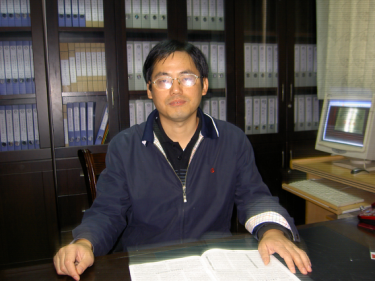 hui Tang (唐晖)合作论文数School of Physical Education, Hunan University of Science and Technology4
 Yanhong Zhu (朱艳红)合作论文数School of Life Science and Technology, Huazhong University of Science and Technology4Huinan Suo合作论文数3Jd Klein合作论文数3QiuXiang Xia (夏秋翔)合作论文数3He Fangfang合作论文数3
 Fan Yi (易凡)合作论文数Department of Pharmaco, School of Basic Medical Sciences, Shandong University3
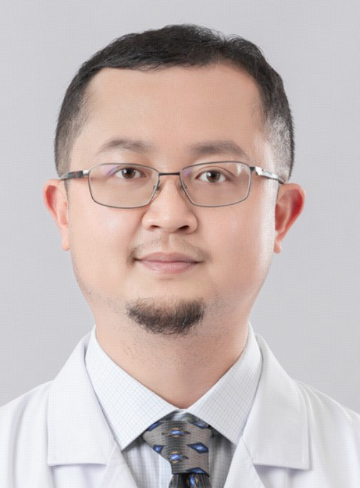 Zhendi Wang (王振迪)合作论文数Union Hospital, Tongji Medical College, Huazhong University of Science and Technology3
 Juan Tao (陶娟)合作论文数Union Hospital, Tongji Medical College, Huazhong University of Science and Technology3
 Zhonghua Zhu (朱忠华)合作论文数Union Hospital, Tongji Medical College, Huazhong University of Science and Technology3
 Jianshe Liu (刘建社)合作论文数Union Hospital, Tongji Medical College, Huazhong University of Science and Technology3
 Yumei Wang (王玉梅)合作论文数Union Hospital, Tongji Medical College, Huazhong University of Science and Technology3Li-Xia Yi合作论文数2Jieyu Zeng合作论文数2Xiaona Chang合作论文数2Liu Yang合作论文数2Yu Chen合作论文数2Xingjie Yin合作论文数2Jingli Ren合作论文数2
 Jun Fan合作论文数2Junjie Zhou合作论文数2Shuo Chen合作论文数2
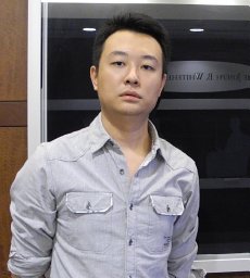 Bing-Chen Liu合作论文数Dept Cardiol, Harbin Med Univ2Xu Gang合作论文数2
 Titilayo Ilori合作论文数2
 He-Ping Ma合作论文数2Lin Jihong合作论文数2
 Lianbin Zhang (张连斌)合作论文数School of Chemistry and Chemical Engineering, Huazhong University of Science and Technology2Luo Dan-Ju合作论文数2
 Agnes B. Fogo合作论文数Department of Pathology, Microbiology and Immunology, Vanderbilt University Medical Center2
 Li Xiang (项莉)合作论文数School of Medicine and Health Management, Tongji Medical College, Huazhong University of Science & Technology2
 Jing Xiong (熊京)合作论文数Union Hospital, Tongji Medical College, Huazhong University of Science and Technology2
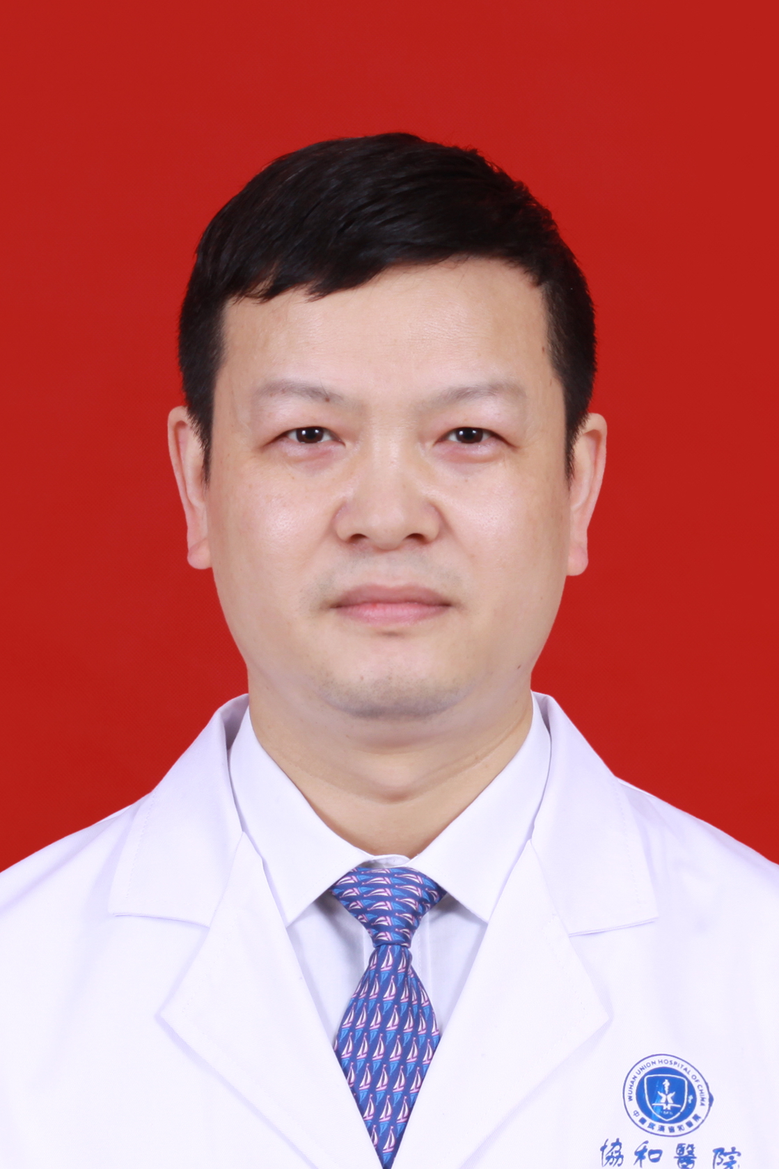 Ming Yang (杨明)合作论文数Union Hospital, Tongji Medical College, Huazhong University of Science and Technology2
 Xiao Yang (杨晓)合作论文数Union Hospital, Tongji Medical College, Huazhong University of Science and Technology2
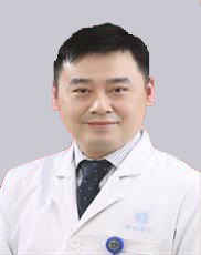 Jing Huang (黄晶)合作论文数Union Hospital, Tongji Medical College, Huazhong University of Science and Technology2
 Bo Huang (黄波)合作论文数Institute of Basic Medical Sciences, Chinese Academy of Medical Sciences2
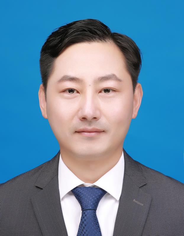 Jintao Zhu (朱锦涛)合作论文数School of Chemistry and Chemical Engineering, Huazhong University of Science and Technology2
 Hai-Chun Yang合作论文数Fu Dan University Medical Center, Shanghai Medical University2




