While typical free fatty acids (FFAs) are essential for maintaining skin barrier homeostasis, non-typical ones can transiently disrupt the skin barrier, which, when properly timed, leads to enhanced drug permeation. Accordingly, we question what structural features of FFAs can be tailored to enhance the transdermal delivery of water-soluble drugs. By systematically tuning acyl chain length, unsaturation, and head group chemistry, we experimentally and computationally investigated FFA-triggered variations in both lipid lamellar organization and drug permeability. Importantly, our findings suggest that the collapse of the ordered lamellar phase does not necessarily ensure increased permeability, challenging the prevailing “disorder-driven permeability” hypothesis. Our experimental observations indicate that transport efficiency appears to be governed by the specific molecular geometry of FFAs, particularly their chain length, unsaturation, and head groups. In addition, our molecular dynamics simulations provide a plausible molecular rationale, suggesting that C18 chains with a mid-leaflet cis-double bond and interfacially exposed carboxylic head groups may represent a favorable configuration for hydrophilic drug delivery. Leveraging this design principle, we engineered photo-switchable, azobenzene-functionalized FFAs as an experimental proof-of-concept to explore our proposed framework. We show that light-induced conformational switching modulates isomer-dependent drug permeability across both artificial skin membranes and ex vivo porcine skin. This molecular-level understanding provides a blueprint for rational design of penetration enhancers and offers a potential foundation for the development of conformation-dependent delivery systems.
Mechanical forces within the tumor microenvironment are increasingly recognized as important regulators of cancer progression, yet their influence on apoptotic regulation in melanoma remains insufficiently defined. Here, we developed a customized stretchable culture platform capable of delivering physiologically relevant cyclic mechanical strain and investigated its effects on intrinsic apoptosis in melanoma cells with different metastatic potentials. Cyclic stretch significantly reduced apoptosis in high metastatic melanoma cells, while low-metastatic counterparts exhibited minimal or differential responses. Mechanistically, mechanical stimulation suppressed intrinsic mitochondrial apoptotic signaling along the BAD-BCL-2 family-BAX axis, leading to reduced caspase-3/7 activation. Pharmacological modulation of mechanosensitive channel activity and intracellular Ca2+ signaling partially restored apoptotic responses under stretch, supporting the involvement of calcium-associated mechanotransduction in this process. Importantly, stretched high metastatic melanoma cells displayed reduced sensitivity to pharmacological apoptosis induction, indicating altered apoptotic responsiveness under mechanical stimulation. In vivo models further demonstrated that prior mechanical conditioning enhanced survival and metastatic behavior in high metastatic melanoma. Together, these findings reveal that cyclic mechanical stress promotes apoptosis resistance in a metastatic potential-dependent manner and highlight the importance of considering tissue mechanics in understanding melanoma progression and therapeutic response.
The development of effective treatment strategies against head and neck cancer (HNC) poses a significant challenge due to the heterogeneity of HNC and the limitations of traditional two-dimensional (2D)in vitrocell cultures. Consequently, there is an urgent need for more advanced HNC models that allow for reliable drug screening. The recent advances in tissue engineering, including the use of digital light processing-based three-dimensional (3D) bioprinters, have enabled the precise fabrication of complex constructs that mimic native tissue environments, exhibiting potential for use in disease modeling and drug discovery. Here, we fabricated a bioprintedin vitroHNC model using the gelatin methacryloyl bioink encapsulating FaDu cells to analyze the drug screening against HNC. The bioprinted tissues showed gradually increased cell viability as well as stable morphology and phenotype expression. The cytotoxicity analyses suggested increased resistance to anti-cancer drugs for cells in 3D-bioprinted tissues than those cultured in 2D, possibly due to the presence of native cell-cell interactions and diffusion barriers. The engineered exosomes with anti-cancer drugs also revealed significantly increased resistance in cells cultured in 3D than in 2D. This study provides a proof-of-concept bioprinted HNC model mimicking some of the native functional and structural components, having the potential for screening novel anti-HNC treatments and paving the way for personalized therapeutic strategies for HNC patients in the future.
Long-term monitoring of plant health is essential in agriculture for managing crop yields. However, sensors with rigid electrodes can harm plants by disrupting their homeostasis and growth. Here, we introduce an electronic plant arrow (EPA), a substrate-free electrode made from photo-crosslinked gelatin methacryloyl (GelMA). GelMA offers flexibility, biocompatibility, and low electrical impedance, allowing electrical plant monitoring. The EPA is rigid when dry for easy insertion but softens and swells upon moisture absorption, minimizing tissue damage while ensuring stable electrical contact. The arrowhead shape enhances stable attachment, while the substrate-free design minimizes growth interference. This system enables continuous, organ-specific monitoring of plants, supporting precision agriculture.
Hybrid nanoassemblies were designed to simultaneously induce apoptosis, disulfidptosis, and ferroptosis in triple-negative breast cancer (TNBC) cells under glucose-deprived conditions. Cystine transport via SLC7A11 is essential for glutathione (GSH) biosynthesis, therefore the concurrent induction of ferroptosis (through GSH depletion) and disulfidptosis (via cystine accumulation) seems to be contradictory. Glucose deprivation markedly reduces intracellular nicotinamide adenine dinucleotide phosphate (NADPH) levels, and this NADPH insufficiency compromises cystine reduction to cysteine, thereby enabling the simultaneous onset of ferroptosis and disulfidptosis in cancer cells. To exploit this metabolic vulnerability, phloretin (PHL) was incorporated into the nanoassemblies as a glucose transporter-1 inhibitor to suppress glucose uptake. In addition, hydrophobic ferrocene (Fc) and D-alpha-tocopherol succinate (TS) were chemically conjugated to a hyaluronic acid (HA) oligomer, serving as an iron-containing ferroptosis inducer and a mitochondria-destabilizing apoptosis trigger, respectively. The resulting amphiphilic hybrid conjugate (Fc-HA-TS) spontaneously self-assembles into nanoscale structures in aqueous environments. The designed Fc-HA-TS/PHL nanoassemblies are expected to orchestrate multiple programmed cell death modalities: (1) disulfidptosis induction, GSH depletion, and GSH-independent ferroptosis suppressor protein 1 deactivation via glucose starvation (PHL), (2) ferroptosis activation through lipid peroxidation (Fc), and (3) apoptosis induction via mitochondrial destabilization (TS), ultimately offering a synergistic and metabolically targeted therapeutic strategy for TNBC.
Uniform dispersion of carbon nanotubes (CNTs) within lithium cobalt oxide (LCO)-based conductive composites is essential for establishing efficient conductive pathways but remains challenging due to CNT agglomeration. Here, we present a gelatin methacryloyl (GelMA)-assisted dispersion strategy for constructing conductive LCO/CNT composites with controlled microstructural organization. LCO particles and CNTs were assembled within GelMA-stabilized emulsions, followed by freeze-drying to preserve the assembled architecture and subsequent pyrolysis to remove the GelMA matrix. X-ray diffraction confirmed that the crystalline structure of LCO was maintained after thermal treatment, while Raman spectroscopy and X-ray photoelectron spectroscopy verified the complete removal of GelMA. SEM observations revealed uniform CNT distribution and intimate CNT-LCO interfacial contact within the composite. Comparison with a pyrolyzed LCO/CNT control prepared without GelMA demonstrated that thermal treatment alone was insufficient to achieve the observed structural and electrical improvements. Instead, the GelMA-assisted dispersion process promoted the formation of an interconnected CNT network throughout the electrode architecture. The resulting composite exhibited reduced electrical resistance and enhanced conductivity, indicating that electrical conductivity is governed primarily by CNT dispersion and network connectivity rather than by porosity alone. This work establishes a practical gel-mediated process for dispersing, assembling, and thermally consolidating LCO/CNT composites, providing a generalizable route to conductive particulate architectures for composite manufacturing.
The osteochondral interface is a finely tuned junction between cartilage and bone, coordinated by gradients in mechanics and metabolism. Recreating this complexity in vitro has remained elusive. Here, we present a developmentally inspired, dual-gradient 3D-printed construct that unites native-like stiffness and metabolic microenvironments to drive spatially resolved regeneration and model osteoarthritis in a phase-specific manner. Human bone marrow-derived mesenchymal stem cell spheroids are placed in soft, hypoxic niches to promote chondrogenesis, and in stiff, vascular-rich regions to induce osteogenesis-preserving cartilage-bone crosstalk within one platform. This integration of gradients amplifies extracellular matrix formation beyond single-cue designs through the synergistic effect of porosity and stiffness. Furthermore, its anisotropic architecture maintains cartilage-bone crosstalk and allows drug response assessment, highlighting its potential as a physiologically relevant platform for osteoarthritis modeling and therapy screening. By bridging functional regeneration with preclinical drug screening, this model offers a physiologically relevant translational platform for advancing both osteochondral repair and disease research.
Accurate, label-free detection of disease-specific biomarkers is essential for early diagnosis and therapeutic monitoring. Here, we present a reusable and ultrasensitive biosensor platform based on van der Waals (vdW) junction field-effect transistor (JFET), integrated with an external metal-insulator-metal (MIM) capacitor. This pseudo-floating gate configuration physically decouples the sensing interface from the transistor's active channel, thereby minimizing charge trapping, eliminating hysteresis, and enabling stable, dynamic step-response measurements and device reusability. This architecture allows for rapid, label-free detection of liver biomarkers-human serum albumin and AFP-L3-with a limit of detection below 0.1 ng mL-1 and a dynamic range from 0.1 ng mL-1 to 1 mg mL-1, addressing the key limitations of conventional ELISA assays. Additionally, we demonstrate its translational potential by evaluating the device with 3D hepatocyte spheroid disease models to detect drug-induced alterations in biomarker secretion in a physiologically relevant context. Notably, the device revealed cell-line-specific biomarker responses to doxorubicin (Dox), highlighting the capability of the device to resolve subtle and physiologically meaningful variations in secretion dynamics. Overall, this work introduces a generalizable sensing strategy for high-performance biosensors, combining electrical robustness, analytical precision, and clinical applicability, paving the way for next-generation technologies in both preclinical research and clinical diagnostics.
Pathological scars are fibrotic disorders arising from aberrant wound healing. PDT is effective but limited by a dense matrix, hypoxia, and post-PDT excessive ROS-induced secondary inflammation. To address this, a NIR-switchable ROS-regulatory liposome (ICC@Lip) with CaO2 NPs (oxygenation to strengthen PDT) and Cu5.4O nanozymes (ROS scavenging) was developed. In addition, inspired by Cyperaceae stems and triangular arrows, a highly penetrable biomimetic microneedle platform loaded with ICC@Lip and collagenase I (ICC@Lip/HPMN) was constructed to realize efficient transdermal cargo delivery and diffusion into a rigid pathological scar matrix. The puncture resistance of ICC@Lip/HPMN was only 56.4% that of conventional rectangular pyramid microneedles, with an effective puncture rate of 96% in mouse skin. In vitro, under NIR irradiation-mediated PDT/PTT, ICC@Lip significantly alleviated intracellular hypoxia and related fibrotic indicators, achieving a cytotoxicity rate of 93.71% against myofibroblasts─1.49-fold higher than that of the IR808 alone group (62.74%)─while reducing M2 macrophage polarization and TGF-β secretion. Notably, timely post-PDT ROS clearance decreased M1 polarization of macrophages and reduced the release of over 50% of inflammatory cytokines. In rabbit ear, a hypertrophic scar model exhibited that ICC@Lip/HPMN reduced the scar elevation index from 3 to 1.7, significantly alleviating collagen deposition and inflammation. The antifibrotic efficacies were majorly exerted through cross-downregulation of TGF-β, IL-4, and toll-like receptor signaling pathways. This study provided a promising adaptive therapeutic strategy of self-strengthened PDT coupling ROS precise regulation for pathological scars.
Engineering functional arterial tissues in vitro requires dynamic cues that recapitulate native mechanical environments. A bidirectional stimulation approach, termed blood and tissue side stretch (BTS), is presented, applying cyclic circumferential stretch to drive maturation of arterial microphysiological systems (aMPSs). These stimuli replicate key biomechanical forces present in vivo—cyclic circumferential stretch—enabling a more physiologically relevant tissue architecture. Human umbilical vein endothelial cells (HUVECs) and human smooth muscle cells (SMCs) are co-cultured within a bilayered vessel structure composed of an elastomeric hydrogel that mimics native vessel geometry and compliance. BTS stimulation enhances alignment of collagen fibers, promotes expression of contractile markers in SMCs, and improves barrier function and junctional protein localization in HUVECs. The matured aMPS exhibits vasomotor responsiveness and biomechanical integrity, validating its physiological relevance. Comparative analysis shows that BTS outperforms static and one-directional controls in promoting vascular tissue maturation. This platform provides a scalable and biomimetic solution for vascular tissue engineering and disease modeling. By integrating orthogonal mechanical cues that mimic the in vivo arterial environment, this approach represents a significant step forward in the development of predictive, functional, and high-fidelity vascular models for drug testing and regenerative medicine applications.
Skin-on-a-chip models provide physiologically relevant platforms for studying diseases and drug evaluation, replicating the native skin structures and functions more accurately than traditional 2D or simple 3D cultures. However, challenges remain in creating models suitable for microneedling applications and monitoring, as well as developing skin cancer models for analysis and targeted therapy. Here, we developed a human skin/skin cancer-on-a-chip platform within a microfluidic device using bioprinting/bioengineering techniques. The fabricated skin models include vascular, dermal, and epidermal layers, demonstrating increased functionalities and maturation of dermal (Collagen I & Fibronectin for 7 days) as well as epidermal (Filaggrin & Keratin 10, 14, and 19 at the air-liquid interface (ALI) for 21 days) layers. Histological analysis confirmed the formation of a differentiated epidermis and ridges at the dermal-epidermal junction in our model, closely resembling native skin tissue. Melanoma cells were embedded approximately 400 μm beneath the epidermis to simulate tumor invasion into the dermis. The platform was further used to test doxorubicin (DOX)-loaded gelatin methacryloyl (GelMA) microneedles (MNs) for localized transdermal drug delivery targeting melanoma. The DOX-loaded MNs penetrated uniformly to a depth of approximately 600 μm, effectively reaching the melanoma cells. Drug delivery via MNs demonstrated significantly higher efficiency than diffusion through media flow, confirming the practicality and robustness of the proposed model for future therapeutic applications.
Advanced Healthcare MaterialsVolume 14, Issue 4 2570025 Inside Back CoverFree Access Pseudo-3D Topological Alignments Regulate Mechanotransduction and Maturation of Smooth Muscle Cells (Adv. Healthcare Mater. 4/2025) Yeji Lee, Yeji LeeSearch for more papers by this authorJihyeon Song, Jihyeon SongSearch for more papers by this authorUlziituya Batjargal, Ulziituya BatjargalSearch for more papers by this authorMin-Seok Kim, Min-Seok KimSearch for more papers by this authorGeonho Lee, Geonho LeeSearch for more papers by this authorGeonwoo Kim, Geonwoo KimSearch for more papers by this authorTaehoon Lee, Taehoon LeeSearch for more papers by this authorRaeHui Kang, RaeHui KangSearch for more papers by this authorYounggyun Kim, Younggyun KimSearch for more papers by this authorHan-Jun Kim, Han-Jun KimSearch for more papers by this authorJunmin Lee, Junmin LeeSearch for more papers by this author Yeji Lee, Yeji LeeSearch for more papers by this authorJihyeon Song, Jihyeon SongSearch for more papers by this authorUlziituya Batjargal, Ulziituya BatjargalSearch for more papers by this authorMin-Seok Kim, Min-Seok KimSearch for more papers by this authorGeonho Lee, Geonho LeeSearch for more papers by this authorGeonwoo Kim, Geonwoo KimSearch for more papers by this authorTaehoon Lee, Taehoon LeeSearch for more papers by this authorRaeHui Kang, RaeHui KangSearch for more papers by this authorYounggyun Kim, Younggyun KimSearch for more papers by this authorHan-Jun Kim, Han-Jun KimSearch for more papers by this authorJunmin Lee, Junmin LeeSearch for more papers by this author First published: 07 February 2025 https://doi.org/10.1002/adhm.202570025AboutPDF ToolsRequest permissionExport citationAdd to favoritesTrack citation ShareShare Give accessShare full text accessShare full-text accessPlease review our Terms and Conditions of Use and check box below to share full-text version of article.I have read and accept the Wiley Online Library Terms and Conditions of UseShareable LinkUse the link below to share a full-text version of this article with your friends and colleagues. Learn more.Copy URL Share a linkShare onEmailFacebookxLinkedInRedditWechat Graphical Abstract Topological Cues Affect Smooth Muscle Cell This cover uses the "Three Little Pigs" analogy to explain how topological cues affect smooth muscle cell mechanotransduction and maturation. On flat surfaces, cells build simple "straw houses," representing less mature growth, while on complex terrains, they construct "brick houses," signifying enhanced maturity. The pseudo-3D muscle mimic model highlights how environmental structure influences mechanotransduction and muscle cell phenotype. More details can be found in article 2402492 by Han-Jun Kim, Junmin Lee, and co-workers. Volume14, Issue4February 7, 20252570025 RelatedInformation
Electrical stimulation (ES) therapy accelerates cartilage healing but faces challenges due to bulky equipment, necessitating the development of a lightweight, implantable, and biocompatible electrical stimulator for improved patient compliance and benefits. Piezoelectric materials have garnered considerable attention for their potential as self-driven bioelectric therapies to treat osteoarthritis (OA) by providing in situ ES. Accordingly, an ES therapy for exercise-driven cartilage regeneration based on hierarchically structured piezoelectric scaffolds is proposed. Piezoelectric scaffolds composed of polyvinylidene fluoride/zinc oxide/polycaprolactone (PZP) are fabricated using combined 3D printing and rolling techniques, showing potential to promote cartilage regeneration as a treatment for OA. The developed PZP scaffolds possess favorable electrical signal-generation capability, good biocompatibility, and strong degradation resistance. Moreover, the PZP scaffolds can promote chondrocyte proliferation, and inhibit inflammation and degradation of the matrix in vitro. Besides, rats with osteochondral defects receiving PZP scaffolds exhibited significant morphological and functional cartilage restoration at 1-2 months after implantation. This study underscores the potential of an efficient and user-friendly piezoelectric scaffold system to generate electrical signals that promote cartilage regeneration.
The skin, as the body's largest organ, plays vital protective and regulatory roles, making it a key target in regenerative medicine. However, current skin models often lack patient specificity and fail to recapitulate native extracellular matrix (ECM) composition, limiting their clinical relevance. This study presents a 3D-bioprinted skin model using a patient-derived decellularized ECM (pddECM) bioink combined with keratin-alginate (KA) bioink, mimicking native skin architecture and function. The pddECM supports high viability of human dermal fibroblasts (HDFs), promoting collagen I production and robust ECM remodeling, while the KA bioink enhances basal keratinocyte activation and cornification. The construct exhibits improved cell migration and angiogenesis, contributing to effective tissue integration and reduced hypoxic stress. Cytokine profiling reveals upregulation of ICAM-1 and complement C5, which are associated with enhanced keratinocyte motility and rapid matrix remodeling, while downregulation of pro-inflammatory cytokines (IL-4 and IL-8) suggests a favorable, fibrosis-suppressive environment. In vivo, GelMA and GelMA+pddECM scaffolds accelerated wound closure without local or systemic toxicity, preserving dermal thickness and inducing migrating epidermal tongue (MET) expression. This patient-specific, bioactive skin model holds strong potential as a next-generation platform for personalized wound healing, drug screening, and high-fidelity skin grafting in translational tissue engineering.
Current tumor models struggle to replicate the complexity of the tumor microenvironment, particularly endothelial sprouting and vascular-tumor interactions. To address these limitations, we developed a vascularized tumors-on-a-chip model by fusing tumor spheroids with HUVEC spheroids to simulate angiogenesis. The model incorporates hypoxia-driven cytokine secretion and dynamic endothelial penetration, enabling accurate recapitulation of angiogenic processes. Spheroids were optimized for size and viability, and four cancer types were studied, with GBM and A549 exhibiting the highest angiogenic potential, as confirmed by Z-stack imaging and qRT-PCR. Encapsulation in GelMA and integration into PDMS-based microfluidic chips provided a dynamic flow environment, mimicking in vivo drug delivery while enabling high-throughput drug screening. This chip-based system allows simultaneous testing of multiple drugs or tumors under physiologically relevant conditions, enhancing its translational potential. The platform was validated using doxorubicin and bevacizumab, revealing reduced VEGF secretion and dynamic cytokine responses, replicating vascular barriers. Further validation in murine models demonstrated its capacity to promote angiogenesis and mimic tumor-vessel interactions. This advanced tumors-on-a-chip model addresses critical shortcomings of conventional 2D and 3D systems and offers a transformative tool for preclinical drug evaluation and the development of precision oncology strategies, bridging the gap between in vitro testing and in vivo relevance.
The interplay between scaffold geometry and mechanical cues is critical in regulating osteogenesis within engineered bone microenvironments. To better mimic native bone physiology and improve regeneration strategies, it is essential to integrate precise topological control with physiologically relevant flow. Here, a bone-on-a-chip (BoC) system coupled with triply periodic minimal surface (TPMS)-based 3D scaffolds is presented to investigate how geometric parameters-pore shape and solidity-govern osteogenic responses under dynamic perfusion. Using Gyroid and Schwarz diamond TPMS architectures, scaffolds with controlled pore geometries are created to modulate wall shear stress (WSS). Under flow conditions in the BoC system, pre-osteoblasts exhibit geometry-dependent behaviors in terms of infiltration, alkaline phosphatase activity, calcium deposition, and collagen formation. Scaffolds with intermediate solidity and curvature optimize WSS distribution and significantly enhance osteogenic differentiation. Additionally, a critical pore size threshold is identified beyond which flow-mediated signaling is attenuated, highlighting the importance of geometric precision. The results demonstrate the synergistic role of scaffold topology and interstitial flow in directing osteogenesis. This integrated platform provides a versatile tool for studying bone mechanobiology and offers a promising strategy for designing biomimetic scaffolds in regenerative medicine and bone tissue engineering.
Spatiotemporally adjustable and multifunctional bioresponsive dual-crosslinked hydrogels (MultiBioGel) were developed by integrating two-dimensional (2D) MXene nanosheets into a peptide-annealed terpolymer matrix via response surface methodology-optimized fabrication. This 2D nanostructure-entangled hydrogel system harnesses the combinatorial contributions of MXene's reactive oxygen species (ROS)-scavenging and photothermal properties, the mechanical durability and pH-responsive nature of the poly(acrylic acid-co-3-acrylamidopropanoic acid-co-acrylamide) network, and daptomycin (DPT)’s antimicrobial activity. Our MultiBioGel hydrogel integrates (i) a mechanically robust dual-crosslinked terpolymer matrix, (ii) MXene, which provides redox-mediated ROS/reactive nitrogen species scavenging and photothermal antibacterial activity, and (iii) DPT, a potent gram-positive membrane-disruptive antibiotic. This design enables rapid gelation, injectability, shape adaptability, sustained drug release, and combinatorial antibacterial performances under near-infrared light irradiation—features that are often absent or underdeveloped in traditional natural polymer-based hydrogels. In addition, the hydrogel promotes tissue regeneration through epidermal renewal, angiogenesis, and macrophage polarization. This hybrid hydrogel platform demonstrated potent therapeutic efficacy in both non-infected and bacteria-infected diabetic wound models, offering a promising solution for advanced combinatorial wound healing therapy.
Cisplatin-loaded hyaluronic acid-dopamine microsphere (MS) is interconnected by metal ion (e.g., calcium ion and iron ion)-catechol coordination and polydopamine linkages to form an MS-aggregated hydrogel (MAH) system to enable convenient peritumoral injection and efficient tumor infiltration in triple-negative breast cancer (TNBC) therapy. Cisplatin (inhibition of DNA replication and cellular H2O2 generation), calcium peroxide (self-generation of H2O2), and ferrous sulfate (conversion of H2O2 to OH radicals) are integrated into the MAH system for chemo/cascade chemodynamic therapy. Reactive oxygen species (ROS) are produced by calcium peroxide and ferrous sulfate, which can assist Ca2+ overload in inducing calcicoptosis in cancer cells. Both iron and calcium ions increase the degree of lipid peroxidation, leading to enhanced ferroptosis in cancer cells. Cisplatin and ROS generation induces apoptosis in cancer cells. Viscoelastic modulation for prolonged therapeutic delivery with enhanced tumor penetration of the jammed MAH system is demonstrated physicochemically and mechanically. Anticancer capabilities are also assessed in 4T1 cells and orthotopic 4T1 tumor mouse models. Remarkably, the MAH system strongly inhibited the recurrence of residual 4T1 tumors in a mouse model. These findings indicate that the MAH system can be applied for the ROS-assisted induction of multiple apoptosis/calcicoptosis/ferroptosis pathways in TNBC therapy.
Interactions between tumors and adjacent blood vessels are critical in the tumor microenvironment (TME) for influencing angiogenesis and hematogenous metastasis. Understanding these interactions within the native TME is vital for targeting various tumors, including brain tumors, due to the complexities of the blood-brain barrier. Developing an accurate tumor model that includes cell-cell and cell-matrix interactions, as well as blood flow-induced shear stress, is essential for high-throughput screening (HTS) of anti-cancer drugs. Here, we developed a glioblastoma (GBM) model surrounded by vascular cells. The arterial model was constructed by encapsulating GBM spheroids with layers of human smooth muscle cells (SMCs) and human umbilical vein endothelial cells (HUVECs), while the capillary cell layered model used only HUVECs. Comparative analysis with tumors from different organs revealed the significant role for platelet endothelial cell adhesion molecule (PECAM) in GBM-blood vascular cell interactions. Cytokine secretion analysis demonstrated PECAM's impact on tumor-specific angiogenic potential. Testing with anti-cancer drugs revealed increased expression of PECAM-associated proteins, drug resistance cytokines, and genes associated with tumor progression and metastasis. Additionally, we developed a HTS platform by encapsulating these tumor models in hydrogels and subjecting them to media circulation, effectively mimicking the dynamic TME, suitable for cancer treatment research and drug development.
 Hanjun Kim合作论文数55
 Ali Khademhosseini合作论文数Omeat;Stealth Startup54
 Mehmet R. Dokmeci合作论文数ECE Department;Center for High Rate Nanomanufacturing;Northeastern University;Center for High Rate Nanomanufacturing, Northeastern University40
 Lee Kang Ju合作论文数School of Mechanical Engineering, Yonsei University39
 Samad Ahadian合作论文数Tohoku University Institute for Materials Research (IMR)36
 Wujin Sun合作论文数University of North Carolina at Chapel Hill, North Carolina State University;UNC Eshelman School of Pharmacy, University of North Carolina at Chapel Hill;University of North Carolina at Chapel Hill, University of North Carolina at Chapel Hill34
 Hyun-Jong Cho合作论文数30
 Shiming Zhang合作论文数Department of Electrical and Electronic Engineering, Faculty of Engineering, The University of Hong Kong26
 Kristopher Alan Kilian (Kris Kilian)合作论文数School of Chemistry, Faculty of Science, The University of New South Wales/School of Materials Science and Engineering, Faculty of Science, The University of New South Wales22
 Nureddin Ashammakhi合作论文数Department of Biomedical Engineering, Henry Samueli School of Engineering, University of California, Los Angeles;Department of Radiological Sciences, David Geffen School of Medicine, University of California, Los Angeles;Center for Minimally Invasive Therapeutics, California NanoSystems Institute, University of California, Los Angeles20
 Amr Abdeen合作论文数Department of Materials Science and Engineering, University of Illinois at Urbana-Champaign19Praveen Bandaru合作论文数14
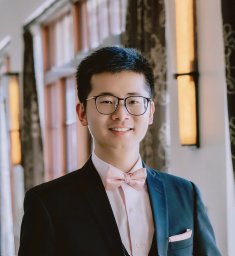 Zhu Yangzhi合作论文数State Key Laboratory of Fine Chemicals, Dalian University of Technology14
 Roberto de Barros Natan合作论文数Biological Sciences Department, Faculty of Language &UNESP),, São Paulo State University;Chemistry Institute, São Paulo State University;São Paulo State University (UNESP),, São Paulo State University13
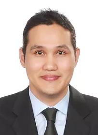 Heemin Kang合作论文数Department of Materials Science and Engineering, College of Engineering, Korea University13
 Martin C. Hartel合作论文数Department of Bioengineering, University of California-Los Angeles10
 Reihaneh Haghniaz合作论文数These authors contributed equally to this work.10
 Peyton Tebon合作论文数Ctr Minimally Invas Therapeut C MIT, Los Angeles, CA 90095 USA10
 Songyi Lee合作论文数Department of Chemistry, Pukyong National University10ChaeRim Hwang合作论文数9
 Yumeng Xue (薛语萌)合作论文数School of Materials Science and Engineering, Northwestern Polytechnical University9
 Yu S Zhang合作论文数Department of Medicine, Harvard Medical School;Harvard Stem Cell Institute;Brigham and Women's Hospital9
 Lee Wooyoung合作论文数Department of Materials Science and Engineering, Yonsei University9
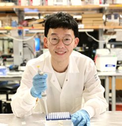 Xingwu Zhou合作论文数Department of Pharmaceutical Sciences, University of Michigan8Da In Jeong合作论文数8Jihyeon Song合作论文数7Sungyun Kim合作论文数7
 Thangam Ramar合作论文数Department of Virology, King Institute of Preventive Medicine & Research7
 Goudie Marcus J合作论文数7
 Su Ryon Shin合作论文数Department of Medicine, Division of Engineering in Medicine, Brigham and Women's Hospital, Harvard Medical School7
 Mohammad A. Darabi合作论文数Department of Mechanical Engineering, University of Guilan7Jin-Seo Noh合作论文数6
 Soufian Lasli合作论文数Henry Samueli School of Engineering and Applied Sciences, University of California-Los Angeles6
 Rohollah Nasiri合作论文数Terasaki Institute for Biomedical Innovation6
 Haonan Ling合作论文数Dept Mech & Aerosp Engn, Univ Calif Los Angeles6
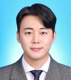 Mingyu Yang合作论文数Coll Pharm, Kangwon Natl Univ6
 Natashya Falcone合作论文数Terasaki Inst Biomed Innovat6Jang Woo Young合作论文数6
 Kim Seong Yeol合作论文数Department of Internal Medicine, Keimyung University School of Medicine6
 Kim Dong Hyun合作论文数Northwestern University Feinberg School of Medicine6Zhang Douglas合作论文数6
 Ramasamy Paulmurugan合作论文数Department of Radiology, School of Medicine, Stanford University;Stanford Cancer Institute, School of Medicine, Stanford University6Chengbin Xue (薛承斌)合作论文数5Aly Ung合作论文数5Moyuan Qu合作论文数5Ji-Hye Seo合作论文数5
 Menekse Ermis Sen合作论文数METU Ctr Excellence Biomat & Tissue Engn, BIOMATEN, Dept Biomed Engn, Ankara, Turkey5
 Sam Emaminejad合作论文数Department of Bioengineering, School of Engineering, University of California, Los Angeles5
 Jong Seung Kim合作论文数Department of Chemistry, College of Science, Korea University5




