There are hundreds of genes typically overexpressed in breast cancer cells and it's often assumed that their overexpression contributes to cancer progression. However, the precise proportion of these overexpressed genes contributing to tumorigenicity remains unclear. To address this gap, we undertook a comprehensive screening of a diverse set of seventy-two genes overexpressed in breast cancer. This systematic screening evaluated their potential for inducing malignant transformation and, concurrently, assessed their impact on breast cancer cell proliferation and viability. Select genes including ALDH3B1, CEACAM5, IL8, PYGO2, and WWTR1, exhibited pronounced activity in promoting tumor formation and establishing gene dependencies critical for tumorigenicity. Subsequent investigations revealed that CEACAM5 overexpression triggered the activation of signaling pathways involving β-catenin, Cdk4, and mTOR. Additionally, it conferred a growth advantage independent of exogenous insulin in defined medium and facilitated spheroid expansion by inducing multiple layers of epithelial cells while preserving a hollow lumen. Furthermore, the silencing of CEACAM5 expression synergized with tamoxifen-induced growth inhibition in breast cancer cells. These findings underscore the potential of screening overexpressed genes for both oncogenic drivers and tumor dependencies to expand the repertoire of therapeutic targets for breast cancer treatment.
BACKGROUND:Glioblastoma is the most common and aggressive primary brain tumor. Large-scale sequencing initiatives have cataloged its mutational landscape in hopes of elucidating mechanisms driving this deadly disease. However, a major bottleneck in harnessing this data for new therapies is deciphering "driver" and "passenger" events amongst the vast volume of information.METHODS:We utilized an autochthonous, in vivo screening approach to identify driver, EGFR variants. RNA-Seq identified unique molecular signatures of mouse gliomas across these variants, which only differ by a single amino acid change. In particular, we identified alterations to lipid metabolism, which we further validated through an unbiased lipidomics screen.RESULTS:Our screen identified A289I as the most potent EGFR variant, which has previously not been characterized. One of the mechanisms through which A289I promotes gliomagenesis is to alter cellular triacylglycerides through MTTP. Knockout of Mttp in mouse gliomas, reduces gliomagenesis in multiple models.CONCLUSIONS:EGFR variants that differ by a single amino acid residue differentially promote gliomagenesis. Among the identified mechanism that drives glioma growth include lipid metabolism through MTTP. Understanding triacylglyceride accumulation may present a prospective therapeutic pathway for this deadly disease.
MCF10A cell growth is dependent on insulin and epidermal growth factor (S1); RPPA analysis (S2).
Fusion genes and their sequences from TCGA (S1-2); Primers for fusion PCR (S3); BRAF mutants and mutagenesis primers (S4); (q)PCR primers for expression detection (S5); Fusion gene construction efficiency and success rate (S6)
PIK3CA mutations in breast cancer from the COSMIC database (S1); Predictions by computational algorithms on the consequences of PIK3CA missense mutations (S2); Mutagenic primer and barcode sequences of PIK3CA constructs and HiTMMoB reaction efficiencies (S3); Representative phenotyping for PIK3CA variants (S4); Barcode representation for PIK3CA variants in each sequenced tumor core (S5).
supplementary Figures S1-S10. S1 growth and regression of iKRAS tumors. S2 culture comparison. S3 immunohistochemistry of p-erk and p-s6 S4 Phospho rtk array S5 Validation of anti AXL antibodies S6 validationn of the AXL inhibitor in vitro S7 validationn of the AXL inhibitor in vivo S8 efficacy of targeting AXL S9 RTK array of iKRAS cells treated with multiple RTK inhibitors S10 P-ERK levels
Fusion expression detection by PCR (S1); MET fusion expression and signaling activation by immunoblot and N-terminal partner control assay (S2); NTRK2 fusion expression and signaling activation by immunoblot and N-terminal partner control assay (S3); Expression of BRAF fusion N-terminal partner controls and BRAF structure-function studies by immunoblot and qPCR (S4)
Abstract There are several hundreds of genes overexpressed in a typical breast cancer cell, and their overexpression is often assumed to have a pro-cancer function, but what percentage of these overexpressed genes have a role in promoting and maintaining tumorigenicity isn’t known. To address this question, we screened an unbiased set of seventy-five genes overexpressed in breast cancer for their ability to induce malignant transformation and for breast cancer cell expression dependency. Barcoded open-reading frames under the control of a constitutive promoter were tested by pooled screening for their ability to promote tumor formation of premalignant mammary epithelial cells and growth in 2D and 3D culture. We found that growth in 3D cell culture was more strongly correlated with tumorigenic growth than was 2D growth, validating a long-standing hypothesis. Genes with high activity in promoting both 3D growth and in vivo tumor formation included CEACAM5, CXCL8, NKX2-5, PYGO2 and WWTR1/TAZ. To screen for corresponding tumor dependencies on these six genes, we performed shRNA screening of breast cancer cell lines and found uniform dependency on expression of the Hippo pathway transcriptional activator WWTR1, selective dependencies on CEACAM5 expression, but no consistent effects for the other three genes. Since breast cancer dependency upon WWTR1 is well established, we focused our follow-up studies on CEACAM5. We confirmed that CEACAM5 overexpression conferred tumorigenicity to pre-malignant mammary epithelial cells and that reduced expression selectively decreased the clonogenicity of luminal breast cancer cell lines. Protein analysis indicated that CEACAM5 activated Cdk4/6 and mTOR signaling. We also determined that CEACAM5 overexpression strongly promoted cell number increase in 3D spheroid culture by a novel mechanism that increased the number of epithelial cell layers from a single layer to multiple layers. These results outline a strategy for functional genomic identification of cancer targets, starting with a set of genes overexpressed in cancer, then using pooled approaches to identify those that promote tumorigenicity and cancer cell dependency, followed by validation experiments and exploration of oncogenic mechanisms. We propose that larger screens would substantially increase the number of potential targets for treating breast cancer.
Fusion genes represent a class of attractive therapeutic targets. Thousands of fusion genes have been identified in patients with cancer, but the functional consequences and therapeutic implications of most of these remain largely unknown. Here, we develop a functional genomic approach that consists of efficient fusion reconstruction and sensitive cell viability and drug response assays. Applying this approach, we characterize ~100 fusion genes detected in patient samples of The Cancer Genome Atlas, revealing a notable fraction of low-frequency fusions with activating effects on tumor growth. Focusing on those in the RTK-RAS pathway, we identify a number of activating fusions that can markedly affect sensitivity to relevant drugs. Last, we propose an integrated, level-of-evidence classification system to prioritize gene fusions systematically. Our study reiterates the urgent clinical need to incorporate similar functional genomic approaches to characterize gene fusions, thereby maximizing the utility of gene fusions for precision oncology.
Germline POT1 mutations are found in a spectrum of cancers and confer increased risk. Recently, we identified a series of novel germline POT1 mutations that predispose carrier families to the development of glioma. Despite these strong associations, how these glioma-associated POT1 mutations contribute to glioma tumorigenesis remains undefined. Here we show that POT1-G95C increases proliferation in glioma-initiating cells in vitro and in progenitor populations in the developing brain. In a native mouse model of glioma, loss of Pot1a/b resulted in decreased survival in females compared with males. These findings were corroborated in human glioma, where low POT1 expression correlated with decreased survival in females. Transcriptomic and IHC profiling of Pot1a/b-deficient glioma revealed that tumors in females exhibited decreased expression of immune markers and increased expression of cell-cycle signatures. Similar sex-dependent trends were observed in human gliomas that had low expression of POT1. Together, our studies demonstrate context-dependent functions for POT1 mutation or loss in driving progenitor proliferation in the developing brain and sexual dimorphism in glioma. SIGNIFICANCE: This study shows that manipulation of POT1 expression in glioma has sex-specific effects on tumorigenesis and associated immune signatures.
The melanoma-associated antigen family A (MAGEA) antigens are expressed in a wide variety of malignant tumors but not in adult somatic cells, rendering them attractive targets for cancer immunotherapy. Here we show that a number of cancer-associated MAGEA mutants that undergo proteasome-dependent degradation in vitro could negatively impact their utility as immunotherapeutic targets. Importantly, in pancreatic ductal adenocarcinoma cell models, MAGEA6 suppresses macroautophagy (autophagy). The inhibition of autophagy is released upon MAGEA6 degradation, which can be induced by nutrient deficiency or by acquisition of cancer-associated mutations. Using xenograft mouse models, we demonstrated that inhibition of autophagy is critical for tumor initiation whereas reinstitution of autophagy as a consequence of MAGEA6 degradation contributes to tumor progression. These findings could inform cancer immunotherapeutic strategies for targeting MAGEA antigens and provide mechanistic insight into the divergent roles of MAGEA6 during pancreatic cancer initiation and progression.
This study identifies a molecular basis for malignant secretion and provides a rationale to test PI4KIIIβ antagonists as an intervention.
Non-small cell lung cancer (NSCLC) is the deadliest form of cancer worldwide, due in part to its proclivity to metastasize. Identifying novel drivers of invasion and metastasis holds therapeutic potential for the disease. We conducted a gain-of-function invasion screen, which identified two separate hits, IMPAD1 and KDELR2, as robust, independent drivers of lung cancer invasion and metastasis. Given that IMPAD1 and KDELR2 are known to be localized to the ER–Golgi pathway, we studied their common mechanism of driving in vitro invasion and in vivo metastasis and demonstrated that they enhance Golgi-mediated function and secretion. Therapeutically inhibiting matrix metalloproteases (MMPs) suppressed both IMPAD1- and KDELR2-mediated invasion. The hits from this unbiased screen and the mechanistic validation highlight Golgi function as one of the key cellular features altered during invasion and metastasis.
Glioblastoma is a universally lethal form of brain cancer that exhibits an array of pathophysiological phenotypes, many of which are mediated by interactions with the neuronal microenvironment1,2. Recent studies have shown that increases in neuronal activity have an important role in the proliferation and progression of glioblastoma3,4. Whether there is reciprocal crosstalk between glioblastoma and neurons remains poorly defined, as the mechanisms that underlie how these tumours remodel the neuronal milieu towards increased activity are unknown. Here, using a native mouse model of glioblastoma, we develop a high-throughput in vivo screening platform and discover several driver variants of PIK3CA. We show that tumours driven by these variants have divergent molecular properties that manifest in selective initiation of brain hyperexcitability and remodelling of the synaptic constituency. Furthermore, secreted members of the glypican (GPC) family are selectively expressed in these tumours, and GPC3 drives gliomagenesis and hyperexcitability. Together, our studies illustrate the importance of functionally interrogating diverse tumour phenotypes driven by individual, yet related, variants and reveal how glioblastoma alters the neuronal microenvironment.
Alternative splicing has been shown to causally contribute to the epithelial–mesenchymal transition (EMT) and tumor metastasis. However, the scope of splicing factors that govern alternative splicing in these processes remains largely unexplored. Here we report the identification of A-Kinase Anchor Protein (AKAP8) as a splicing regulatory factor that impedes EMT and breast cancer metastasis. AKAP8 not only is capable of inhibiting splicing activity of the EMT-promoting splicing regulator hnRNPM through protein–protein interaction, it also directly binds to RNA and alters splicing outcomes. Genome-wide analysis shows that AKAP8 promotes an epithelial cell state splicing program. Experimental manipulation of an AKAP8 splicing target CLSTN1 revealed that splice isoform switching of CLSTN1 is crucial for EMT. Moreover, AKAP8 expression and the alternative splicing of CLSTN1 predict breast cancer patient survival. Together, our work demonstrates the essentiality of RNA metabolism that impinges on metastatic breast cancer.
DNA damage provokes mutations and cancer and results from external carcinogens or endogenous cellular processes. However, the intrinsic instigators of endogenous DNA damage are poorly understood. Here, we identify proteins that promote endogenous DNA damage when overproduced: the DNA "damage-up" proteins (DDPs). We discover a large network of DDPs in Escherichia coli and deconvolute them into six function clusters, demonstrating DDP mechanisms in three: reactive oxygen increase by transmembrane transporters, chromosome loss by replisome binding, and replication stalling by transcription factors. Their 284 human homologs are over-represented among known cancer drivers, and their RNAs in tumors predict heavy mutagenesis and a poor prognosis. Half of the tested human homologs promote DNA damage and mutation when overproduced in human cells, with DNA damage elevating mechanisms like those in E. coli. Our work identifies networks of DDPs that provoke endogenous DNA damage and may reveal DNA damage-associated functions of many human known and newly implicated cancer-promoting proteins.
 Gordon Mills合作论文数School of Medicine, Oregon Health Sciences University34
 Chad Creighton合作论文数Dan L Duncan Comprehensive Cancer Center, Baylor College of Medicine;Human Genome Sequencing Center, Baylor College of Medicine25
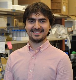 Turgut Dogruluk合作论文数24
 Yiu Huen Tsang合作论文数Dept Mol & Human Genet, Baylor Coll Med23
 Caitlin L Grzeskowiak合作论文数Baylor Coll Med, Dept Mol & Human Genet, Houston, TX 77030 USA15Lu Hengyu合作论文数15
 Lynda Chin合作论文数Apricity Health15
 Han Liang (梁晗)合作论文数Department of Bioinformatics and Computational Biology, Division of Basic Science Research, The University of Texas MD Anderson Cancer Center;Department of Systems Biology, Division of Basic Science Research, The University of Texas MD Anderson Cancer Center12Kong Kathleen合作论文数11Rosalba Minelli合作论文数10
 Don L. Gibbons合作论文数Department of Thoracic-Head & Neck Med Onc, Division of Cancer Medicine, The University of Texas MD Anderson Cancer Center10
 Ken Chen合作论文数Department of Bioinformatics and Computational Biology, Division of Discovery Science, The University of Texas MD Anderson Cancer Center9
 Kang Jin Jeong合作论文数Univ Texas MD Anderson Canc Ctr, Dept Syst Biol, Houston, TX 77030 USA8Ng Patrick Kwok-Shing合作论文数8Yiling Lu合作论文数7
 Angeliki Pantazi合作论文数Departments of Genetics and7Chen Fengju合作论文数7
 Ignacio I. Wistuba合作论文数Department of Translational Molecular Pathology, The University of Texas MD Anderson Cancer Center;Department of Pathology, The University of Texas MD Anderson Cancer Center7
 Ronald DePinho合作论文数Department of Cancer Biology, Division of Basic Science Research, The University of Texas MD Anderson Cancer Center7
 Raju Kucherlapati合作论文数Division of Medical Sciences, Harvard Medical School;Brigham and Women's Hospital;KEW Group7Nikitha Nair合作论文数6Armel Dogruluk合作论文数6
 Penelope E Bonnen合作论文数Human Genome Sequencing Center, Baylor College of Medicine6
 Kundu Samrat T合作论文数Department of Molecular, Cellular and Development Biology, Yale University6Wu Ping合作论文数6
 Karina Eterovic合作论文数Eurofins Viracor Biopharma6Timothy P. Heffernan合作论文数6Zhenlin Ju合作论文数5Ali Jalali合作论文数Department of Neurosurgery, Baylor College of Medicine5Christopher A. Bristow合作论文数University of Texas MD Anderson Cancer Center5Sahil Seth合作论文数Applied Cancer Science Institute, The University of Texas MD Anderson Cancer Center5
 Zechen Chong合作论文数Chong Lab, Department of Biomedical Informatics and Data Science, The University of Alabama at Birmingham;School of Medicine, The University of Alabama at Birmingham5
 Peter J Park合作论文数Department of Biomedical Informatics, Harvard Medical School;Division of Genetics, Brigham and Women’s Hospital5Carmen Behrens合作论文数Department of Thoracic Head & Neck Medical Oncology, The University of Texas MD Anderson Cancer Center;Center for Cancer Epigenetics, The University of Texas MD Anderson Cancer Center5
 Jonathan M. Kurie合作论文数Department of Thoracic-Head & Neck Med Onc, Division of Cancer Medicine, University of Texas MD Anderson Cancer Center5Yonghong Xiao合作论文数5
 Haoqiang Ying合作论文数Department of Molecular and Cellular Oncology, Division of Basic Science Research, The University of Texas MD Anderson Cancer Center5
 Shuangxing Yu合作论文数5
 Oksana Zagorodna合作论文数The University of Iowa5Maribel Espitia合作论文数4Nicole Villafane合作论文数4Daniela Moreno合作论文数Baylor Coll Med, Dept Mol & Human Genet, Houston, TX 77030 USA4
 Yumeng Wang合作论文数Department of Bioinformatics and Computational Biology, The University of Texas MD Anderson Cancer Center, Houston, TX 77030, USA4
 Jared J Fradette合作论文数Univ Texas MD Anderson Canc Ctr, Dept Thorac Head & Neck Med Oncol, Houston, TX 77030 USA4
 Lixing Yang合作论文数Center for Biomedical Informatics, Harvard Medical School;Center for Biomedical Informatics, Harvard Medical School4Shaw Kenna R合作论文数4Jianhua Zhang合作论文数4
 Huang-Hobbs Emmet合作论文数The University of Texas MD Anderson Cancer Center, The University of Texas MD Anderson Cancer Center4Ip Carman Ka Man合作论文数4




