Abstract: L. J. Thénard and J. L. Gay-Lussac discovered hydrogen peroxide in 1818. Later, Thénard noticed that animal and plant tissues decompose hydrogen peroxide. The substance which is responsible for this reaction was named as catalase by O. Loew in 1900. The catalase enzyme was regarded as a diagnostic and a tumour marker in the late years of the 19th century and in the early years of the 20th century. Acatalasemia, an inherited deficiency of enzyme catalase, was studied in Japan, Switzerland and Hungary. The recent findings on catalase are focusing on the effects of reactive oxygen species and on the association of acatalasemia and diabetes mellitus. Orv Hetil. 2018; 159(24): 959–964.
Diabetes mellitus is a risk factor for atherosclerosis/cardiovascular diseases. Atherosclerosis in diabetic patients has been linked to increased oxidative stress and intima-media thickness is used to detect its presence. Catalase is involved in hydrogen peroxide catabolism and is important in defense against oxidative stress, which contributes significantly to atherosclerotic processes. There are no data on association of catalase gene genotypes of rs769217 polymorphism and carotid artery intima-media thickness. Therefore, this study investigated the potential association of catalase rs769217 polymorphism and intima media thickness in diabetic patients. Right and left carotid intima-media thickness increased (P≤0.001) in type 1 and type 2 diabetes when it was compared to those of controls. Blood catalase activities of CC genotypes of rs769217 polymorphism in catalase gene were associated with cITT (P<0.043) in type 1 diabetics, type 2 diabetics and in controls. This interesting new finding could suggest that diabetics and controls with CC genotype of rs769217 polymorphism may have higher carotid intima media thickness and higher risks for cardiovascular events when they blood catalase is high. This higher catalase could destroy hydrogen peroxide more effectively thus preventing the signaling action of hydrogen peroxide.
We checked our simple screening technique for detection of the known polymorphism of rs769217, and the two acatalasemic mutations in exon 9 of the catalase gene. This fast and inexpensive method yielded better resolution than those of the standard SSCP. We suppose that the method detects the spontaneously formed single stranded DNAs.
Introduction: Catalase decomposes hydrogen peroxide into oxygen and water. Its low concentration could be involved in signaling while its high concentration is toxic.
A previous study of 25 252 hospitalized patients yielded decreased (bellow the reference range) blood catalase activities in 2 884 (11.4 %) anemic patients, in 130 (0.5%) patients with different tumors, in 1 237 (4,9%) patients with atherosclerosis, and in 275 (1.1%) patients with schizophrenia. The decreased blood catalase could be attributed to the low number of erythrocytes in the anemic patients and partly in those with tumors. Genetic or regulatory factors could be responsible for the other 1642 (36.3%) cases [3].
In blood, the hydrogen peroxide concentration is regulated by catalase. Decreased activity of catalase may lead to increased hydrogen peroxide concentration, which may contribute to the manifestation of age-related disease. The aim of this study is to examine association of decreased blood catalase activity and catalase exon mutations in patients (n = 617) with diabetes (n = 380), microcytic anemia (n = 58), beta-thalassemia (n = 43) and presbycusis (n = 136) and in controls (n = 295). Overall, 51 patients (8.3%) had less than half of normal blood catalase activity. Their genomic DNA was used for mutation screening of all exons and exon/intron boundaries with polymerase chain reaction-single-strand conformation polymorphism (PCR-SSCP) and PCR-heteroduplex analyses, and mutations were verified with nucleotide sequencing. Seven patients (type 2 diabetes (n = 3), gestational diabetes (n = 1), microcytic anemia (n = 2)) had four novel catalase exon mutations namely, c.106_107insC, p.G36Afs*5(n = 3, Hungarian type G1), c.379C>T, p.R127Y (n = 2, Hungarian type H1), c.390T>C, p.R129L, (n = 1, Hungarian type H2) and c.431A>T, p.N143V (n = 1, Hungarian type H3). In patients with decreased blood catalase, the incidence of acatalasemia mutations was significantly high (P < 0.0002) in microcytic anemia, type 2 and gestational diabetes. The four novel mutations were probably responsible for low blood catalase activity in 7/51 patients. In the remainder of the cases, other polymorphisms and epigenetic/regulatory factors may be involved.
The catalase enzyme decomposes the toxic concentrations of hydrogen peroxide into oxygen and water. Hydrogen peroxide is a highly reactive small molecule and its excessive concentration may cause significant damages to proteins, deoxyribonucleic acid, ribonucleic acid and lipids. Acatalasemia refers to inherited deficiency of the catalase enzyme. In this review the authors discuss the possible role of the human catalase enzyme, the metabolism of hydrogen peroxide, and the phenomenon of hydrogen peroxide paradox. In addition, they review data obtained from Hungarian acatalasemic patients indicating an increased frequency of type 2 diabetes mellitus, especially in female patients, and an early onset of type 2 diabetes in these patients. There are 10 catalase gene variants which appear to be responsible for decreased blood catalase activity in acatalasemic patients with type 2 diabetes. It is assumed that low levels of blood catalase may cause an increased concentration of hydrogen peroxide which may contribute to the pathogenesis of type 2 diabetes mellitus.
Hydrogen peroxide was - and is still - considered toxic for a wide range of living organisms. Oxidative stress occurs when there is an excess of pro-oxidants over antioxidants and it has been implicated in several diseases. Catalase is involved in hydrogen peroxide catabolism and is important in defense against oxidative stress. Acatalasemia means the inherited near-total deficiency of catalase activity, usually in reference to red cell catalase. Acatalasemia was thought at first to be an asymptotic disorder. In the absence of catalase, neither the Japanese, or Hungarian acatalasemics nor acatalasemic mice had significantly increased blood glutathione peroxidase activity. In animal models, catalase deficient tissues show much slower rates of removal of extracellular hydrogen peroxide. In catalase knock-out mice, a decreased hydrogen peroxide removing capacity and increased reactive oxygen species formation were reported. Hydrogen peroxide may cause methemoglobinemia in patients with catalase deficiency. During anesthesia for a Japanese acatalasemic patient the disinfection with hydrogen peroxide solution caused severe methemoglobinemia. Patients with inherited catalase deficiency, who are treated with uric acid oxidase (rasburicase) may experience very high concentrations of hydrogen peroxide and may suffer from methemoglobinemia and hemolysis. The high (18.5%) prevalence of diabetes mellitus in inherited catalase deficient individuals and the earlier (10 years) manifestation of the disease may be attributed to the oxidative damage of oxidant sensitive, insulin producing pancreatic beta-cells. Ninety-seven of 114 acatalasemics had diseases related to oxidative stress and aging. The oxidative stress due to catalase deficiency could contribute to the manifestation of diabetes while for the other diseases it may be one of the factors in their causations. In summary, inherited catalase deficiency is associated with clinical features, pathologic laboratory test results, age and oxidative stress related disorders. Rather than considering it a benign condition, it should be considered as a complicating condition for aging and oxidative stress. (C) 2013 Elsevier B.V. All rights reserved.
The enzyme catalase catalyzes the breakdown of hydrogen peroxide into oxygen and water. It is the main regulator of hydrogen peroxide metabolism. Hydrogen peroxide is a highly reactive small molecule formed as a natural byproducts of energy metabolism. Excessive concentrations may cause significant damages to protein, DNA, RNA and lipids. Low levels in muscle cells, facilitate insulin signaling. Acatalasemia is a result of the homozygous mutations in the catalase gene, has a worldwide distribution with 12 known mutations. Increased hydrogen peroxide, due to catalase deficiency, plays a role in the pathogenesis of several diseases such as diabetes mellitus. Diabetes mellitus is a disorder caused by multiple genetic and environmental factors. Examination of Hungarian diabetic and acatalasemic patients showed that an increased frequency of catalase gene mutations exists among diabetes patients. Inherited catalase deficiency may increase the risk of type 2 diabetes mellitus, especially for females. Early onset of type 2 diabetes occurs with inherited catalase deficiency. Low levels of SOD and glutathione peroxidase could contribute to complications caused by increased oxidative stress.
Oxidative stress and deficiency of the enzyme catalase, which is the primary scavenger of the oxidant H2O2, may contribute to diabetes. The current study examined two polymorphisms in the catalase gene, -262C > T in the promoter and 111C > T in exon 9, and their eff ects on blood catalase activity as well as on concentrations of blood glucose, haemoglobin A1c, triglyceride, cholesterol, HDL, LDL, ApoA-I and ApoB. Subjects were type-1 and type-2 diabetics. We evaluated PCR-single strand conformational polymorphism for 111C > T and PCR-restriction fragment length polymorphism for -262C > T. TT genotype frequency of 111C > T polymorphism was increased in type-1 diabetes. Type-2 diabetics with the CC or CT genotypes had decreased catalase and increased glucose, hemoglobinA1c and ApoB. Type-2 diabetics who have TT genotype in -262C > may have elevated risk for diabetes complications; these patients had the lowest mean catalase and HDL, as well as the highest glucose, haemoglobin A1c, cholesterol and ApoB.
Summary Introduction: Thalassemia erythrocytes are exposed to oxidative stress especially to hydrogen peroxide, which is regulated with the enzyme catalase. The aim of this study was to examine blood catalase activity and the relationship of blood catalase and beta‐thalassemia gene mutations. Methods: Blood catalase activity, hemoglobin, HbA 2 , HbF, and beta‐globin gene mutations were determined in 43 Hungarian patients with beta‐thalassemia trait. Results: Compared to controls, the beta‐thalassemia trait patients showed a low mean ( P < 0.001) of blood catalase (men: 84 ± 29 MU/L vs. sex‐matched controls: 118 ± 18 MU/L and women: 74 ± 18 MU/L vs. 108 ± 114 MU/L) and a low mean of blood catalase‐to‐blood hemoglobin ratio (men: 0.72 ± 0.22 MU/g vs. 0.85 ± 0.12 MU/g, women: 0.77 ± 0.26 MU/g vs. 0.84 ± 0.11 MU/g). The HbA 2 determination showed high sensitivity and specificity for the detection of beta‐thalassemia trait patients. Mutation analyses revealed 13 beta‐thalassemia trait mutations, of which six have not been reported before in Hungarian beta‐thalassemia trait patients. Each group of mutations revealed decreased ( P < 0.01) mean of blood catalase and catalase‐to‐hemoglobin ratio. Acatalasemia mutations were not found in beta‐thalassemia trait patients. Conclusion: The decrease in blood catalase activity might be due to the damaging effects of free radicals on the catalase protein. Consequently, these beta‐thalassemia trait patients may be relatively susceptible to damage caused by oxidative stress.
Catalase decreases the high, toxic concentrations of hydrogen peroxide but it lets the physiological, low concentrations in the cells mainly for signaling purposes. Its decreased activity may contribute to development of several pathological conditions. Catalase mutations occur frequently in exon 9, these were examined with different, complicated and costly methods. The aim of the current study was to evaluate a method for screening of polymorphisms in catalase exon 9. We used the slab gel electrophoresis of PCR amplicons without denaturation and silver staining for visualization of the DNA bands. We detected extra DNA bands in the 400-800 bp region of the catalase exon 9. Their single stranded nature was proved with nucleotide sequence analyses, comparison with the standard SSCP, staining with Sybr Green II and Sybr Green I, ethidium bromide, no digestion with RFLP (BstX I), and digestion with plant nuclease. We used this method for examination of polymorphisms of catalase exon 9 in microcytic anemia and beta-thalassemia patients. The lowest blood catalase activities were detected in microcytic anemia and beta-thalassemia patients with the TT genotypes of the C111T polymorphism. This method was sensitive for detection of G113A acatalasemia mutation, but poorly detected C37T and G5A acatalasemia mutations.
An early event in the pathogenesis of vitiligo may be the chronic oxidative stress via elevated hydrogen peroxide due to decreased blood catalase. There are controversial reports on association of vitiligo and +22348C→T polymorphism. We examined this polymorphism and blood catalase in Hungarian vitiligo patients (n: 78) and controls (n: 201). Genomic DNA was extracted from leukocytes and a PCR/SSCP method was used for genetic examination. Blood catalase activity, genotype and allele frequencies did not change in Hungarian vitiligo patients. The mutant CT and TT genotypes yielded decreased (p < 0.05) blood catalase activities in females. The association of the +22348C→T polymorphism and vitiligo in Hungary is non-significant (p > 0.0813), contrary to the significant (p < 0.05) increase of mutant CT genotype and T allele frequencies in US/Canada and in UK. Our results based on blood catalase determinations and genetic examination of +22348C→T polymorphism suggest that they may provide a weak contribution to the vitiligo pathology in female vitiligo patients by the increased oxidative stress. It may be caused by the increased hydrogen peroxide due to the decreased blood catalase.
Catalase is the main regulator of hydrogen peroxide metabolism. In vitiligo patients there are conflicting data on its activity and no data on the effect of −262C>T polymorphism in the catalase gene. Blood catalase activity, −262C>T polymorphism and acatalasemia mutations were examined in 75 vitiligo patients and in 162 controls, in Hungary. We measured blood catalase activity and conducted analyses with PCR-SSCP, polyacrylamide gel electrophoresis and silver staining in combination with RFLP and nucleotide sequencing. Comparison of the wild (CC) genotype and the mutant (TT) genotype in the vitiligo patients revealed a non significant (P > 0.19) increase in blood catalase. Male controls with the CT genotype had significantly (P < 0.04) lower blood catalase activity than CC genotype controls. Female vitiligo patients with CC genotype had lower (P < 0.04) blood catalase than female controls. The frequency of wild genotype (CC) and C alleles is significantly (P < 0.04) decreased in Hungarian controls when compared to controls in Slovenia, Morocco, UK, Greece, Turkey, USA, China. The detection of a novel acatalasemia mutation (37C>T in exon 9) and the 113G>A (exon 9) mutation in Hungary are further proofs of genetic heterogeneity origin of acatalasemia mutations. In conclusion, the −262 C>T polymorphism has a reverse effect on blood catalase in vitiligo patients and in controls. In controls the mutant genotypes and alleles are more frequent in Hungary than in several other populations. The new acatalasemia mutations are further examples of heterogeneity of acatalasemia.
Glycated proteins are formed during the nonenzymatic reaction of glucose and amino groups of proteins. Hemoglobin A1c is formed by the condensation of glucose with the N-terminal valine residue of each beta-chain of hemoglobin A. The amount of glycated hemoglobin in blood depends on both life-span of red blood cells and blood glucose concentration. As the rate of formation of hemoglobin A1c is directly proportional to the concentration of glucose in the blood, it represent the integrated values for glucose over the preceding 6 to 8 weeks. Hemoglobin A1c determination is widely used for monitoring long-term glycemic control, and it is a risk factor for complications of diabetes. The concentration of blood hemoglobin A1c depends on further factors such as half-life of hemoglobin, blood carbohydrates, blood analytes, methods of determination and calibration. Committees were established under the auspices of the American Association of Clinical Chemistry, American Diabetes Association, International Federation of Clinical Chemistry (IFCC) to standardize HbA1c assays (DCCT: Diabetes Control and Complications Trial, NGSP: National Glycohemoglobin Standardization Program, IFCC reference method for measurement of HbA1c). The NGSP recommends to report HbA1c result in % (g HbA1c/g hemoglobin) while IFCC suggests mmol HbA1c/mol hemoglobin A. Reports are presenting mathematical relationship between HbA1c and average glucose concentration in blood, however, the clinical usefulness of estimating average serum glucose from HbA1c level is under discussion.
Background: Serum LD activity is increased in several diseases. The clinical significance of elevated LD may be evaluated by isoenzyme analysis. Here, we report on a long-term elevation of serum LD activity with a rare isoenzyme pattern caused by LD-IgG complexes. Patient, methods: A 75-year old female with atherosclerosis and implantations of two stents showed increased LD activity and an unusual isoenzyme pattern. LD activity, inhibition and immunoglobulins were measured with Roche tests; immunofixation was performed with a Helena test. LD isoenzymes were separated in agarose gel and quantified by the color formed by NADH, reduced phenazine methosulfate and nitro blue tetrazolium. Results: LD isoenzyme analyses revealed one band near the slowly migrating LD-4 position. The band contained LD complexed with immunoglobulins (IgG with κ light chains), The long-term increased serum LD enzyme activity in this patient may have been due to abnormally slow clearance of these large macro-LD complexes from circula...
Hyperuricemia contributes to the pathomechanism of diseases such as renal failure, gout, tumor lysis syndrome and metabolic syndrome. Tumor lysis syndrome is a complication of malignancies caused by massive tumor cell lysis due to either spontaneous tumor cell lysis or to different therapies and it may cause hyperuricemia. Recently, for treatment of hyperuricemia the recombinant urate oxidase (rasburicase) therapy has been used. This enzyme converts uric acid with high affinity into soluble allantoin which is eliminated by the kidneys. In this reaction high concentration of hydrogen peroxide is generated. This hydrogen peroxide could cause hemolysis and especially methemoglobin formation, in case of glucose-6-phosphate-dehydrogenase and catalase deficiencies. Therefore it is recommended that these enzymes are determined before therapy. For monitoring of rasburicase therapy the determination of serum uric acid concentration is used. More than 95 per cent of Hungarian clinical laboratories are using the uricate oxidase/peroxidase reactions and hydrogen peroxide measurements in the uric acid assays. These assays may be interfered by ascorbic acid and hydrogen peroxide which is generated by rasburicase either in vivo or in vitro.
 Nagy Terez合作论文数9
 Ilona Mészáros合作论文数Department of Botany;University of Debrecen;Department of Botany, University of Debrecen7
 M. Vitai合作论文数6
 Káplár Miklós合作论文数Anyagcsere Betegségek Tanszék, Debreceni Egyetem6Ildikó Tarnai合作论文数5eszter sukei合作论文数4Kósa Zsuzsanna合作论文数4Csordás Melinda合作论文数4Németh H合作论文数4Enikő Simics合作论文数4
 Tibor Kalmar合作论文数3
 Shemirani Amir-Houshang合作论文数University of Debrecen, University of Debrecen3Bigler William N合作论文数3W. N. Bigler合作论文数3P Rass合作论文数2
 Zsolt Fejes合作论文数2
 Harjit Pal Bhattoa合作论文数Laboratory Endocrinology and Andrology Unit, Department of Laboratory Medicine, University of Debrecen2Péter Rass合作论文数2
 Aniko Pay合作论文数2Anikó Páy合作论文数1R Czehelnik合作论文数1Z Kósa合作论文数1András Gorzsás合作论文数Umea Univ, Dept Chem, Umea, Sweden1
 Balazs Dezso合作论文数Clin Ctr, Univ Debrecen1Fazakas F合作论文数1Lenkey Agota合作论文数1Scheller G合作论文数1Paszti Erika合作论文数1
 Nagy Tibor合作论文数Department of Applied Chemistry, University of Debrecen1
 Gábor Vattay合作论文数Eotvos University1
 Zsolt Toth合作论文数Research Group on Laser Physics, the Hungarian Academy of Sciences1
 Marta Vitai合作论文数1
 György Paragh合作论文数Department of Internal Medicine, Faculty of Medicine, University of Debrecen;Department of Metabolic Diseases, University of Debrecen1
 Török Péter合作论文数university of debrecen1A. Lenkey合作论文数1
 Éva Remenyik合作论文数Clinical Centre, University of Debrecen1
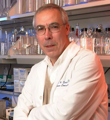 John W. Eaton合作论文数Departments of Medicine, and Pharmacology & Toxicology, School of Medicine, University of Louisville1
 Laszlo Asztalos合作论文数Institute of Surgery, Medical and Health Science Center, University of Debrecen1




