PURPOSE:The MRI safety standard IEC 60601-2-33 limits the maximum dB/dt and E-field induced by MRI gradients to protect patients from cardiac stimulation (CS). Those limits were set in the early 2000s based on a compilation of electrode stimulation measurements in animals as well as electromagnetic calculations in a homogeneous ellipsoid. We re-evaluate CS in MRI using state-of-the-art electromagnetic and electrophysiological modeling in realistic body models and gradient coils. METHODS:We predicted CS thresholds in 56 realistic body models and 13 commercial gradient systems for trapezoidal waveforms with rise times (the time during which the gradient amplitude is ramped from zero to maximum) ranging from 0.1 to 5.0 ms. We used threshold predictions in individual body models to estimate the gradient field amplitude below which the probability of occurrence of a CS adverse event is < 2 ppb, the historically agreed upon probability used in IEC 60601-2-33. RESULTS:We found that the variability of CS thresholds due to differences in anatomy and body habitus was 10%-36%, depending on rise time, while the CS threshold variability across gradient coils was 27%. In the range of typically used MRI rise times (≤ 2.5 ms), the predicted gradient amplitude associated with a CS probability of 2 ppb is > 5.5X higher than the IEC CS limit. CONCLUSION:Detailed simulations in a large population of realistic body models and gradient coils indicate that the IEC CS limit is substantially lower (more conservative) than the predicted gradient amplitude associated with a 2-ppb probability for CS.
PURPOSE:The IEC 60601-2-33 standard provides consensus-based safety provisions for MRI equipment. Protection of patients against cardiac stimulation (CS) is based on limiting the maximum E-field induced by MRI gradient coils. In practice, this is achieved by imposing a conservative dB/dt threshold on any gradient waveform. The dB/dt-over-E-field conversion ratio currently used in IEC 60601-2-33 was derived in a homogeneous ellipsoid exposed to a uniform B-field and is 10 (T/s)*(V/m)-1. This limit is becoming increasingly restrictive in high performance clinical systems. We therefore evaluate dB/dt-over-E-field ratios in realistic body models and coils using state-of-the-art electromagnetic simulations. METHODS:We performed two independent simulation studies in a total of 75 realistic body models and 13 commercial gradient systems and derived dB/dt-over-E-field ratios in the heart. We thresholded the E-field maps to mitigate the impact of staircasing artifacts in boundary voxels between the myocardium and the lungs. RESULTS:Thresholding the E-field maps at the 99th percentile E-field value (E99) eliminates staircasing artifacts in both simulation studies. Study #1 predicts a larger range of dB/dt-over-E99 ratios (13-53 (T/s)*(V/m)-1) than study #2 (12-35 (T/s)*(V/m)-1). Despite differences in EM solvers, body models, coils, mesh resolution, and post-processing, both studies find similar worst-case ratios of dB/dt-over-E99 of 12-13 (T/s)*(V/m)-1. CONCLUSION:Our simulations of dB/dt-over-E-field ratios for cardiac safety in MRI cover a large range of realistic clinical scenarios. An increase of the allowable dB/dt beyond the current CS limit in IEC 60601-2-33 may be feasible.
Rapidly switching the gradient coils during MRI induces electric fields (E-fields) in the human body strong enough to evoke action potentials (AP) in peripheral nerves which are perceived by the subject. This peripheral nerve stimulation (PNS) has emerged as one of the primary constraints to full use of the parameter space ($\mathrm{G}_{\text {max }}$ and $\text{SR}_{\text {max }}$) of modern MRI gradients, yet is not typically considered during the design phase of new gradient systems. Instead, PNS is typically only measured after the coil design phase using a constructed prototype coil. To inform the iterative design process, we developed a coupled electromagnetic (EM) and neurodynamic PNS model capable of predicting thresholds and sites of AP initiation in computational body models exposed to the switching gradient B-fields. Assessing the stimulation due to generalized current basis elements on a design surface allows us to laboriously precompute each nerve's response in this general basis but quickly compute a candidate design's nerve response. While initially computed on a specialized design surface a transformation can be quickly found to the basis of another design surface (such as a cylinder) via Huygen's principle. The linear metric allows each nerve's response to a given candidate winding pattern's to be assessed as a simple vector dot product. In this work, we summarize the method and show validation of this model by comparing its predicted and experimentally measured PNS thresholds in multiple gradient systems.
Magnetic fields switching at kilohertz frequencies induce electric fields in the body, which can cause peripheral nerve stimulation (PNS). Although magnetostimulation has been extensively studied below 10 kHz, the behavior of PNS at higher frequencies remains poorly understood. This study aims to investigate PNS thresholds at frequencies up to 88.1 kHz and to explore deviations from the widely accepted hyperbolic strength-duration curve (SDC).PNS thresholds were measured in the head of 8 human volunteers using a solenoidal coil at 16 distinct frequencies, ranging from 200 Hz to 88.1 kHz. A hyperbolic SDC was used as a reference to compare the frequency-dependent behavior of PNS thresholds.Contrary to the predictions of the hyperbolic SDC, PNS thresholds did not decrease monotonically with frequency. Instead, thresholds reached a minimum near 25 kHz, after which they increased by an average of 39% from 25 kHz to 88.1 kHz across subjects. This pattern indicates a significant deviation from previously observed behavior at lower frequencies.Our results suggest that PNS thresholds exhibit a non-monotonic frequency dependence at higher frequencies, diverging from the traditional hyperbolic SDC. These findings offer critical data for refining neurodynamic models and provide insights for setting PNS safety limits in applications like MRI gradient coils and magnetic particle imaging (MPI). Further investigation is needed to understand the biological mechanisms driving these deviations beyond 25 kHz.Clinical impact—These findings call for further basic research into biological mechanisms underlying high frequency PNS threshold trends, and supports refinement of safety guidelines for MRI and MPI systems for clinical implementation.
PURPOSE:Peripheral nerve stimulation (PNS) remains a physiologic limitation to boosting spatiotemporal resolution with more powerful gradients. We investigate discrepancies in previous measurements and model predictions from PNS experienced by volunteers scanned with the investigational "Impulse" gradient coil on the NexGen 7T scanner. METHODS:Twenty-nine volunteers (18 males, mean ± standard deviation age 52.2 ± 17.1 years) underwent PNS characterizations in the scanner. The process was repeated after the subject positions were moved by 2 and 4 cm toward the feet, away from isocenter. These new data were compared with prior experimental data acquired at the factory (32 volunteers, 16 males, mean ± standard deviation age 58.3 ± 13.5 years) and to modeling results initially used to guide the gradient winding pattern. RESULTS:The PNS threshold for the x-axis (left-right) was significantly below the threshold level predicted by the model used to optimize the wiring pattern and thresholds measured in the factory, whereas there was closer agreement for the y-axis (anterior-posterior) and z-axis (superior-inferior). The x-axis threshold increased as the subject was moved in the Z-direction toward the foot end of the magnet, at the expense of gradient nonlinearity distortions. Sensitivity of the threshold for the x-axis was measured as 20 mT/m per centimeter Z-offset. CONCLUSION:The PNS threshold of the x-axis measured in the scanner was much lower than predicted by the optimization model and as measured at the factory. Our measurements verified that PNS thresholds of asymmetric head gradient coils were sensitive to head position, subject variability, and age. The discrepancy of the PNS prediction model remains to be elucidated.
Defining the connectome, the complete matrix of structural connections between the nervous system nodes, is a challenge for human systems neuroscience due to the range of scales that must be bridged. Here we report the design of the Connectome 2.0 human magnetic resonance imaging (MRI) scanner to perform connectomics at the mesoscopic and microscopic scales with strong gradients for in vivo human imaging. We construct a 3-layer head-only gradient coil optimized to minimize peripheral nerve stimulation while achieving a gradient strength of 500 mT m−1 and a slew rate of 600 T m−1 s−1, corresponding to a 5-fold greater gradient performance than state-of-the-art research gradient systems, including the original Connectome (Connectome 1.0) scanner. We find that gains in sensitivity of up to two times were achieved by integrating a 72-channel in vivo head coil and a 64-channel ex vivo whole-brain radiofrequency coil with built-in field monitoring for data fidelity. We demonstrate mapping of fine white matter pathways and inferences of cellular and axonal size and morphology approaching the single-micron level, with at least a 30
We report experimental PNS threshold measurements of an asymmetric PNS optimized whole-body gradient coil and compare it to a standard symmetric coil designed without PNS optimization. Stimulation thresholds were measured in 10 healthy adult subjects for five clinically relevant scan positions. The optimized design raised thresholds by up to 47% in four out of the five studied scan positions (head, cardiac, pelvic, and knee imaging positions). These results support the potential value of PNS-optimized asymmetric whole-body gradients for maximizing image encoding performance
PurposePeripheral nerve stimulation (PNS) limits the usability of state-of-the-art whole-body and head-only MRI gradient coils. We used detailed electromagnetic and neurodynamic modeling to set an explicit PNS constraint during the design of a whole-body gradient coil and constructed it to compare the predicted and experimentally measured PNS thresholds to those of a matched design without PNS constraints.MethodsWe designed, constructed, and tested two actively shielded whole-body Y-axis gradient coil winding patterns: YG1 is a conventional symmetric design without PNS-optimization, whereas YG2's design used an additional constraint on the allowable PNS threshold in the head-imaging landmark, yielding an asymmetric winding pattern. We measured PNS thresholds in 18 healthy subjects at five landmark positions (head, cardiac, abdominal, pelvic, and knee).ResultsThe PNS-optimized design YG2 achieved 46% higher average experimental thresholds for a head-imaging landmark than YG1 while incurring a 15% inductance penalty. For cardiac, pelvic, and knee imaging landmarks, the PNS thresholds increased between +22% and +35%. For abdominal imaging, PNS thresholds did not change significantly between YG1 and YG2 (-3.6%). The agreement between predicted and experimental PNS thresholds was within 11.4% normalized root mean square error for both coils and all landmarks. The PNS model also produced plausible predictions of the stimulation sites when compared to the sites of perception reported by the subjects.ConclusionThe PNS-optimization improved the PNS thresholds for the target scan landmark as well as most other studied landmarks, potentially yielding a significant improvement in image encoding performance that can be safely used in humans.
Objective. Rapid switching of magnetic resonance imaging (MRI) gradient fields induces electric fields that can cause peripheral nerve stimulation (PNS) and so accurate characterization of PNS is required to maintain patient safety and comfort while maximizing MRI performance. The minimum magnetic gradient amplitude that causes stimulation, the PNS threshold, depends on intrinsic axon properties and the spatial and temporal properties of the induced electric field. The PNS strength-duration curve is widely used to characterize simulation thresholds for periodic waveforms and is parameterized by the chronaxie and rheobase. Safety limits to avoid unwanted PNS in MRI rely on a single chronaxie value to characterize the response of all nerves. However, experimental magnetostimulation peripheral nerve chronaxie values vary by an order of magnitude. Given the diverse range of chronaxies observed and the importance of this number in MRI safety models, we seek a deeper understanding of the mechanisms contributing to chronaxie variability. Approach. We use a coupled electromagnetic-neurodynamic PNS model to assess geometric sources of chronaxie variability. We study the impact of the position of the stimulating magnetic field coil relative to the body, along with the effect of local anatomical features and nerve trajectories on the driving function and the resulting chronaxie. Main results. We find realistic variation of local axon and tissue geometry can modulate a given axon's chronaxie by up to two-fold. Our results identify the temporal rate of charge redistribution as the underlying determinant of the chronaxie. Significance. This charge distribution is a function of both intrinsic axon properties and the spatial stimulus along the nerve; thus, examination of the local tissue topology, which shapes the electric fields, as well as the nerve trajectory, are critical for better understanding chronaxie variations and defining more biologically informed MRI safety guidelines.
We calculate the impact of peripheral nerve geometry (bend angle, radius of curvature, and axon diameter) and electric field characteristics (hot-spot amplitude and extent) on the chronaxie of nerve stimulation to better understand the variability in experimental chronaxie values seen with MRI gradient coils.
We describe the process of designing and analyzing two body gradient coils with and without PNS optimization suitable for prototype construction and experimental validation. The optimized coil achieves a 51% increase in PNS thresholds at a 15% inductance penalty. Both coils are construction-ready (single continuous wire path) and have realistic and matched design characteristics (actively shielded, torque/force balanced, high field linearity in 40 cm ROL). We are in the process of constructing coil prototypes, with the ultimate goal of experimentally validating their PNS differences.
We measured cardiac magnetostimulation thresholds in ten healthy pigs by discharging a 220-µF capacitor into a flat spiral coil placed close to the pigs’ torso. We used MR Dixon images to locate the porcine heart and determine the relative coil position to calculate the B-field in the heart (Biot-Savart). The average threshold for cardiac magnetostimulation during diastole was dB/dt≈1570±320 T/s at the center of the heart. This value is >10X greater than the IEC 60601-2-33 cardiac dB/dt limit for the effective stimulus duration of the magnetic stimulator system used in the experiments (0.45 ms).
PURPOSE:In MRI, the magnetization of nuclear spins is spatially encoded with linear gradients and radiofrequency receivers sensitivity profiles to produce images, which inherently leads to a long scan time. Cartesian MRI, as widely adopted for clinical scans, can be accelerated with parallel imaging and rapid magnetic field modulation during signal readout. Here, by using an 8-channel local B 0 $$ {\mathrm{B}}_0 $$ coil array, the modulation scheme optimized for sampling efficiency is investigated to speed up 2D Cartesian scans. THEORY AND METHODS:An 8-channel local B 0 $$ {\mathrm{B}}_0 $$ coil array is made to carry sinusoidal currents during signal readout to accelerate 2D Cartesian scans. An MRI sampling theory based on reproducing kernel Hilbert space is exploited to visualize the efficiency of nonlinear encoding in arbitrary sampling duration. A field calibration method using current monitors for local B 0 $$ {\mathrm{B}}_0 $$ coils and the ESPIRiT algorithm is proposed to facilitate image reconstruction. Image acceleration with various modulation field shapes, aliasing control, and distinct modulation frequencies are scrutinized to find an optimized modulation scheme. A safety evaluation is conducted. In vivo 2D Cartesian scans are accelerated by the local B 0 $$ {\mathrm{B}}_0 $$ coils. RESULTS:For 2D Cartesian MRI, the optimal modulation field by this local B 0 $$ {\mathrm{B}}_0 $$ array converges to a nearly linear gradient field. With the field calibration technique, it accelerates the in vivo scans (i.e., proved safe) by threefold and eightfold free of visible artifacts, without and with SENSE, respectively. CONCLUSION:The nonlinear encoding analysis tool, the field calibration method, the safety evaluation procedures, and the in vivo reconstructed scans make significant steps to push MRI speed further with the local B 0 $$ {\mathrm{B}}_0 $$ coil array.
Purpose: Modern high-amplitude gradient systems can be limited by the International Electrotechnical Commission 60601-2-33 cardiac stimulation (CS) limit, which was set in a conservative manner based on electrode experiments and E-field simulations in uniform ellipsoidal body models. Here, we show that coupled electromagnetic-electrophysiological modeling in detailed body and heart models can predict CS thresholds, suggesting that such modeling might lead to more detailed threshold estimates in humans. Specifically, we compare measured and predicted CS thresholds in eight pigs. Methods: We created individualized porcine bodymodels using MRI (Dixon for thewhole body, CINE for the heart) that replicate the anatomy and posture of the animals used in our previous experimental CS study. Wemodel the electric fields induced along cardiac Purkinje and ventricular muscle fibers and predict the electrophysiological response of these fibers, yielding CS threshold predictions in absolute units for each animal. Additionally, we assess the total modeling uncertainty through a variability analysis of the 25 main model parameters. Results: Predicted and experimental CS thresholds agreewithin 19% on average (normalized RMS error), which is smaller than the 27% modeling uncertainty. No significant difference was found between the modeling predictions and experiments (p < 0.05, paired t-test). Conclusion: Predicted thresholds matched the experimental data within the modeling uncertainty, supporting the model validity. We believe that our modeling approach can be applied to study CS thresholds in humans for various gradient coils, body shapes/postures, and waveforms, which is difficult to do experimentally.
To increase granularity in human neuroimaging science, we designed and built a next-generation 7 Tesla magnetic resonance imaging scanner to reach ultra-high resolution by implementing several advances in hardware. To improve spatial encoding and increase the image signal-to-noise ratio, we developed a head-only asymmetric gradient coil (200 mT m-1, 900 T m-1s-1) with an additional third layer of windings. We integrated a 128-channel receiver system with 64- and 96-channel receiver coil arrays to boost signal in the cerebral cortex while reducing g-factor noise to enable higher accelerations. A 16-channel transmit system reduced power deposition and improved image uniformity. The scanner routinely performs functional imaging studies at 0.35-0.45 mm isotropic spatial resolution to reveal cortical layer functional activity, achieves high angular resolution in diffusion imaging and reduces acquisition time for both functional and structural imaging.
Peripheral nerve stimulation (PNS) limits the image encoding performance of both body gradient coils and the latest generation of head gradients. We analyze a variety of head gradient design aspects using a detailed PNS model to guide the design process of a new high‐performance asymmetric head gradient to raise PNS thresholds and maximize the usable image‐encoding performance.
Purpose Peripheral nerve stimulation (PNS) modeling has a potential role in designing and operating MRI gradient coils but requires computationally demanding simulations of electromagnetic fields and neural responses. We demonstrate compression of an electromagnetic and neurodynamic model into a single versatile PNS matrix (P-matrix) defined on an intermediary Huygens' surface to allow fast PNS characterization of arbitrary coil geometries and body positions. Methods The Huygens' surface approach divides PNS prediction into an extensive pre-computation phase of the electromagnetic and neurodynamic responses, which is independent of coil geometry and patient position, and a fast coil-specific linear projection step connecting this information to a specific coil geometry. We validate the Huygens' approach by performing PNS characterizations for 21 body and head gradients and comparing them with full electromagnetic-neurodynamic modeling. We demonstrate the value of Huygens' surface-based PNS modeling by characterizing PNS-optimized coil windings for a wide range of patient positions and poses in two body models. Results The PNS prediction using the Huygens' P-matrix takes less than a minute (instead of hours to days) without compromising numerical accuracy (error <= 0.1%) compared to the full simulation. Using this tool, we demonstrate that coils optimized for PNS at the brain landmark using a male model can also improve PNS for other imaging applications (cardiac, abdominal, pelvic, and knee imaging) in both male and female models. Conclusion Representing PNS information on a Huygens' surface extended the approach's ability to assess PNS across body positions and models and test the robustness of PNS optimization in gradient design.
Powerful MRI gradient systems can surpass the International Electrotechnical Commission (IEC) 60601‐2‐33 limit for cardiac stimulation (CS), which was determined by simple electromagnetic simulations and electrode stimulation experiments. Only a few canine studies measured magnetically induced CS thresholds in vivo and extrapolating them to human safety limits can be challenging.
 Lawrence Wald合作论文数Athinoula A. Martinos Center for Biomedical Imaging, Massachusetts General Hospital, Harvard Medical School36
 Bastien Guerin合作论文数Harvard Medical School;Massachusetts General Hospital33
 Valerie Klein合作论文数Department of Radiology, Massachusetts General Hospital;Harvard Medical School25
 Lothar R. Schad合作论文数Medizinische Fakultät Mannheim, Universität Heidelberg16
 Natalie Ferris合作论文数12Livia Vendramini合作论文数8Peter Dietz合作论文数6
 Christopher Nguyen合作论文数Cedars-Sinai Medical Center, Biomedical Imaging Research Institute;Department of Bioengineering, University of California Los Angeles;Biomedical Imaging Research Institute, University of California Los Angeles5
 Bruce Rosen合作论文数Harvard Medical School;Athinoula A. Martinos Center for Biomedical Imaging, Massachusetts General Hospital;Division of Health Sciences and Technology, Harvard-MIT5David E. Sosnovik合作论文数5
 Boris Keil合作论文数Klinik für Strahlendiagnostik, Philipps-Universität Marburg5
 Thomas Witzel合作论文数School of Medicine, University of California-San Diego5Natalie G Ferris合作论文数4
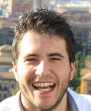 Gabriel Ramos Llordén合作论文数Athinoula A. Martinos Center for Biomedical Imaging, Massachusetts General Hospital4
 Susie Huang合作论文数Division of Neuroradiology, Massachusetts General Hospital;Harvard Medical School;Athinoula A. Martinos Center4
 Anastasia Yendiki合作论文数Harvard Medical School, Massachusetts General Hospital4
 Berkin Bilgic合作论文数Athinoula A. Martinos Center for Biomedical Imaging, Massachusetts General Hospital;Harvard Medical School4
 Kawin Setsompop合作论文数Department of Radiology, School of Medicine, Stanford University;Stanford Cancer Institute, Stanford University;Harvard-MIT Division of Health Sciences and Technology;Department of Electrical Engineering, School of Engineering, Stanford University;Athinoula A. Martinos Center for Biomedical Imaging4Monika Sliwiak合作论文数Dept Radiol, AA Martinos Ctr Biomed Imaging3John Kirsch合作论文数3
 Chiara Maffei合作论文数Athinoula A. Martinos Center for Biomedical Imaging, Massachusetts General Hospital and Harvard Medical School;Center for Neurotechnology and Neurorecovery, Massachusetts General Hospital;Massachusetts General Hospital, Massachusetts General Hospital and Harvard Medical School3
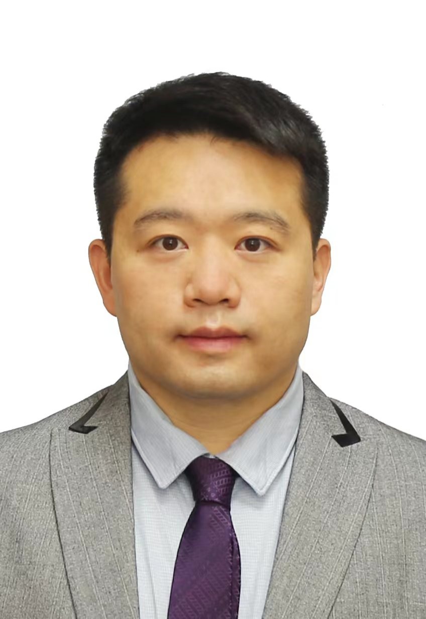 Qiyuan Tian (田启源)合作论文数School of Biomedical Engineering, Tsinghua University3
 David A Feinberg合作论文数University of California, Berkeley3
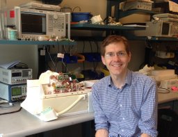 Stockmann Jason P合作论文数Massachusetts General Hospital Martinos Center for Imaging3Ruttorf Michaela合作论文数3
 Matthias Malzacher合作论文数3
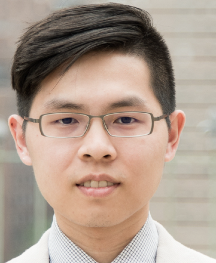 Hong-Hsi Lee合作论文数Department of Radiology, Massachusetts General Hospital3
 Aapo Nummenmaa合作论文数3
 Jaume Coll-Font合作论文数B-Spiral Group, Northeastern University3Andreas Potthast合作论文数3
 Peter J. Basser合作论文数Section on Quantitative Imaging and Tissue Sciences, Eunice Kennedy Shriver National Institute of Child Health and Human Development, National Institutes of Health3Mirsad Mahmutovic合作论文数2Eli Mattingly合作论文数2Alina Scholz合作论文数2Matthias Gebhardt合作论文数2Thomas Benner合作论文数2Rui Tian合作论文数2
 Frank Gerrit Zöllner合作论文数Mannheimer Institut for Intelligent Systems in Medicine, Medical Faculty Mannheim, Heidelberg University2
 Klaus Scheffler合作论文数Department High-field Magnetic Resonance, Max-Planck-Institute for Biological Cybernetics;Department of Biomedical Magnetic Resonance, University of Tubingen2
 Azma Mareyam合作论文数2
 Clifford Bryan合作论文数2
 Qiuyun Fan (范秋筠)合作论文数Medical School of Tianjin University2
 Chacón-Caldera Jorge合作论文数2
 Samantha J. Ma合作论文数2
 Johan van den Brink合作论文数2Kai Buckenmaier合作论文数2
 Jonathan Polimeni合作论文数Harvard Medical School;Athinoula A. Martinos Center for Biomedical Imaging, Massachusetts General Hospital;Division of Health Sciences and Technology, Harvard-MIT2
 Axel Thielscher合作论文数Department of Psychiatry;University of Ulm;Department of Psychiatry, University of Ulm2




