BACKGROUND:Intracranial atherosclerotic disease (ICAD) is a leading cause of ischemic stroke, yet how post-stenotic cerebral hemodynamics regulate endothelial phenotype remains poorly defined. We tested whether focal post-stenotic low wall shear stress (WSS) in patient-specific middle cerebral artery (MCA) stenoses associates with endothelial proliferation and pro-thrombotic activation. METHODS:CTA-derived geometries from SAMMPRIS participants were reconstructed for computational fluid dynamics (CFD) analysis (n=33 paired MCAs). A subset of stenotic and contralateral control models was 3D-printed, endothelialized with Human Umbilical Vein Endothelial Cells (HUVECs), and perfused under physiologic flow (n=8 pairs). Anatomically matched regions were analyzed for proliferation (Ki-67), biglycan (BGN), cell morphology, and pro-thrombotic mediators (LPCAT2, PAI1) using confocal imaging and automated segmentation. RESULTS:Stenotic MCAs exhibited significantly greater post-stenotic low-WSS area ratios than paired controls (p<0.0001). Distal low-WSS area correlated with increased proliferation (r=0.688, p=0.013) and BGN expression (r=0.580, p=0.046), confirmed by bootstrapping (p<0.001). Low-WSS regions demonstrated reduced cell area (0.85-fold, p=0.0013) and elevated LPCAT2 (1.34-fold) and PAI1 (1.41-fold) expression (both p<0.0001), which inversely correlated with particle-flow linearity (p≤0.031). CONCLUSIONS:Patient-specific cerebral stenoses generate focal low-WSS environments that can induce endothelial proliferation and pro-thrombotic signaling, supporting a mechanistic link between intracranial hemodynamics and endothelial dysfunction in ICAD.
Transcatheter aortic valve replacement (TAVR) is an established treatment for severe aortic stenosis; however, it carries the risk of periprocedural strokes. Current cerebral embolic protection (CEP) devices, such as the Sentinel, provide partial protection but are limited by inadequate anatomical coverage and inability to capture smaller emboli effectively. This study aimed to evaluate the effectiveness of a novel CEP device, the F2 filter with a 28 μm pore size and full cervical vessel coverage, in preventing emboli from entering the cerebral circulation. The Sentinel and F2 filter were evaluated for the ability to prevent embolic particles of various sizes (45-300 µm) from entering cerebral arteries using two in vitro flow models, incorporating standard and tortuous aortic anatomies. Additionally flow rates were also measured to confirm that normal perfusion was maintained while the devices were in place. The F2 filter maintained normal cerebral arterial flow and significantly reduced the number of particles across all sizes compared to the Sentinel and control groups. This reduction was observed in all four cerebral branches and across both standard and tortuous aorta models. The F2 filter showed superior neuroprotective effectiveness to prevent embolic debris from entering the cerebral circulation in the in vitro models. By offering comprehensive coverage to all cervical arteries and with a smaller mesh size, this filter has the potential to improve cerebral protection during TAVR.
Background and Purpose: In the current era, where mechanical thrombectomy (MT) has become the gold standard for treating large vessel occlusions in acute ischemic stroke, the role of intravenous tissue plasminogen activator (tPA) remains pivotal. It is well-established that pre-thrombectomy administration of tPA can enhance recanalization rates and excellent outcomes, leading to an increased adoption of this combined treatment approach. Understanding how tPA alters clots occluding large vessels is of crucial clinical importance for improving outcomes in thrombectomy when tPA is combined. This study seeks to elucidate the temporal changes in clots characteristics post-tPA administration, providing insights that could optimize the timing and strategy of subsequent mechanical intervention. Methods: We utilized a modified Chandler loop technique to generate RBC-rich clots from ovine blood for simulating clot analogs. The clots were treated with 10 μg/ml concentration of Alteplase to evaluate clot dissolution. An in-vitro MCA occlusion model, replicating human neurovascular anatomy, was used to assess the impact of clot treatment under dynamic flow conditions. Clots were analyzed for weight changes before and after treatment, and detailed pathological analyses were conducted using advanced imaging techniques. Statistical significance was determined using Student t test and 2-way repeated-measures ANOVA, with p-values <0.05 considered significant. All analyses were performed using SPSS software. Results: In the tPA group, clot weight decreased over time, with an average reduction to 46.7% of the initial value after 1 hour of solution exposure. In contrast, the control group showed relatively stable clot weight, remaining around 80% throughout the 1-hour exposure (P<0.001). Figure showing fhe fibrin content of the clot in the tPA group decreased significantly from 10.3% to 4.5% over time (P=0.038), whereas in the control group, it remained relatively unchanged, going from 7.3% to 8.1% (P=0.868). Conclusion: This study confirmed that tPA effectively reduces clot weight and decreases the fibrin content within clots. Our results suggest that tPA administration not only reduces clot mass but also modifies its structural integrity, thereby potentially increasing the success rate of subsequent mechanical thrombectomy procedures.
Objective Neuroendovascular procedures rely on successful navigation and stable access to the target vessel. The Stabilizer is a 300 cm long exchange wire with a 0.014 diameter and a soft, flexible stent at the distal end designed to assist with navigation and device delivery. This study aims to assess the efficacy of the Stabilizer for navigation in a variety of challenging environments. Methods The efficacy of the Stabilizer was evaluated using three challenging vascular models: a giant aneurysm model, a severe tortuosity model, and an M1 stenosis model. The Stabilizer was compared with a conventional wire during navigation in each model. Results In the giant aneurysm model, there was no significant difference of success during straightening of a looped wire and significantly higher success rates when advancing an intermediate catheter with the Stabilizer beyond the aneurysm neck compared to a conventional guidewire. The Stabilizer also significantly increased success rates when advancing an intermediate catheter through a model with severe tortuosity compared to a conventional guidewire, as well as exchange maneuver for intracranial stenting in a stenosis model compared to an exchange wire. Conclusions In our experimental model, the Stabilizer significantly improved navigation and device delivery in a variety of challenging settings compared to conventional wires.
OBJECTIVE:Fragile soft clots and stiff clots remain challenging in the treatment of acute ischemic stroke. This study aims to investigate the impact of clot stiffness on the efficacy of thrombectomy devices and a new aspiration catheter with a hydro-separator.METHODS:The Neurostar aspiration catheter has a novel hydro-separator technology that macerates clots by a stream of saline inside the catheter. The Neurostar catheter and two commercially available devices, the SOFIA aspiration catheter and Solitaire stent retriever, were tested in this study. We evaluated the efficacy of each device on clots with various stiffness in a simple in vitro model. We also assessed single-pass recanalization performance in challenging situations with large erythrocyte-rich clots and fibrin-rich clots in a realistic vascular model.RESULTS:We observed an inverse association between the clot stiffness and recanalization rates. The aspiration catheter, SOFIA ingested soft clots but not moderately stiff clots. When removing soft clots with the stent retriever, fragmentation was observed, although relatively stiff clots were well-integrated and removed. The Neurostar ingested soft clots similar to the aspiration catheter, and also aspirated stiff clots by continuous suction with hydro-separator. In the experiments with challenging clots, the Neurostar led to significantly higher recanalization rates than the stent retriever and aspiration catheter.CONCLUSIONS:The stiffness of the clots affected the efficacy of endovascular thrombectomy based on the type of device. The Neurostar catheter with hydro-separator resulted in better success rates than a commercially available aspiration catheter and stent retriever in this experimental model.
Introduction: 3D printed human vascular in vitro models of aneurysms and acute stroke have been utilized for training, simulation and device development. However, there are no realistic in vitro arteriovenous malformation (AVM) models. Current experimental models analyzing the efficacy of embolic materials or flow conditions are limited by their simplistic design, lacking complex AVM nidus anatomic features. The purpose of this study is to develop a new in vitro AVM model for embolic material testing and flow analysis. Methods: 3D images of the AVM nidus were extracted from 3D rotational angiography from a patient. Artificial feeders and drainers were added to the nidus and an inner vascular mold was printed using a 3D printer. The inner mold was coated with polydimethylsiloxanes. The inner plastic mold was removed by acetone, leaving a hollow AVM model. ONYX injection and 4DFlow MRI (Phase Contrast MRA) were performed using the AVM models. In addition, computational fluid dynamics (CFD) analysis was performed to compare flow rate with 4DFlow MRI. Results: An in vitro AVM model with realistic representation of nidus vasculature and complexity was successfully created. Liquid onyx injection performed in the in vitro model successfully replicated real-life treatment conditions. The model effectively simulated plug and push technique before penetration of the ONYX into the AVM nidus. 4DFlow MRI flow rates were similar to the CFD analysis. Conclusions: An in vitro AVM model using 3D printing technology was successfully created. The model demonstrated realistic pliability during ONYX injection. This in vitro AVM model may represent a useful tool for training and development of new materials, and have potential of highly-resolved flow quantifications.
Background In vitro vascular models for brain aneurysms and acute stroke have been used for training, simulation and research purpose. However, the use of realistic in vitro models for arteriovenous malformation (AVM) have not been reported. Current in vitro AVM models analyzing the efficacy of embolic materials or flow conditions are limited due to a lack of realistic anatomical and dynamic features of complex nidus. Materials and Methods 3D AVM nidus images were extracted and segmented from 3D rotational angiography from a patient. Additional artificial feeders and drainers were attached to the AVM nidus. The inner vascular mold was printed using a plastic 3D printer. The inner mold was coated with silicone and then removed with acetone, leaving a hollow AVM model. Injections of liuid embolic material and 4D flow MRI were performed using the 3D in vitro AVM model. Computational fluid dynamics (CFD) analysis was also performed to compare the flow volume and velocity to 4D flow MRI Results The created in vitro AVM models had realistic representation of nidus vasculature and complexity derived from patients. The injection of liquid embolic material performed in this model replicated real-life treatment conditions. The plug and push technique was successfully simulated to penetratreliquid embolic material into the AVM nidus. The flow data from 4D flow MRI were comparable to CFD analysis. Conclusions An in vitro human brain AVM model with realistic complexities of nidus was successfully manufactured using 3D printing technology. The model demonstrated realistic pliability during the liquid embolic material injection and also feasibility of flow analysis. This in vitro AVM model may represent a valuable tool for simulation, flow research and development of new materials or technique. Disclosures N. Kaneko: None. H. Ullman: None. F. Ali: None. P. Berg: 1; C; German Research Foundation, Federal Ministry of Education and Research within the Forschungscampus STIMULATE. Y. Ooi: None. S. Tateshima: 2; C; Cerenovus, Medtronic, Stryker. G. Colby: 2; C; Medtronic, Microvention, Stryker. Y. Komuro: None. P. Hu: None. V. Szeder: None. M. Nour: None. L. Guo: None. A. Chien: None. F. Vinuela: None. S. Nemoto: None. J. Hinman: None. G. Duckwiler: 1; C; Tarsadia Foundation. 2; C; Medtronic. R. Jahan: None.
Background Large clot burden and fibrin clots remain challenging for mechanical thrombectomy. The aim of this study is to assess a new-generation aspiration system with hydro-separator technology that works regardless of clot size or composition. Materials and Methods The Neurostar thrombectomy system is comprised of a 6F Neurostar catheter and a Saline Drive Unit (SDU) with a peristaltic pump, which creates a stream of saline on the tip of the catheter to macerate the thrombus during aspiration (hydro-separator). The Neurostar catheter as well as two commercially available thrombectomy devices, the Solitaire FR stent retriever and Sofia Plus aspiration catheter, were tested to compare single-pass recanalization performance in challenging situations. Fibrous clots and cohesive erythrocyte-rich clots were produced from porcine blood. A segment of fibrous clot was placed in the MCA distal to M1 and proximal to M2 in a tortuous in-vitro intracranial vascular model, or a relatively long (20 mm) erythrocyte-rich clot was placed in the middle segment of the M1. Mechanical thrombectomy using each device was repeated 10 times with fibrous clots and erythrocyte-rich clots. Success recanalization was defined as clot removal without visible fragmentation or migration. Results The Neurostar catheter led to significantly better single-pass recanalization performance (18/20) compared to the Sofia Plus aspiration catheter (11/20) and Solitaire FR stent retriever (10/20). All of the recanalization procedures with the Sofia Plus required complete removal of the catheter because the clot was stuck at the tip or could not be ingested entirely. In contrast, the Neurostar catheter could ingest the clot and remain at the site of occlusion in all of the successful single pass recanalization procedures. Conclusions Mechanical thrombectomy with the new aspiration system with hydro-separator resulted in higher success rates than a commercially available stent retriever and aspiration catheter in this experimental model. Further in vivo studies should be performed to confirm its performance. Disclosures N. Kaneko: 2; C; Walk Vascular. L. Guo: None. Y. Komuro: None. S. Tateshima: 2; C; Walk Vascular, Medtronic.
BACKGROUND: Current in vitro models for human brain arteriovenous malformation (AVM) analyzing the efficacy of embolic materials or flow conditions are limited by a lack of realistic anatomic features of complex AVM nidus. The purpose of this study was to evaluate a newly developed in vitro AVM model for embolic material testing, preclinical training, and flow analysis. METHODS: Three-dimensional (3D) images of the AVM nidus were extracted from 3D rotational angiography from a patient. Inner vascular mold was printed using a 3D printer, coated with polydimethylsiloxanes, and then was removed by acetone, leaving a hollow AVM model. Injections of liquid embolic material and 4-dimensional (4D) flow magnetic resonance imaging (MRI) were performed using the AVM models. Additionally, computational fluid dynamics analysis was performed to examine the flow volume rate as compared with 4D flow MRI. RESULTS: The manufacture of 3D in vitro AVM models delivers a realistic representation of human nidus vasculature and complexity derived from patients. The injection of liquid embolic agents performed in the in vitro model successfully replicated real-life treatment conditions. The model simulated the plug and push technique before penetration of the liquid embolic material into the AVM nidus. The 4D flow MRI results were comparable to computational fluid dynamics analysis. CONCLUSIONS: An in vitro human brain AVM model with realistic geometric complexities of nidus was successfully created using 3D printing technology. This AVM model offers a useful tool for training of embolization techniques and analysis of hemodynamics analysis, and development of new devices and materials.
Introduction: The clinical syndrome associated with Sars-CoV-2 infection is associated with an increased rate of cerebrovascular events, predominantly ischemic stroke. Direct viral invasion of the cerebral endothelium could be a significant driver of Sars-CoV-2-mediated stroke through expression of ACE2 and viral entry co-factors including TMPRSS2. Prior work indicates that ACE2 mRNA and protein are expressed in the endothelial cells of the brain, yet the effect of flow-mediated phenomena on ACE2 expression and the susceptibility of human cerebral endothelia to Sars-CoV-2 infection is unknown. Methods: 3D printed models of the human middle cerebral artery were endothelialized with either human umbilical vein endothelial cells (HUVECs) or human brain microvascular endothelial cells (HBMECs). Endothelialized models were subjected to 3D rotational perfusional culture at variable pulsatile flow rates (0-22.5 dynes/cm 2 ). ACE2 and TMPRSS2 expression were measured by Taqman qPCR. Biotinylated recombinant Sars-CoV-2 S protein was used to judge regional vessel binding of virus using confocal microscopy. Results: In 3D models endothelialized with either HUVECs or HBMECs, ACE2 mRNA expression is significantly modulated by vessel diameter and the presence of of flow (15 dynes/cm 2 ) (p<0.0001 by two-way ANOVA). Pulsatile flow drives ACE2 expression more robustly in HUVECs than in HBMECs (11.5-fold vs. 4.9-fold; p<0.001). Higher flow rates (22.5 dynes/cm 2 ) produced further increases in ACE2 expression in both cell types. Gene expression analysis demonstrated no detection of the Sars-CoV-2 viral entry co-factor TMPRSS2 in HBMECs. Flow-mediated ACE2 expression was associated with binding of recombinant Sars-CoV-2 S protein to the vessel wall suggesting direct cerebrovascular susceptibility to Sars-CoV-2. Conclusion: The Sars-CoV-2 cellular receptor ACE2 is expressed by human cerebral endothelia in a flow-dependent manner. Cerebral vessels are susceptible to Sars-CoV-2 infection and bind viral S protein under flow conditions but likely requires unique co-factors for viral entry. Identification of the molecular mechanisms triggered by Sars-CoV-2 infection in brain vasculature can alleviate the burden of stroke associated with COVID-19.
BACKGROUND AND PURPOSE:Severe acute respiratory syndrome coronavirus-2 (SARS-CoV-2) infection is associated with an increased rate of cerebrovascular events including ischemic stroke and intracerebral hemorrhage. The mechanisms underlying cerebral endothelial susceptibility and response to SARS-CoV-2 are unknown yet critical to understanding the association of SARS-CoV-2 infection with cerebrovascular events.METHODS:Endothelial cells were isolated from human brain and analyzed by RNA sequencing. Human umbilical vein and human brain microvascular cells were used in both monolayer culture and endothelialized within a 3-dimensional printed vascular model of the middle cerebral artery. Gene expression levels were measured by quantitative polymerase chain reaction and direct RNA hybridization. Recombinant SARS-CoV-2 S protein and S protein-containing liposomes were used to measure endothelial binding by immunocytochemistry.RESULTS:ACE2 (angiotensin-converting enzyme-2) mRNA levels were low in human brain and monolayer endothelial cell culture. Within the 3-dimensional printed vascular model, ACE2 gene expression and protein levels were progressively increased by vessel size and flow rates. SARS-CoV-2 S protein-containing liposomes were detected in human umbilical vein endothelial cells and human brain microvascular endothelial cells in 3-dimensional middle cerebral artery models but not in monolayer culture consistent with flow dependency of ACE2 expression. Binding of SARS-CoV-2 S protein triggered 83 unique genes in human brain endothelial cells including upregulation of complement component C3.CONCLUSIONS:Brain endothelial cells are susceptible to direct SARS-CoV-2 infection through flow-dependent expression of ACE2. Viral S protein binding triggers a unique gene expression profile in brain endothelia that may explain the association of SARS-CoV-2 infection with cerebrovascular events.
Abstract Histone deacetylase inhibitors (HDACi’s) have emerged as a promising class of drugs for treatment of malignancies such as glioblastoma (GBM). Several studies have demonstrated the anti-tumor property of HDACi’s against GBM in both in vitro and in vivo experiments. Nonetheless, in clinical trials, HDACi only marginally increased overall survival of patients with GBM. The mixed results of trials with HDACi’s in glioma have prompted us to hypothesize that improved selection of patients by tumor characteristics could enhance the efficacy of therapy. We specifically tested the effects of valproic acid (VPA), a HDACi and an antiepileptic drug against IDH mutant gliomas. We have previously demonstrated that our IDH mutant glioma cell lines have gene expression and methylation patterns highly similar to IDH mutant tumors in situ. Mutant IDH1 alters the epigenetic landscape of gliomas leading to the hypermethylation phenotype and transcriptional repression of genes. This repression of genes may contribute to tumorigenesis and progression of IDH mutant gliomas. We found that VPA inhibits the growth of patient-derived IDH1 mutant glioma lines. In addition, RNA sequencing analysis of vehicle and VPA-treated IDH1 mutant glioma cells showed de-repression of several genes previously shown to be downregulated in IDH1 mutant glioma cell lines. We also treated cells with another HDACi LBH589 and found that both VPA and LBH589 upregulates similar gene sets suggesting that HDAC inhibition promotes de-repression of previously repressed genes. Ongoing studies are aimed at determining the molecular mechanism by which VPA regulates the growth of IDH1 mutant tumors.
Summary Glioblastoma (GBM) metabolism has traditionally been characterized by a primary dependence on aerobic glycolysis, prompting the use of the ketogenic diet (KD) as a potential therapy. In this study we evaluated the effectiveness of the KD in GBM and assessed the role of fatty acid oxidation (FAO) in promoting GBM propagation. In vitro assays revealed FA utilization throughout the GBM metabolome, and growth inhibition in nearly every cell line in a broad spectrum of patient-derived glioma cells treated with FAO inhibitors. In vivo assessments revealed that knockdown of carnitine palmitoyltransferase 1A (CPT1A), the rate limiting enzyme for FAO, reduced the rate of tumor growth and increased survival. However, the unrestricted ketogenic diet did not reduce tumor growth, and for some models significantly reduced survival. Altogether, these data highlight important roles for FA and ketone body metabolism that could serve to improve targeted therapies in GBM.
Angiogenesis plays a critical role in progression of malignant gliomas. The development of glioma-specific labeling molecules that can aid detection and visualization of angiogenesis can help surgical planning and improve treatment outcome. The aim of this study was to evaluate if two peptides (GX1 and RGD-GX1) linked to angiogenesis can be used as an MR-imaging markers of angiogenesis. MR imaging was performed in U87 glioblastoma-bearing NOD-SCID mice at different time points between 15 and 120 min post-injection to visualize particle distribution. GX1 and RGD-GX1 exhibited the highest accumulation in U87 glioblastoma at 120 min post i.v. administration. GX1-conjugated agents lead to higher decrease in transverse relaxation time (T 2) (i.e., stronger contrast enhancement) than RGD-GX1-conjugated agents in U87 glioblastoma tumor model. In addition, we tested if U87-IDH1R132 mutated cell line had different pattern of GX1 or RGD-GX1 particle accumulation. Responses in U87-IDH1WT followed a similar pattern with GX1 contrast agents; however, lower contrast enhancement was observed with RGD-GX1 agents. The specific binding of these peptides to human glioblastoma xenograft in the brain was confirmed by magnetic resonance imaging. The contrast enhancement following injection of magnetonanoparticles conjugated to GX1 peptide matched well with CD31 staining and iron staining.
BACKGROUND:Nitroxoline is an FDA-approved antibiotic with potential antitumor activity. Here we evaluated whether nitroxoline has antiproliferative properties on glioma cell growth in vitro and in vivo using glioma cell lines and a genetically engineered PTEN/KRAS mouse glioma model.METHODS:The effect of nitroxoline treatment on U87 and/or U251 glioma cell proliferation, cell-cycle arrest, invasion, and ability to induce an apoptotic cascade was determined in vitro. Magnetic resonance imaging was used to measure glioma volumes in genetically engineered PTEN/KRAS mice prior to and after nitroxoline therapy. Induction of apoptosis by nitroxoline was evaluated at the end of treatment using terminal deoxyribonucleotidyl transferase (TDT)-mediated dUTP-digoxigenin nick end labeling (TUNEL).RESULTS:Nitroxoline inhibited the proliferation and invasion of glioblastoma cells in a time- and dose-dependent manner in vitro. Growth inhibition was associated with cell-cycle arrest in G1/G0 phase and induction of apoptosis via caspase 3 and cleaved poly(ADP-ribose) polymerase. In vivo, nitroxoline-treated mice had no increase in tumor volume after 14 days of treatment, whereas tumor volumes doubled in control mice. Histological examination revealed 15%-20% TUNEL-positive cells in nitroxoline-treated mice, compared with ∼5% in the control group.CONCLUSION:Nitroxoline induces apoptosis and inhibits glioma growth in vivo and in vitro. As an already FDA-approved treatment for urinary tract infections with a known safety profile, nitroxoline could move quickly into clinical trials pending confirmatory studies.
Malignant gliomas are the most common human primary brain tumors. Point mutation of amino acid arginine 132 to histidine (R132H) in the IDH1 protein leads to an enzymatic gain-of-function and is thought to promote gliomagenesis. Little is known about the downstream effects of the IDH1 mutation on protein expression and how and whether changes in protein expression are involved in tumor formation or propagation. In the current study, we used 2D DIGE (difference gel electrophoresis) and mass spectrometry to analyze differences in protein expression between IDH1R132H mutant and wild type anaplastic (grade III) astrocytoma from human brain cancer tissues. We show that expression levels of many proteins are altered in IDH1R132H mutant anaplastic astrocytoma. Some of the most over-expressed proteins in the mutants include several forms of αB-crystallin, a small heat-shock and anti-apoptotic protein. αB-crystallin proteins are elevated up to 22-fold in IDH1R132H mutant tumors, and αB-crystallin expression appears to be controlled at the post-translational level. We identified the most abundant form of αB-crystallin as a low molecular weight species that is C-terminally truncated. We also found that overexpression of αB-crystallin can be induced by transfecting U251 human glioblastoma cell lines with the IDH1R132H mutation. In conclusion, the association of a C-terminally truncated form of αB-crystallin protein with the IDH1R132H mutation is a novel finding that could impact apoptosis and stress response in IDH1 mutant glioma.
Mammalian target of rapamycin (mTOR) is a serine/threonine kinase that regulates a variety of cellular functions such as growth, proliferation and autophagy. In a variety of cancer cells, overactivation of mTOR has been reported. In addition, mTOR inhibitors, such as rapamycin and its derivatives, are being evaluated in clinical trials as anticancer drugs. However, no active mutants of mTOR have been identified in human cancer. Here, we report that two different point mutations, S2215Y and R2505P, identified in human cancer genome database confer constitutive activation of mTOR signaling even under nutrient starvation conditions. S2215Y was identified in large intestine adenocarcinoma whereas R2505P was identified in renal cell carcinoma. mTOR complex 1 prepared from cells expressing the mutant mTOR after nutrient starvation still retains the activity to phosphorylate 4E-BP1 in vitro. The cells expressing the mTOR mutant show increased percentage of S-phase cells and exhibit resistance to cell size decrease by amino-acid starvation. The activated mutants are still sensitive to rapamycin. However, they show increased resistance to 1-butanol. Our study points to the idea that mTOR activating mutations can be identified in a wide range of human cancer.
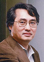 Fuyuhiko Tamanoi合作论文数Molecular Biology Institute, University of California, Los Angeles14
 Naoki Kaneko合作论文数Department of Neurosurgery, Jichi Medical University11
 Jason Hinman合作论文数Department of Neurology, David Geffen School of Medicine, University of California, Los Angeles;Mary S. Easton Center for Alzheimer's Research and Care, University of California, Los Angeles7
 Yutaro Komuro合作论文数MSL, Tokyo Tech7
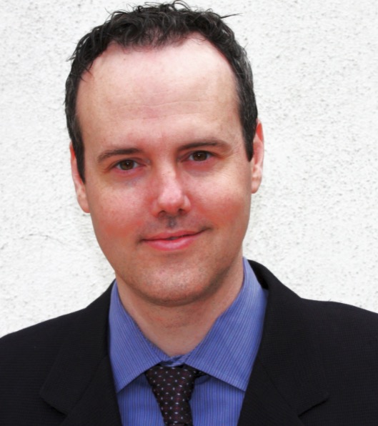 Whitney B. Pope合作论文数Department of Radiology, David Geffen School of Medicine, University of California5
 Satoshi Tateshima合作论文数David Geffen School of Medicine, University of California5
 Mahsa Ghovvati合作论文数4
 Juran Kato合作论文数4
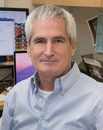 Harley I. Kornblum合作论文数Department of Molecular and Medical Pharmacology, University of California Los Angeles4Jennifer An合作论文数3Fadil Ali合作论文数Dept Radiol Sci, Univ Calif Los Angeles3
 Yinn Cher Ooi合作论文数Department of Radiological Sciences, University of California Los Angeles3Henrik Ullman合作论文数3
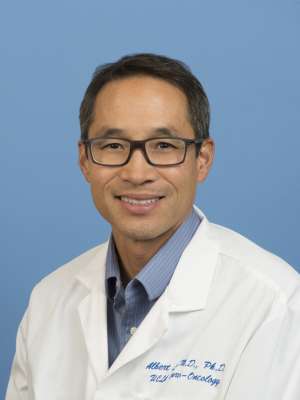 Albert Lai合作论文数Department of Neurology, David Geffen School of Medicine, University of California, Los Angeles3Taichiro Imahori (今堀太一郎)合作论文数Kobe University;David Geffen School of Medicine, University of California, Los Angeles3
 Kenichi Sakuta合作论文数3
 Philipp Berg合作论文数University of Magdeburg, Otto-von-Guericke-Univ. Magdeburg3
 Gary Ross Duckwiler合作论文数David Geffen School of Medicine at UCLA3
 Reza Jahan合作论文数Division of Interventional Neuroradiology, School of Medicine, UCLA;Department of Radiology, School of Medicine, UCLA;Department of Neurosurgery, School of Medicine, UCLA;UCLA Stroke Center3Geoffrey P. Colby合作论文数Department of Neurological Surgery, Columbia University3N Kaneko合作论文数2N. Thapar合作论文数2S Tateshima合作论文数2M Martinez合作论文数2
 Chia-Ling Gau合作论文数2Ph Patel合作论文数2Lucido Ponce Mejia合作论文数2Jantzen Sperry合作论文数2Visesha Kakarla合作论文数2
 Kasra Khatibi合作论文数Div Intervent Neuroradiol, Univ Calif Los Angeles2Viktor Szeder合作论文数2
 Linda Liau合作论文数Jonsson Comprehensive Cancer Center, University of California, Los Angeles;Department of Neurosurgery, University of California, Los Angeles2
 Peng Hu合作论文数Department of Bioengineering, School of Engineering, University of California, Los Angeles2
 Lai Chan合作论文数Department of Laboratory Medicine and2
 Fanny Elahi合作论文数Department of Neurology, Icahn School of Medicine at Mount Sinai;Friedman Brain Institute, Icahn School of Medicine at Mount Sinai;Ronald M. Loeb Center for Alzheimer’s Disease, Icahn School of Medicine at Mount Sinai2Nour May合作论文数2
 Michael C Condro合作论文数Molecular, Cellular &Integrative Physiology Interdepartmental Program, University of California;Molecular, Cellular & Integrative Physiology Interdepartmental Program, University of California2
 Mirsadraei Leili合作论文数Department of Radiological Sciences, Ronald Reagan UCLA Medical Center;David Geffen School of Medicine, University of California at Los Angeles;Department of Pathology, University of California San Diego Medical Center;David Geffen School of Medicine, University of California at Los Angeles;David Geffen School of Medicine, UCLA;University of California San Diego Medical Center, University of California at Los Angeles2
 Fernando Vinuela合作论文数Department of Radiology, University of California, Los Angeles2
 Erica Golemis合作论文数Department of Cancer and Cellular Biology, Lewis Katz School of Medicine, Temple University;Fox Chase Cancer Center2
 David S. Liebeskind合作论文数Department of Neurology, University of California, Los Angeles2Angel P. Tabancay合作论文数2Jelena Lazovic合作论文数Department of Biology, California Institute of Technology2Vladimir Khazak合作论文数2
 Hamidreza Saber合作论文数UCLA Health;Ascension2Jun Urano合作论文数2
 Aichi Chien合作论文数Division of Interventional Neuroradiology, University of California Los Angeles;Department of Radiology, David Geffen School of Medicine, University of California Los Angeles2Shigeru Nemoto合作论文数2
 Hironori Edamatsu合作论文数Faculty of Bioscience and Biotechnology, Tokyo Institute of Technology2




