To broaden our understanding of bradyarrhythmias and conduction disease, we performed common variant genome-wide association analyses in up to 1.3 million individuals and rare variant burden testing in 460,000 individuals for sinus node dysfunction (SND), distal conduction disease (DCD) and pacemaker (PM) implantation. We identified 13, 31 and 21 common variant loci for SND, DCD and PM, respectively. Four well-known loci (SCN5A/SCN10A, CCDC141, TBX20 and CAMK2D) were shared for SND and DCD, while others were more specific for SND or DCD. SND and DCD showed a moderate genetic correlation (rg = 0.63). Cardiomyocyte-expressed genes were enriched for contributions to DCD heritability. Rare-variant analyses implicated LMNA for all bradyarrhythmia phenotypes, SMAD6 and SCN5A for DCD and TTN, MYBPC3 and SCN5A for PM. These results show that variation in multiple genetic pathways (for example, ion channel function, cardiac developmental programs, sarcomeric structure and cellular homeostasis) appear critical to the development of bradyarrhythmias. Genome-wide analyses identify variants associated with sinus node dysfunction, distal conduction disease and pacemaker implantation, implicating ion channel function, cardiac developmental programs and sarcomeric structure in bradyarrhythmia susceptibility.
Human induced pluripotent stem cell-derived cardiomyocytes (hiPSC-CMs) are increasingly used for disease modeling, drug discovery, and precision medicine, yet their utility is often limited by their immature phenotype. One promising maturation strategy involves using micropatterned substrates that mimic native cardiomyocytes' organizational growth and stiffness. However, the maturity of this model has not fully been assessed, and there is currently no method to extract proteins from micropatterned hiPSC-CMs for top-down proteomic analysis. Herein, we present a high-sensitivity protein extraction protocol for top-down proteomic analysis of hiPSC-CMs. Through this method, we assessed the maturation of micropatterned hiPSC-CMs compared to traditional monoculture and coculture monolayers at the proteoform level. We found that micropatterned hiPSC-CMs display molecular signatures of cardiomyocyte maturation including increased expression of ventricular myosin light chain isoforms, reduced expression of the fetal troponin T isoform, and decreased phosphorylation of alpha-tropomyosin. This high-sensitivity approach enables robust top-down proteomics from limited, heterogeneous cell populations and identifies the micropattern hiPSC-CM as a more adult-like CM model, broadening the utility of structured culture systems for cardiac disease modeling and translational research. Source data for this manuscript are available via MassIVE repository at massive.ucsd.edu with identifier: MSV000097864.
The hERG1 potassium channel conducts the cardiac repolarizing current, IKr. hERG1 has emerged as a therapeutic target for cardiac diseases marked by prolonged action potential duration (APD). Unfortunately, many hERG1 activators display off-target and proarrhythmic effects that limit their therapeutic potential. A Per-Arnt-Sim (PAS) domain in the hERG1 N-terminus reduces IKr by slowing channel activation and promoting inactivation. Disrupting PAS activity increases IKr and shortens APD in human induced pluripotent stem cell-derived cardiomyocytes (hiPSC-CMs). We thus hypothesized that the hERG1 PAS domain could represent a therapeutic target to reduce arrhythmogenic potential in a long QT syndrome (LQTS) background. To test this, we measured the antiarrhythmic capacity of a PAS-disabling single-chain variable fragment antibody, scFv2.10, in a hiPSC-CM line derived from a patient with Jervell and Lange Nielsen (JLN) syndrome. JLN is a severe form of LQTS caused by autosomal recessive mutations in KCNQ1. The patient in this study carried compound heterozygous mutations in KCNQ1. Corresponding JLN hiPSC-CMs displayed prolonged APD and early afterdepolarizations (EADs). Disrupting PAS with scFv2.10 increased IKr, shortened APD, and reduced the incidence of EADs. These data demonstrate that the hERG1 PAS domain could serve as a therapeutic target to treat disorders of cardiac electrical dysfunction.
KCNJ2 encodes the inward rectifying potassium channel (Kir2.1) that underlies IK1 which maintains the cardiac resting membrane potential and regulates excitability. Mutations in KCNJ2 have been linked to several clinical phenotypes associated with life-threatening ventricular arrhythmia and sudden death including Andersen-Tawil syndrome (ATS) from loss of function mutations, and Short QT Syndrome 3 from gain of function mutations. Detailed structural-functional relationships to explain the arrhythmia phenotypes are understudied and limit the capacity to provide precision medicine. Here, we combine in-depth and complementary computational molecular modeling techniques with functional analysis from three patients with ATS that harbor KCNJ2 mutations R67Q, R218L, and G300D. Whole-cell patch-clamp experiments revealed loss of function in homomeric mutant channels. Full-length Kir2.1 models were developed for structure-based investigation, and mutations were introduced in both open and closed conformations. Site-directed mutagenesis identified altered interaction profiles contributing to structural perturbations. Molecular dynamics simulations assessed the impact of each mutation on overall channel conformation and stability. Principal component analysis and normal mode analysis revealed mutation-specific structural perturbations. The findings afford atomic mechanistic underpinnings of mutation-specific perturbations. Our multifaceted approach provides first atomic-level insights into the molecular mechanisms underlying ATS, paving the way for structure-guided targeted therapeutic strategies for ATS and related channelopathies.
Human induced pluripotent stem cells (iPSCs) have emerged as a transformative platform for modeling inherited cardiac arrhythmia syndromes and uncovering human-specific disease mechanisms. However, the promise of iPSC-derived cardiomyocytes lies beyond the recapitulation of arrhythmogenic phenotypes and channelopathies. In this review, we explore recent works which have enabled mechanistic interrogation and therapeutic insight for inherited arrhythmia syndromes, beyond the capabilities of traditional animal models. Such studies have leveraged iPSCs to elucidate the role of splice variants, transcriptional regulation, and mitochondrial stress in arrhythmogenesis. Further, iPSC systems have proven important for reclassifying variants of uncertain significance and in modeling idiopathic arrhythmias where genotype-phenotype links are elusive. Advances in directed differentiation now permit chamber-specific cardiac cell generation, allowing for atrial and ventricular disease modeling and revealing critical cell-cell interactions. iPSCs also serve as high-fidelity precursor platforms for drug testing, offering predictive insight into mutation-specific responses to pharmacologic and genetic therapies. Though limitations in maturation and scalability persist, ongoing efforts for integration with tissue engineering, multi-cellular models, and computational frameworks are evolving to improve model reliability. iPSC-based systems now occupy a critical role in arrhythmia research, bridging basic discovery with translational applications, thereby contributing to personalizing care and advancing therapeutics in inherited and idiopathic arrhythmic syndromes.
Introduction:Cardiac fibroblasts deposit and turnover the extracellular matrix in the heart, as well as secrete soluble factors that play critical roles in development, homeostasis, and disease. Coculture of CFs and human induced pluripotent stem cell (hiPSC)-derived cardiomyocytes (CMs) enhances CM mechanical output, yet the mechanism remains unclear. Methods:Here, we use an in vitro engineered platform to compare the effects on CM mechanical function of direct CM-CF Coculture and soluble signaling alone through CF Conditioned Medium to a CM Only monoculture. Mechanical analysis is performed using digital image correlation and custom software to quantify the coordination and organization of CM contractile behavior. Results:CM-CF Coculture induces larger CM contractile strains, and an increased rate of spontaneous contraction compared to CM Only. Additionally, CM-CF Cocultures have increased contractile anisotropy and myofibril alignment and faster kinetics. The paracrine effects of fibroblast conditioned medium (FCM) are sufficient to induce larger contractile strains and faster contraction kinetics with these effects remaining after the removal of FCM. However, FCM does not influence CM spontaneous rate, contractile alignment, anisotropy, or relaxation kinetics compared to CM Only control. Discussion:These data suggest that hiPSC-CFs exert dynamic and multifactorial effects on the mechanical function of hiPSC-CMs and highlight the importance of CFs in both the native heart and in vitro cardiac models. Further, this work demonstrates the applicability of the coculture-conditioned medium-monoculture paradigm to decouple the effects of paracrine factor and cell-cell signaling on hiPSC-CM mechanical function and maturation.
Background: Kir2.1 channels maintain the cardiac resting membrane potential and assist in phase 3 repolarization. PKA-mediated phosphorylation modulates Kir2.1 function, impacting heart excitability and rhythm. Using as mass-spetometry based integrated top-down and bottom up proteomic approach, we identified five novel and confirmed a 6 th phosphorylation site. However, which of these residues impact channel function has not been previously identified. Methods: HEK293 cells were engineered to stably express either WT-Kir2.1. Ablation of the phosphorylation site was achieved with an alanine substitution of the PKA phosphorylation residue (S13A, S14A, S313A,Y326A, T347A, and S425A). Whole-cell patch clamp experiments of WT-Kir2.1 or the phosphorylation mutants were performed using a standard protocol. Following the initial recording, cells were perfused with PKA-stimulating cocktail (20µM Forskolin and 10µ IBMX) for 4 minutes and the protocol was repeated. Clampfit 10.7, GraphPad Prism, and Origin 20 were used for Data Analysis. Results: HEK293 cells stabling expressing Kir2.1 demonstrate typical inward rectification with a maximum outward current at -50mV and responds to PKA-cocktail with 2.0 pA/pF increase in maximum outward current. Both S13A and S313A had a normal response PKA stimulation. In contrast, ablation of S425 and S14 resulted in no PKA response. T347A showed little response to PKA; however, the current density is very low at baseline, indicating that this residue is necessary for normal Kir2.1 current density. The Y326 is known to be critical for normal protein folding, and ablation of the site resulted in a complete loss of function. Discussion: Of the 6 PKA sites identified by a proteomic approach, only S425 and S14 functionally influence Kir2.1 response to PKA stimulation. Further molecular dynamics simulation investigation will provide deeper insight into the conformational changes of Kir2.1 in response to phosphorylation at these sites and the relationship of channel phosphorylation with clinical channel mutation residues.
BACKGROUND:Substantial data support a heritable basis for supraventricular tachycardias, but the genetic determinants and molecular mechanisms of these arrhythmias are poorly understood. We sought to identify genetic loci associated with atrioventricular nodal reentrant tachycardia (AVNRT) and atrioventricular accessory pathways or atrioventricular reciprocating tachycardia (AVAPs/AVRT).METHODS:We performed multiancestry meta-analyses of genome-wide association studies to identify genetic loci for AVNRT (4 studies) and AVAP/AVRT (7 studies). We assessed evidence supporting the potential causal effects of candidate genes by analyzing relations between associated variants and cardiac gene expression, performing transcriptome-wide analyses, and examining prior genome-wide association studies.RESULTS:Analyses comprised 2384 AVNRT cases and 106 489 referents, and 2811 AVAP/AVRT cases and 1,483 093 referents. We identified 2 significant loci for AVNRT, which implicate NKX2-5 and TTN as disease susceptibility genes. A transcriptome-wide association analysis supported an association between reduced predicted cardiac expression of NKX2-5 and AVNRT. We identified 3 significant loci for AVAP/AVRT, which implicate SCN5A, SCN10A, and TTN/CCDC141. Variant associations at several loci have been previously reported for cardiac phenotypes, including atrial fibrillation, stroke, Brugada syndrome, and electrocardiographic intervals.CONCLUSIONS:Our findings highlight gene regions associated with ion channel function (AVAP/AVRT), as well as cardiac development and the sarcomere (AVAP/AVRT and AVNRT) as important potential effectors of supraventricular tachycardia susceptibility.
 Timothy J. Kamp合作论文数Department of Medicine, School of Medicine and Public Health, University of Wisconsin-Madison;Department of Cell and Regenerative Biology, School of Medicine and Public Health, University of Wisconsin-Madison29
 Louise Reilly合作论文数25
 Corey L. Anderson合作论文数University of Wisconsin-Madison25
 Jonathan C. Makielski合作论文数Department of Medicine, School of Medicine and Public Health, University of Wisconsin–Madison;University of Wisconsin - Oshkosh;University of Wisconsin - Stout23
 Orland Kate M合作论文数19
 Vaidyanathan R合作论文数19
 Ryan Kipp合作论文数Department of Medicine, School of Medicine and Public Health, University of Wisconsin-Madison14
 Jianhua Zhang合作论文数13Matthew Kalscheur合作论文数12
 Miguel Leal合作论文数11
 Wendy C. Crone合作论文数Departments of Biomedical Engineering and Materials Science and Engineering, University of Wisconsin-Madison10
 Michael J. Ackerman合作论文数Children's Center, Mayo Clinic;Division of Heart Rhythm Services, Department of Cardiovascular Medicine, Mayo Clinic10
 Craig January合作论文数Division of Cardiovascular Medicine, Department of Medicine, School of Medicine and Public Health, University of Wisconsin–Madison10
 Alexey Glukhov合作论文数Department of Medicine, School of Medicine and Public Health, University of Wisconsin-Madison9
 Mina Chung合作论文数The Robert and Suzanne Tomsich Department of Cardiovascular Medicine, Cleveland Clinic;Section of Pacing and Electrophysiology, Cleveland Clinic9Di Lang合作论文数8
 Saba Munawar合作论文数1] Institute of Biomedical and Genetic Engineering (IBGE),, National University of Sciences and Technology (NUST),8
 Kathleen Maginot合作论文数Department of Pediatrics, School of Medicine and Public Health, University of Wisconsin-Madison8Abozeid Sara合作论文数8
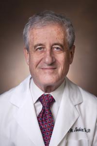 Dan Mark Rode合作论文数Division of Clinical Pharmacology, Department of Medicin, Vanderbilt University Medical Center8
 Jason D. Roberts合作论文数Arrhythmia Research Laboratory, University of Ottawa Heart Institute7
 Emelia J. Benjamin合作论文数Chobanian and Avedisian School of Medicine, Boston University;School of Public Health, Boston University7
 Eleonora Grandi合作论文数6Hanora Van Ert合作论文数6Jennifer Wright合作论文数6
 Gopinathannair Rakesh合作论文数Division of Cardiovascular Medicine, University of Louisville6
 Marco V Perez合作论文数6Yogananda S. Markandeya合作论文数University of Wisconsin6
 Ying Ge合作论文数5
 Michael Eugene Field合作论文数Department of Medicine, College of Medicine, Medical University of South Carolina5
 Arthur Wilde合作论文数Academic Cardiovascular Science Institute, Amsterdam UMC;Department of Clinical and Experimental Cardiology, University of Amsterdam;Academic Medical Centre, University of Amsterdam;Irving Medical Centre, Columbia University5
 Andrew Krahn合作论文数Department of Medicine, Faculty of Medicine, University of British Columbia5
 Andrea Russo合作论文数Cooper Medical School, Rowan University;Cooper University Hospital5
 Paul J. Wang合作论文数Department of Medicine, Stanford University;Cardiovascular Institute, Stanford University;Maternal & Child Health Research Institute, Stanford University5Qing Zhou合作论文数5
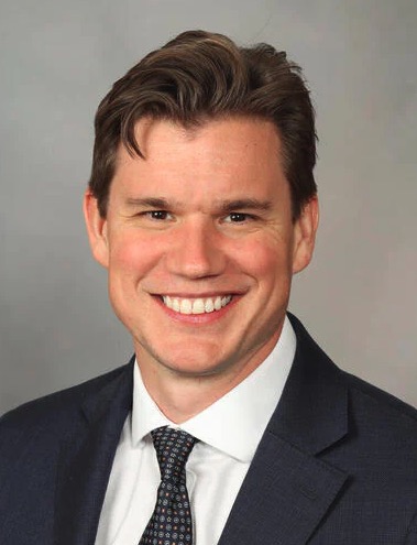 Peter A. Noseworthy合作论文数Department of Cardiovascular Medicine, Mayo Clinic;Department of Business Development, Mayo Clinic5Jose A. Joglar合作论文数5Steven A. Lubitz合作论文数Division of Preventive Medicine, Brigham and Women's Hospital, Boston, Massachusetts5Nipavan Chiamvimonvat合作论文数UC Davis, University of California5
 Michael J Cutler合作论文数Heart and Vascular Research Center, Case Western Reserve University5Samuel Esch合作论文数4Elise C McCune合作论文数4amanda l vega合作论文数4Jarrett Warden合作论文数4Blake Fleeman合作论文数4S Rajamani合作论文数4Fang Liu合作论文数4M Benjamin Shoemaker合作论文数4




