Most genetic variants associated with complex traits are hypothesized to regulate gene expression. To understand the genetics underlying gene expression variability, we characterized 14,324 RNA-sequencing samples from the Trans-Omics for Precision Medicine program and performed expression and splicing quantitative trait locus (e/sQTL) analyses in six tissues and cell types, including whole blood (n = 6454) and lung (n = 1291). We detected tens of thousands of secondary cis-e/sQTLs, showing that secondary cis-e/sQTL discovery remains unsaturated. We fine-mapped UK Biobank-derived genome-wide association study (GWAS) signals from 164 traits and identified e/sQTL colocalizations for 10,611 GWAS signals, including 7096 that colocalize with secondary e/sQTLs. Our results suggest that even larger e/sQTL analyses will uncover additional secondary e/sQTLs, further benefiting GWAS interpretation.
Background & Aims Metabolic dysfunction-associated steatotic liver disease (MASLD) spans from simple steatosis to metabolic dysfunction-associated steatohepatitis (MASH) and can progress to cirrhosis or hepatocellular carcinoma. Despite its prevalence, effective therapies are lacking. Recent genome-wide association studies identified a common missense variant (rs2642438) in the Mitochondrial Amidoxime Reducing Component 1 (MTARC1) gene that protects against liver cirrhosis without increasing cardiovascular disease risk. Biochemical and disease risk signatures associated with carriers of this missense variant also aligned with those of a known loss-of-function MTARC1 variant, suggesting mARC1 inhibition as a potential MASLD treatment.Methods To validate mARC1 loss-of-function as protective against MASLD, we generated Mtarc1 knockout (KO) mice and placed them on a choline-deficient, L-amino acid-defined, high-fat diet (CDAHFD). Effects of Mtarc1 KO on obesity and type 2 diabetes were explored using a high-fat diet. Hepatocytes from Mtarc1 KO mice were isolated to explore the molecular mechanisms by which Mtarc1 KO impacts lipid metabolism.Results Mtarc1 KO mice exhibited no vital growth or development defects. With a high-fat diet-induced obesity model, obese Mtarc1 KO mice exhibited reduced liver mass and lower cholesterol levels, with no effect on glucose homeostasis. In a CDAHFD-induced MASLD model, mARC1 deficiency significantly reduced liver steatosis, profibrosis, and inflammation. Untargeted metabolomics profiling further showed hepatic enrichment of phospholipids in Mtarc1 KO mice. Primary hepatocytes isolated from Mtarc1 KO mice exhibited reduced lipid droplet accumulation, decreased fatty acid uptake, and increased lipid secretion.Conclusions These findings support mARC1 inhibition as a promising therapeutic strategy for MASLD/MASH.
BACKGROUND:Heart failure with preserved ejection fraction (HFpEF) is a poorly understood, multisystem disease with high morbidity and mortality. To improve understanding of its pathobiology, we analyzed single-nucleus RNA sequencing in human HFpEF myocardium versus controls. METHODS:Septal myocardial biopsies from 19 HFpEF and 24 nonfailing controls were analyzed using the 10× Genomics Chromium platform, with nuclei isolated from combined samples (6 patients/pool). Genotype-based demultiplexing was performed with souporcell, and gene expression was quantified with CellRanger and CellBender. After quality control, nuclei were annotated by cell types, and differential expression was performed between HFpEF versus controls using limma-voom. Functional analysis was performed using Gene Set Enrichment Analysis. Data were compared with prior single-nucleus RNA sequencing in dilated cardiomyopathy versus controls. RESULTS:We successfully demultiplexed pooled myocardial biopsies, assigning >70% of nuclei to individuals. After quality control, we recovered 48 886 nuclei and identified 14 cell types. Many differentially expressed genes across cell types were detected in HFpEF versus controls (fibroblasts, 5905; cardiomyocytes, 5159; endothelial cells, 2143; pericytes, 1812; and macrophages, 1405). Enriched pathways common to multiple cell types included immune activation, transcription/translation, metabolism, and protein quality control. They were particularly shared between cardiomyocytes and fibroblasts. Vascular smooth muscle cells had a more synthetic, proliferative phenotype. Immune cell analyses suggested enhanced T-cell activation and reduced macrophage clearance programs. Comparative analysis between HFpEF and dilated cardiomyopathy identified transcriptional differences primarily in cardiomyocytes. Two of 3 cardiomyocyte differential expression genes unique to HFpEF were validated to have concordant protein expression changes in HFpEF (MAP2K6 and PLPP3). CONCLUSIONS:Our findings reveal a distinct, cell-type-specific transcriptomic landscape in the human HFpEF myocardium. While HFpEF and dilated cardiomyopathy share significant molecular pathways across most cell types, the profound divergence within cardiomyocytes suggests a unique pathological driver for HFpEF. These signatures may provide a high-resolution roadmap for identifying precision therapeutic targets in HFpEF.
Cardiovascular disease remains the leading cause of global mortality. Understanding its complexity requires dissecting the heart's cellular landscape. Here we present HeartMap, a comprehensive single-nucleus RNA sequencing atlas of the adult human heart. This resource integrates data from nine studies, encompassing over 2.4 million nuclei, 209 individuals, eight anatomical regions and seven disease and healthy states. After rigorous data harmonization and method comparison of batch correction methods, we characterized transcriptional diversity across 14 cell types. To demonstrate the utility of HeartMap, we identified robust disease-associated gene signatures in dilated cardiomyopathy by comparing multiple studies. Notably, we identified distinct activated fibroblast populations, enriched for COL22A1 or TNC, that display variable prevalence across cardiomyopathies. HeartMap provides a valuable tool for exploring cardiac disease at the single-cell level, facilitating both fundamental research and potential therapeutic development.
Rare coding genetic variants may exert large effects on risk of common disease, yet their contribution to disease architecture and their utility in gene prioritization remain limited by inadequate sample sizes. Here, we performed a massive-scale rare variant association study (RVAS), analyzing over 1.1 million sequenced participants among which 130,000 had atrial fibrillation (AF). Through a multi-mask burden testing approach, we identified 15 genes significantly associated with AF through rare large-effect variation. Integrative analyses revealed strong convergence between genes implicated by rare and common variation, and highlighted instances where RVAS data may aid in GWAS prioritization. Nevertheless, several RVAS genes were not among GWAS loci ( FAM189A2 , ACTC1 , FNIP1 , FBN1 ), or were not nominated through contemporary GWAS prioritization ( KDM5B , ZFP36L2 ). Finally, we observed that ultra-rare protein-disrupting variants - concentrated in a small number of large-effect size genes - explained at least 2% of AF susceptibility across European and African ancestry groups. These findings refine the genetic architecture of AF, while highlighting the value and cost of RVAS for genomic discovery in common disease.
To broaden our understanding of bradyarrhythmias and conduction disease, we performed common variant genome-wide association analyses in up to 1.3 million individuals and rare variant burden testing in 460,000 individuals for sinus node dysfunction (SND), distal conduction disease (DCD) and pacemaker (PM) implantation. We identified 13, 31 and 21 common variant loci for SND, DCD and PM, respectively. Four well-known loci (SCN5A/SCN10A, CCDC141, TBX20 and CAMK2D) were shared for SND and DCD, while others were more specific for SND or DCD. SND and DCD showed a moderate genetic correlation (rg = 0.63). Cardiomyocyte-expressed genes were enriched for contributions to DCD heritability. Rare-variant analyses implicated LMNA for all bradyarrhythmia phenotypes, SMAD6 and SCN5A for DCD and TTN, MYBPC3 and SCN5A for PM. These results show that variation in multiple genetic pathways (for example, ion channel function, cardiac developmental programs, sarcomeric structure and cellular homeostasis) appear critical to the development of bradyarrhythmias. Genome-wide analyses identify variants associated with sinus node dysfunction, distal conduction disease and pacemaker implantation, implicating ion channel function, cardiac developmental programs and sarcomeric structure in bradyarrhythmia susceptibility.
Atrial fibrillation (AF) is a prevalent and morbid abnormality of the heart rhythm with a strong genetic component. Here, we meta-analyzed genome and exome sequencing data from 36 studies that included 52,416 AF cases and 277,762 controls. In burden tests of rare coding variation, we identified novel associations between AF and the genes MYBPC3, LMNA, PKP2, FAM189A2 and KDM5B. We further identified associations between AF and rare structural variants owing to deletions in CTNNA3 and duplications of GATA4. We broadly replicated our findings in independent samples from MyCode, deCODE and UK Biobank. Finally, we found that CRISPR knockout of KDM5B in stem-cell-derived atrial cardiomyocytes led to a shortening of the action potential duration and widespread transcriptomic dysregulation of genes relevant to atrial homeostasis and conduction. Our results highlight the contribution of rare coding and structural variants to AF, including genetic links between AF and cardiomyopathies, and expand our understanding of the rare variant architecture for this common arrhythmia.
The 9p21.3 locus is the strongest genetic association with coronary artery disease (CAD), yet its causal mechanisms remain unresolved. We map the regulatory architecture of 9p21.3 in disease-relevant vascular cells, identifying 12 enhancers within the CAD risk haplotype that respond dynamically to inflammatory and metabolic stress in fibroblasts and smooth muscle cells. These activated states are enriched for CAD heritability, implicating stress-responsive vascular wall cells in disease pathophysiology. Dense CRISPRi tiling integrated with fine-mapping and genomic constraint across >500,000 individuals nominates MTAP as the effector gene, with rs1537371 as a likely causal variant. Perturbation and multi-modal analyses show that MTAP loss induces pro-fibrotic and angiogenic programs and sensitizes vascular cells to TGF-β-driven pathological transitions. Our findings reveal a vascular-specific enhancer network through which noncoding variation at 9p21.3 modulates CAD risk via MTAP-a previously unrecognized regulator of vascular remodeling located 269 kb from the risk haplotype.
BACKGROUND:Heart failure with preserved ejection fraction (HFpEF) is a poorly understood, multi-system disease with high morbidity and mortality. To improve our understanding of its underlying biology, we used single-nucleus RNA sequencing (snRNA-seq) to characterize cell-specific gene expression patterns in human HFpEF myocardium. METHODS:Septal myocardial biopsies (2-3 mg) from 30 HFpEF patients and 29 non-failing donor controls were analyzed using the 10X Genomics platform, with nuclei isolated from combined samples (6 patients/pool). Genotype-based demultiplexing was performed with souporcell, and gene expression quantified with CellRanger and CellBender. After quality control, nuclei were clustered and annotated by cell types based on specific marker genes. Differential expression (DE) by cell-type in HFpEF vs controls was performed using limma-voom and functional analysis performed using Gene Set Enrichment Analysis. Data were compared to dilated cardiomyopathy (DCM) using prior snRNA-seq in DCM vs respective controls. RESULTS:We successfully demultiplexed pooled myocardial biopsies, assigning >75% of droplets to individual patients. From eight pooled samples (19 HFpEF, 24 controls), we recovered 48,886 nuclei and identified 14 cell types. Cardiomyocytes (5159 differentially expressed [DE] genes, 36%) and fibroblasts (5905 DE genes, 49%) showed the most DE genes, while endothelial cells (2143), pericytes (1812), and macrophages (1405) had fewer. Enriched pathways common to multiple cell types included transcription/translation, immune activation, metabolism, and protein quality control. Of 7848 DE genes identified via pseudo-bulk snRNA-seq, 51% were DE in fibroblasts and 47% in cardiomyocytes, compared to <20% in other cell types. Unlike dilated cardiomyopathy (DCM), sub-clustering fibroblasts did not reveal an activated fibroblast population in HFpEF. Comparative analysis between HFpEF and DCM identified transcriptional differences primarily in cardiomyocytes. CONCLUSIONS:This study demonstrates the power of genotype-based demultiplexing for single-cell transcriptomic analyses of small endomyocardial biopsies and identifies cardiomyocytes as the principal cell type with distinct transcriptional changes in HFpEF versus DCM. These findings, coupled with differential gene expression and functional pathway analyses, illuminate HFpEF pathways and may nominate compelling targets for future mechanistic studies and therapeutic efforts for HFpEF.
Heart failure (HF) is a major contributor to global morbidity and mortality. While distinct clinical subtypes, defined by etiology and left ventricular ejection fraction, are well recognized, their genetic determinants remain inadequately understood. In this study, we report a genome-wide association study of HF and its subtypes in a sample of 1.9 million individuals. A total of 153,174 individuals had HF, of whom 44,012 had a nonischemic etiology (ni-HF). A subset of patients with ni-HF were stratified based on left ventricular systolic function, where data were available, identifying 5,406 individuals with reduced ejection fraction and 3,841 with preserved ejection fraction. We identify 66 genetic loci associated with HF and its subtypes, 37 of which have not previously been reported. Using functionally informed gene prioritization methods, we predict effector genes for each identified locus, and map these to etiologic disease clusters through phenome-wide association analysis, network analysis and colocalization. Through heritability enrichment analysis, we highlight the role of extracardiac tissues in disease etiology. We then examine the differential associations of upstream risk factors with HF subtypes using Mendelian randomization. These findings extend our understanding of the mechanisms underlying HF etiology and may inform future approaches to prevention and treatment.
Atrial fibrillation (AF) is the most common heart rhythm abnormality and is a leading cause of heart failure and stroke. This large-scale meta-analysis of genome-wide association studies increased the power to detect single-nucleotide variant associations and found more than 350 AF-associated genetic loci. We identified candidate genes related to muscle contractility, cardiac muscle development and cell-cell communication at 139 loci. Furthermore, we assayed chromatin accessibility using assay for transposase-accessible chromatin with sequencing and histone H3 lysine 4 trimethylation in stem cell-derived atrial cardiomyocytes. We observed a marked increase in chromatin accessibility for our sentinel variants and prioritized genes in atrial cardiomyocytes. Finally, a polygenic risk score (PRS) based on our updated effect estimates improved AF risk prediction compared to the CHARGE-AF clinical risk score and a previously reported PRS for AF. The doubling of known risk loci will facilitate a greater understanding of the pathways underlying AF.
Cardiovascular disease remains the leading cause of global mortality. Understanding its complexity requires dissecting the heart's cellular landscape. We present HeartMap, a comprehensive single-nucleus RNA sequencing atlas of the adult human heart. This resource integrates data from nine studies, encompassing over 2.4 million nuclei, 209 individuals, eight anatomical regions, and seven disease and healthy states. After rigorous data harmonization and benchmarking of batch-correction methods, we characterized transcriptional diversity across 14 cell types. To demonstrate the utility of HeartMap, we identified robust disease-associated gene signatures in dilated cardiomyopathy by comparing across multiple studies. Notably, we identified distinct activated fibroblast populations, with COL22A1 or TNC enrichment, revealing potential differences between inherited and ischemic cardiomyopathies. HeartMap provides a valuable tool for exploring cardiac disease at the single-cell level, facilitating both fundamental research and potential therapeutic development.
Atrial fibrillation (AF) is the most common sustained arrhythmia in humans, yet the molecular basis of AF remains incompletely understood. To determine the cell type-specific transcriptional changes underlying AF, we perform single-nucleus RNA-seq (snRNA-seq) on left atrial (LA) samples from patients with AF and controls. From more than 175,000 nuclei we find that only cardiomyocytes (CMs) and macrophages (M Phi s) have a significant number of differentially expressed genes in patients with AF. Attractin Like 1 (ATRNL1) was overexpressed in CMs among patients with AF and localized to the intercalated disks. Further, in both knockdown and overexpression experiments we identify a potent role for ATRNL1 in cell stress response, and in the modulation of the cardiac action potential. Finally, we detect an unexpected expression pattern for a leading AF candidate gene, KCNN3. In sum, we uncover a role for ATRNL1 which may serve as potential therapeutic target for this common arrhythmia. Characterizing atrial fibrillation (AF) at the single cell level is challenging. Here, the authors perform snRNA-seq on 18 patients with AF to investigate the cell composition, and gene expression shifts associated with this common arrhythmia.
Large-scale sequencing has enabled unparalleled opportunities to investigate the role of rare coding variation in human phenotypic variability. Here, we present a pan-ancestry analysis of sequencing data from three large biobanks, including the All of Us research program. Using mixed-effects models, we performed gene-based rare variant testing for 601 diseases across 748,879 individuals, including 155,236 with ancestry dissimilar to European. We identified 363 significant associations, which highlighted core genes for the human disease phenome and identified potential novel associations, including UBR3 for cardiometabolic disease and YLPM1 for psychiatric disease. Pan-ancestry burden testing represented an inclusive and useful approach for discovery in diverse datasets, although we also highlight the importance of ancestry-specific sensitivity analyses in this setting. Finally, we found that effect sizes for rare protein-disrupting variants were concordant between samples similar to European ancestry and other genetic ancestries (βDeming = 0.7-1.0). Our results have implications for multi-ancestry and cross-biobank approaches in sequencing association studies for human disease.
Background: Single-nucleus RNA sequencing (snRNA-seq) is a powerful tool to define the cell-type specific transcriptional signature in health and disease. Although it has been applied to study many forms of cardiovascular disease, few resources exist to compare results across studies and disease states. Methods: We identified 9 studies from the literature with > 10k nuclei, individuals > 18 years, and non-diseased controls. Existing data was downloaded, similarly re-processed, aligned to the GRCh38 reference transcriptome, and aggregated for quality control. Sixteen versions of data integration methods were benchmarked with the single-cell integration benchmarking (scIB) package. Clustering analysis was conducted in SCANPY using established methods to identify global cell types and distinct cell states within cell types. Cell type and cell state labels were assigned to clusters based on differential expression testing using limma-voom. Compositional analysis was conducted using scCODA. Results: HeartMap contains 2,600,078 cells across 36,601 genes from 9 studies, 260 individuals, 7 disease states and normal controls, and 8 anatomical regions. The median age was 54 years (IQR: 17) and 40% were female. Benchmarking selected single cell annotated variational inference as the optimal integration method. Global clustering of all nuclei revealed 14 major cell types. At the sample level, after controlling for differences between studies, the strongest sources of variation in transcription included disease status, patient sex, and anatomical region of sample. To demonstrate the utility of this resource, we identified 10 distinct fibroblast sub-populations among ~580,000 fibroblast cells. Two sub-populations of activated fibroblasts show POSTN+COL22A1+ and POSTN+TNC+ enrichment in primary (DCM/HCM) and ischemic (ICM/AMI/CAD) cardiomyopathies, respectively. Validation using RNA-scope and immunofluorescence staining revealed peri-vascular patterns of TNC+ fibroblasts and interstitial patterns of COL22A1+ fibroblasts in diseased tissues. Conclusions: HeartMap provides a unique resource to the cardiovascular research community and provides additional insights into the transcriptional diversity in health and disease.
HomeCirculationVol. 149, No. 14Distinct Plasma Extracellular Vesicle Transcriptomes in Acute Decompensated Heart Failure Subtypes: A Liquid Biopsy Approach No AccessLetterRequest AccessFull TextAboutView Full TextView PDFView EPUBSections ToolsAdd to favoritesDownload citationsTrack citationsPermissions ShareShare onFacebookTwitterLinked InMendeleyReddit Jump toNo AccessLetterRequest AccessFull TextDistinct Plasma Extracellular Vesicle Transcriptomes in Acute Decompensated Heart Failure Subtypes: A Liquid Biopsy Approach Priyanka Gokulnath, Michail Spanos, H. Immo Lehmann, Quanhu Sheng, Rodosthenis Rodosthenous, Mark Chaffin, Dimitrios Varrias, Emeli Chatterjee, Elizabeth Hutchins, Guoping Li, George Daaboul, Farhan Rana, Ashley Mingyi Wang, Kendall Van Keuren-Jensen, Patrick T. Ellinor, Ravi Shah and Saumya Das Priyanka GokulnathPriyanka Gokulnath https://orcid.org/0000-0003-2813-330X Cardiovascular Research Center, Massachusetts General Hospital and Harvard Medical School, Boston, MA (P.G., M.S., H.I.L., D.V., E.C., G.L., F.R., A.M.W., P.T.E., S.D.). , Michail SpanosMichail Spanos https://orcid.org/0000-0003-1205-4201 Cardiovascular Research Center, Massachusetts General Hospital and Harvard Medical School, Boston, MA (P.G., M.S., H.I.L., D.V., E.C., G.L., F.R., A.M.W., P.T.E., S.D.). , H. Immo LehmannH. Immo Lehmann https://orcid.org/0000-0003-4379-283X Cardiovascular Research Center, Massachusetts General Hospital and Harvard Medical School, Boston, MA (P.G., M.S., H.I.L., D.V., E.C., G.L., F.R., A.M.W., P.T.E., S.D.). , Quanhu ShengQuanhu Sheng https://orcid.org/0000-0001-8951-9295 Department of Biostatistics (Q.S.), Vanderbilt University Medical Center, Nashville, TN. , Rodosthenis RodosthenousRodosthenis Rodosthenous Institute for Molecular Medicine Finland (FIMM), HiLIFE, University of Helsinki, Finland (R.R.). , Mark ChaffinMark Chaffin https://orcid.org/0000-0002-1234-5562 Cardiovascular Disease Initiative, The Broad Institute of MIT and Harvard, Cambridge, MA (M.C., P.T.E.). , Dimitrios VarriasDimitrios Varrias https://orcid.org/0000-0001-9778-1728 Cardiovascular Research Center, Massachusetts General Hospital and Harvard Medical School, Boston, MA (P.G., M.S., H.I.L., D.V., E.C., G.L., F.R., A.M.W., P.T.E., S.D.). , Emeli ChatterjeeEmeli Chatterjee Cardiovascular Research Center, Massachusetts General Hospital and Harvard Medical School, Boston, MA (P.G., M.S., H.I.L., D.V., E.C., G.L., F.R., A.M.W., P.T.E., S.D.). , Elizabeth HutchinsElizabeth Hutchins https://orcid.org/0000-0003-2543-0798 Division of Neurogenomics, Translational Genomics Research Institute, Phoenix, AZ (E.H., K.V.K.-J.). , Guoping LiGuoping Li https://orcid.org/0000-0003-3874-0744 Cardiovascular Research Center, Massachusetts General Hospital and Harvard Medical School, Boston, MA (P.G., M.S., H.I.L., D.V., E.C., G.L., F.R., A.M.W., P.T.E., S.D.). , George DaaboulGeorge Daaboul NanoView Biosciences, Boston, MA (G.D.). , Farhan RanaFarhan Rana Cardiovascular Research Center, Massachusetts General Hospital and Harvard Medical School, Boston, MA (P.G., M.S., H.I.L., D.V., E.C., G.L., F.R., A.M.W., P.T.E., S.D.). , Ashley Mingyi WangAshley Mingyi Wang Cardiovascular Research Center, Massachusetts General Hospital and Harvard Medical School, Boston, MA (P.G., M.S., H.I.L., D.V., E.C., G.L., F.R., A.M.W., P.T.E., S.D.). , Kendall Van Keuren-JensenKendall Van Keuren-Jensen Division of Neurogenomics, Translational Genomics Research Institute, Phoenix, AZ (E.H., K.V.K.-J.). , Patrick T. EllinorPatrick T. Ellinor https://orcid.org/0000-0002-2067-0533 Cardiovascular Research Center, Massachusetts General Hospital and Harvard Medical School, Boston, MA (P.G., M.S., H.I.L., D.V., E.C., G.L., F.R., A.M.W., P.T.E., S.D.). Cardiovascular Disease Initiative, The Broad Institute of MIT and Harvard, Cambridge, MA (M.C., P.T.E.). , Ravi ShahRavi Shah https://orcid.org/0000-0002-4471-7156 Vanderbilt Translational and Clinical Research Center (R.S.), Vanderbilt University Medical Center, Nashville, TN. and Saumya DasSaumya Das Correspondence to: Saumya Das, MD, PhD, Cardiovascular Research Center, Massachusetts General Hospital, 185 Cambridge St, Boston, MA 02114. Email E-mail Address: [email protected] https://orcid.org/0000-0002-4521-4606 Cardiovascular Research Center, Massachusetts General Hospital and Harvard Medical School, Boston, MA (P.G., M.S., H.I.L., D.V., E.C., G.L., F.R., A.M.W., P.T.E., S.D.). Originally published1 Apr 2024https://doi.org/10.1161/CIRCULATIONAHA.123.065513Circulation. 2024;149:1147–1149Footnotes*P. Gokulnath, M. Spanos, and H.I. Lehmann contributed equally.†R. Shah and S. Das contributed equally.For Sources of Funding and Disclosures, see page 1149.Circulation is available at www.ahajournals.org/journal/circCorrespondence to: Saumya Das, MD, PhD, Cardiovascular Research Center, Massachusetts General Hospital, 185 Cambridge St, Boston, MA 02114. Email sdas@mgh.harvard.eduREFERENCES1. Hahn VS, Knutsdottir H, Luo X, Bedi K, Margulies KB, Haldar SM, Stolina M, Yin J, Khakoo AY, Vaishnav J, et al. Myocardial gene expression signatures in human heart failure with preserved ejection fraction.Circulation. 2021; 143:120–134. doi: 10.1161/CIRCULATIONAHA.120.050498LinkGoogle Scholar2. Rodosthenous RS, Hutchins E, Reiman R, Yeri AS, Srinivasan S, Whitsett TG, Ghiran I, Silverman MG, Laurent LC, Van Keuren-Jensen K, et al. Profiling extracellular long RNA transcriptome in human plasma and extracellular vesicles for biomarker discovery.iScience. 2020; 23:101182. doi: 10.1016/j.isci.2020.101182CrossrefMedlineGoogle Scholar3. Chaffin M, Papangeli I, Simonson B, Akkad A-D, Hill MC, Arduini A, Fleming SJ, Melanson M, Hayat S, Kost-Alimova M, et al. Single-nucleus profiling of human dilated and hypertrophic cardiomyopathy.Nature. 2022; 608:174–180. doi: 10.1038/s41586-022-04817-8CrossrefMedlineGoogle Scholar4. Srinivasan S, Yeri A, Cheah PS, Chung A, Danielson K, De Hoff P, Filant J, Laurent CD, Laurent LD, Magee R, et al. Small RNA sequencing across diverse biofluids identifies optimal methods for exRNA isolation.Cell. 2019; 177:446–462.e16. doi: 10.1016/j.cell.2019.03.024CrossrefMedlineGoogle Scholar5. Kumarswamy R, Bauters C, Volkmann I, Maury F, Fetisch J, Holzmann A, Lemesle G, De Groote P, Pinet F, Thum T. Circulating long noncoding RNA, LIPCAR, predicts survival in patients with heart failure.Circ Res. 2014; 114:1569–1575. doi: 10.1161/CIRCRESAHA.114.303915LinkGoogle Scholar eLetters(0)eLetters should relate to an article recently published in the journal and are not a forum for providing unpublished data. Comments are reviewed for appropriate use of tone and language. Comments are not peer-reviewed. Acceptable comments are posted to the journal website only. Comments are not published in an issue and are not indexed in PubMed. Comments should be no longer than 500 words and will only be posted online. References are limited to 10. Authors of the article cited in the comment will be invited to reply, as appropriate.Comments and feedback on AHA/ASA Scientific Statements and Guidelines should be directed to the AHA/ASA Manuscript Oversight Committee via its Correspondence page.Sign In to Submit a Response to This Article Previous Back to top Next FiguresReferencesRelatedDetails April 2, 2024Vol 149, Issue 14 Advertisement Article InformationMetrics © 2024 American Heart Association, Inc.https://doi.org/10.1161/CIRCULATIONAHA.123.065513PMID: 38557125 Originally publishedApril 1, 2024 Keywordscell-free nucleic acidsextracellular vesiclesheart failureheart failure, diastolicheart failure, systolicRNAPDF download Advertisement SubjectsBiomarkersHeart FailureTranslational Studies
 Patrick Ellinor合作论文数Cardiovascular Research Center, Massachusetts General Hospital;Harvard Medical School66
 Sekar Kathiresan合作论文数Eli Lilly and Company;Verve Therapeutics;Department of Medicine, Massachusetts General Hospital50
 Steven A. Lubitz合作论文数Division of Preventive Medicine, Brigham and Women's Hospital, Boston, Massachusetts41
 Roselli C合作论文数35Krishna G Aragam合作论文数34
 Seung Hoan Choi合作论文数School of Public Health, Boston University32Lu-Chen Weng合作论文数Massachusetts General Hospital28
 Jerome I Rotter合作论文数Institute for Translational Genomics and Population Sciences, The Lundquist Institute;Division of Genomic Outcomes, Departments of Pediatrics and Medicine, Harbor-UCLA Medical Center;David Geffen School of Medicine, University of California, Los Angeles24
 Pradeep Natarajan合作论文数Mass General Research Institute, Massachusetts General Hospital;Broad Institute of MIT and Harvard;Harvard Medical School23
 Honghuang Lin合作论文数23
 Kathryn L. Lunetta合作论文数Department of Biostatistics, School of Public Health, Boston University22
 Bruce M. Psaty合作论文数Department of Epidemiology, School of Public Health, University of Washington22
 Tucker Nathan R合作论文数20
 Amit V. Khera合作论文数Center for Genomic Medicine, Massachusetts General Hospital;Verve Therapeutics;Harvard Medical School20
 Kenneth B. Margulies合作论文数Margulies Laboratory, Perelman School of Medicine, University of Pennsylvania19
 Emelia J. Benjamin合作论文数Chobanian and Avedisian School of Medicine, Boston University;School of Public Health, Boston University19J. Gustav Smith合作论文数19Derek Klarin合作论文数Malcolm Randall VA Med Ctr, Gainesville, FL USA18
 James P. Pirruccello合作论文数18
 Amelia Weber Hall合作论文数Gene Regulation Observatory, Broad Institute of Harvard and the Massachusetts Institute of Technology17
 Dan E. Arking合作论文数Claude D. Pepper Older Americans Independence Center, Johns Hopkins University;Division of Cardiology, Mckusick-Nathans Institute and the Department of Genetic Medicine, School of Medicine, Johns Hopkins University15
 Daniel J. Rader合作论文数Division of Translational Medicine and Human Genetics, University of Pennsylvania;Department of Genetics, University of Pennsylvania;Institute for Translational Medicine and Therapeutics, University of Pennsylvania15
 Nilesh Samani合作论文数Department of Cardiovascular Sciences, University of Leicester15
 Kent Taylor合作论文数The Lundquist Institute;Department of Pediatrics, Harbor-UCLA Medical Center14Sean J Jurgens合作论文数Cardiovascular Disease Initiative, Broad Institute of MIT and Harvard14
 Scott M. Damrauer合作论文数Surgery Faculty, Perelman School of Medicine, University of Pennsylvania14
 Haggerty Christopher M合作论文数14
 Ruth Loos合作论文数Faculty of Health and Medical Sciences, Novo Nordisk Foundation Center for Basic Metabolic Research, University of Copenhagen;Charles Bronfman Institute for Personalized Medicine, Icahn School of Medicine at Mount Sinai14
 Vasan Ramachandran合作论文数Department of Quantitative and Qualitative Health Sciences, School of Public Health, The University of Texas Health Science Center at San Antonio;College of Health, Community and Policy, The University of Texas at San Antonio14
 Daniel I. Chasman合作论文数Brigham and Women’s Hospital14
 Pim van der Harst合作论文数14
 Alessandro Arduini合作论文数Department of Physiology, University of Valencia14
 Xiuqing Guo合作论文数The Lundquist Institute13Danish Saleheen合作论文数13Sotoodehnia Nona合作论文数13
 Alonso Alvaro合作论文数Rollins School of Public Health, Emory University13
 Eric Boerwinkle合作论文数Department of Epidemiology, Human Genetics and Environmental Sciences, School of Public Health, The University of Texas Health Science Center at Houston;Baylor College of Medicine Human Genome Sequencing Center13Olle Melander合作论文数13
 Namrata Gupta合作论文数Broad Clinical Labs;The Broad Institute13
 Elsayed Z Soliman合作论文数School of Medicine, Wake Forest University;Wake Forest Baptist Health12
 Svati Hasmukh Shah合作论文数Department of Medicine, Duke University School of Medicine12
 Mary Haas合作论文数Department of Emergency Medicine, Medical School, University of Michigan12
 Stephen S. Rich合作论文数Department of Genome Sciences, School of Medicine, University of Virginia12
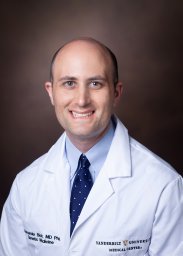 Alexander Bick合作论文数Department of Medicine, Vanderbilt University Medical Center12Simonson Bridget合作论文数12
 Susan R. Heckbert合作论文数Department of Epidemiology, School of Public Health, University of Washington12
 Dan Mark Rode合作论文数Division of Clinical Pharmacology, Department of Medicin, Vanderbilt University Medical Center12
 Stacey Gabriel合作论文数The Broad Institute of Harvard and Massachusetts Institute of Technology12Niek Verweij合作论文数11




