Plasmodium, the causative agent of malaria, infects hepatocytes prior to establishing a symptomatic blood stage infection. During this liver stage development, parasites reside in a parasitophorous vacuole (PV), whose membrane acts as the critical interface between the parasite and the host cell. It is well-established that host cell autophagy-related processes significantly impact the development of Plasmodium liver stages. Expression of genes related to autophagy and lysosomal biogenesis is orchestrated by transcription factor EB (TFEB). In this study, we explored the activation of host cell TFEB in Plasmodium berghei-infected cells during the liver stage of the parasite. Our results unveiled a critical role of proteins belonging to the Gamma-aminobutyric acid receptor-associated protein subfamily (GABARAP) of ATG8 proteins (GABARAP/L1/L2 and LC3A/B/C) in recruiting the TFEB-blocking FLCN-FNIP (Folliculin-Folliculin-interacting protein) complex to the PVM. Remarkably, the sequestration of FLCN-FNIP resulted in a robust activation of TFEB, reliant on conjugation of ATG8 proteins to single membranes (CASM) and GABARAP proteins. Our findings provide novel mechanistic insights into host cell signaling occurring at the PVM, shedding light on the complex interplay between Plasmodium parasites and the host cell during the liver stage of infection. Plasmodium berghei activates transcription factor EB by sequestering the host cell FLCN/FNIP complex to its parasitophorous vacuole membrane depending on host cell GABARAP proteins in a CASM dependent mechanism.
BACKGROUND & AIMS: In the classic form of a 1-antitrypsin deficiency fi ciency (ATD), the misfolded a 1-antitrypsin Z (ATZ) variant accumulates in the endoplasmic reticulum (ER) of liver cells. A gain-of-function proteotoxic mechanism is responsible for chronic liver disease in a subgroup of homozygotes. Proteostatic response pathways, including conventional endoplasmic reticulum-associated - associated degradation and autophagy, have been proposed as the mechanisms that allow cellular adaptation and presumably protection from the liver disease phenotype. Recent studies have concluded that a distinct lysosomal pathway called endoplasmic reticulum-to-lysosome - to-lysosome completely supplants the role of the conventional macroautophagy pathway in degradation of ATZ. Here, we used several state-ofthe-art approaches to characterize the proteostatic responses more fully in cellular systems that model ATD. METHODS: We used clustered regularly interspaced short palindromic repeats (CRISPR)-mediated genome editing coupled to a cell selection step by fl uorescence-activated cell sorter to perform screening for proteostasis genes that regulate ATZ accumulation and combined that with selective genome editing in 2 other model systems. RESULTS: Endoplasmic reticulum-associated - associated degradation genes are key early regulators and multiple autophagy genes, from classic as well as from ER-to-lysosome and other newly described ER-phagy pathways, participate in degradation of ATZ in a manner that is temporally regulated and evolves as ATZ accumulation persists. Time-dependent changes in gene expression are accompanied by specific fi c ultrastructural changes including dilation of the ER, formation of globular inclusions, budding of autophagic vesicles, and alterations in the overall shape and component parts of mitochondria. CONCLUSIONS: Macroautophagy is a critical component of the proteostasis response to cellular ATZ accumulation and it becomes more important over time as ATZ synthesis continues unabated. Multiple subtypes of macroautophagy and nonautophagic lysosomal degradative pathways are needed to respond to the high concentrations of misfolded protein that characterizes ATD and these pathways are attractive candidates for genetic variants that predispose to the hepatic phenotype. (Cell Mol Gastroenterol Hepatol 2024;17:1007-1024; - 1024; https:// doi.org/10.1016/j.jcmgh.2024.02.006)
Induction of lipid-laden foamy macrophages is a cellular hallmark of tuberculosis (TB) disease, which involves the transformation of infected phagolysosomes from a site of killing into a nutrient-rich replicative niche. Here, we show that a terpenyl nucleoside shed from Mycobacterium tuberculosis, 1-tuberculosinyladenosine (1-TbAd), caused lysosomal maturation arrest and autophagy blockade, leading to lipid storage in M1 macrophages. Pure 1-TbAd, or infection with terpenyl nucleoside-producing M. tuberculosis, caused intralysosomal and peribacillary lipid storage patterns that matched both the molecules and subcellular locations known in foamy macrophages. Lipidomics showed that 1-TbAd induced storage of triacylglycerides and cholesterylesters and that 1-TbAd increased M. tuberculosis growth under conditions of restricted lipid access in macrophages. Furthermore, lipidomics identified 1-TbAd-induced lipid substrates that define Gaucher's disease, Wolman's disease, and other inborn lysosomal storage diseases. These data identify genetic and molecular causes of M. tuberculosis-induced lysosomal failure, leading to successful testing of an agonist of TRPML1 calcium channels that reverses lipid storage in cells. These data establish the host-directed cellular functions of an orphan effector molecule that promotes survival in macrophages, providing both an upstream cause and detailed picture of lysosome failure in foamy macrophages.
The allosteric inhibitor of the mechanistic target of rapamycin (mTOR) everolimus reduces seizures in tuberous sclerosis complex (TSC) patients through partial inhibition of mTOR functions. Due to its limited brain permeability, we sought to develop a catalytic mTOR inhibitor optimized for central nervous system (CNS) indications. We recently reported an mTOR inhibitor (1) that is able to block mTOR functions in the mouse brain and extend the survival of mice with neuronal-specific ablation of the Tsc1 gene. However, 1 showed the risk of genotoxicity in vitro. Through structure-activity relationship (SAR) optimization, we identified compounds 9 and 11 without genotoxicity risk. In neuronal cell-based models of mTOR hyperactivity, both corrected aberrant mTOR activity and significantly improved the survival rate of mice in the Tsc1 gene knockout model. Unfortunately, 9 and 11 showed limited oral exposures in higher species and dose-limiting toxicities in cynomolgus macaque, respectively. However, they remain optimal tools to explore mTOR hyperactivity in CNS disease models.
Supplementary Fig from Identification and characterization of NVP-BEZ235, a new orally available dual phosphatidylinositol 3-kinase/mammalian target of rapamycin inhibitor with potent in vivo antitumor activity
As a maturing field that continues to provide fundamental insights into cell physiology, autophagy is also beginning to attract considerable interest from the biotechnology/pharmaceutical sector. For this Editor's corner, I thought it would be both useful and interesting to talk with somebody who has spent a lot of time in the commercial sphere, working on autophagy and related processes. I was fortunate that Dr. Leon Murphy, Chief Scientific Officer at Casma therapeutics, was willing and able to answer my questions. In addition to his insights on the commercial interest for autophagy, Dr. Murphy also shared his personal experience on the scientific life working in large and small pharmaceutical companies.
Adaptive changes in lysosomal capacity are driven by the transcription factors TFEB and TFE3 in response to increased autophagic flux and endolysosomal stress, yet the molecular details of their activation are unclear. LC3 and GABARAP members of the ATG8 protein family are required for selective autophagy and sensing perturbation within the endolysosomal system. Here, we show that during the conjugation of ATG8 to single membranes (CASM), Parkin-dependent mitophagy, and Salmonella-induced xenophagy, the membrane conjugation of GABARAP, but not LC3, is required for activation of TFEB/TFE3 to control lysosomal capacity. GABARAP directly binds to a previously unidentified LC3-interacting motif (LIR) in the FLCN/FNIP tumor suppressor complex and mediates sequestration to GABARAP-conjugated membrane compartments. This disrupts FLCN/FNIP GAP function toward RagC/D, resulting in impaired substrate-specific mTOR-dependent phosphorylation of TFEB. Thus, the GABARAP-FLCN/FNIP-TFEB axis serves as a molecular sensor that coordinates lysosomal homeostasis with perturbations and cargo flux within the autophagy-lysosomal network.
Adaptive changes in lysosomal capacity are driven by the transcription factors TFEB and TFE3 in response to increased autophagic flux and endolysosomal stress, yet the molecular details of their activation are unclear. LC3 and GABARAP members of the ATG8 protein family are required for selective autophagy and sensing perturbation within the endolysosomal system. Here we show that during single membrane ATG8 conjugation (SMAC), Parkin-dependent mitophagy, and Salmonella -induced xenophagy, the membrane conjugation of GABARAP, but not LC3, is required for activation of TFEB/TFE3 to control lysosomal homeostasis and capacity. GABARAP directly binds to a novel LC3-interacting motif (LIR) in the FLCN/FNIP tumor suppressor complex with picomolar affinity and regulates its relocalization to these GABARAP-conjugated membrane compartments. This disrupts the regulation of RagC/D by the FLCN/FNIP GAP complex, resulting in impaired mTOR-dependent phosphorylation of TFEB without changing mTOR activity towards other substrates. Thus, the GABARAP-FLCN/FNIP-TFEB axis serves as a universal molecular sensor that coordinates lysosomal homeostasis with perturbations and cargo flux within the autophagy-lysosomal network.
Recent clinical evaluation of everolimus for seizure reduction in patients with tuberous sclerosis complex (TSC), a disease with overactivated mechanistic target of rapamycin (mTOR) signaling, has demonstrated the therapeutic value of mTOR inhibitors for central nervous system (CNS) indications. Given that everolimus is an incomplete inhibitor of the mTOR function, we sought to develop a new mTOR inhibitor that has improved properties and is suitable for CNS disorders. Starting from an in-house purine-based compound, optimization of the physicochemical properties of a thiazolopyrimidine series led to the discovery of the small molecule 7, a potent and selective brain-penetrant ATP-competitive mTOR inhibitor. In neuronal cell-based models of mTOR hyperactivity, 7 corrected the mTOR pathway activity and the resulting neuronal overgrowth phenotype. The new mTOR inhibitor 7 showed good brain exposure and significantly improved the survival rate of mice with neuronal-specific ablation of the Tsc1 gene. These results demonstrate the potential utility of this tool compound to test therapeutic hypotheses that depend on mTOR hyperactivity in the CNS.
Autophagy is a lysosome-dependent intracellular degradation system required for various physiological processes and can be dysregulated in human disease. To understand its biological significance and underlying mechanisms, measuring autophagic activity (i.e., autophagic flux) is critical. However, navigating which assays to use, and when, is complicated and at times the results are often interpreted inappropriately. This review will summarize both advantages and disadvantages of currently available methods to monitor autophagy. In addition, we discuss how these assays should be used in high-throughput screens to identify autophagy-modulating drugs and genes and the general features needed for biomarkers to assess autophagy in humans.
Zhenyi An, Andrea Ballabio, Lynda Bennett, Patricia Boya, Francesco Cecconi, Wei-Chung Chiang, Patrice Codogno, Maria Isabel Colombo, Ana Maria Cuervo, Jayanta Debnath, Vojo Deretic, Ivan Dikic, Keith Dionne, Xiaonan Dong, Zvulun Elazar, Lorenzo Galluzzi, Frank Gentile, Diane E. Griffin, Malene Hansen, J. Marie Hardwick, Congcong He, Shu-Yi Huang, James Hurley, William T. Jackson, Cindy Jozefiak, Richard N. Kitsis, Daniel J. Klionsky, Guido Kroemer, Alfred J. Meijer, Alicia Meléndez, Gerry Melino, Noboru Mizushima, Leon O. Murphy, Ralph Nixon, Anthony Orvedahl, Sophie Pattingre, Mauro Piacentini, Fulvio Reggiori, Theodora Ross, David C. Rubinsztein, Kevin Ryan, Junichi Sadoshima, Stuart L. Schreiber, Frederick Scott, Salwa Sebti, Michael Shiloh, Sanae Shoji, Anne Simonsen, Haley Smith, Kathryn M. Sumpter, Craig B. Thompson, Andrew Thorburn, Michael Thumm, Sharon Tooze, Maria I. Vaccaro, Herbert W. Virgin, Fei Wang, Eileen White, Ramnik J. Xavier, Tamotsu Yoshimori, Junying Yuan, Zhenyu Yue, Qing Zhong.
VPS34 is a key regulator of endomembrane dynamics and cargo trafficking, and is essential in cultured cell lines and in mice. To better characterize the role of VPS34 in cell growth, we performed unbiased cell line profiling studies with the selective VPS34 inhibitor PIK-III and identified RKO as a VPS34-dependent cellular model. Pooled CRISPR screen in the presence of PIK-III revealed endolysosomal genes as genetic suppressors. Dissecting VPS34-dependent alterations with transcriptional profiling, we found the induction of hypoxia response and cholesterol biosynthesis as key signatures. Mechanistically, acute VPS34 inhibition enhanced lysosomal degradation of transferrin and low-density lipoprotein receptors leading to impaired iron and cholesterol uptake. Excess soluble iron, but not cholesterol, was sufficient to partially rescue the effects of VPS34 inhibition on mitochondrial respiration and cell growth, indicating that iron limitation is the primary driver of VPS34-dependency in RKO cells. Loss of RAB7A, an endolysosomal marker and top suppressor in our genetic screen, blocked transferrin receptor degradation, restored iron homeostasis and reversed the growth defect as well as metabolic alterations due to VPS34 inhibition. Altogether, our findings suggest that impaired iron mobilization via the VPS34-RAB7A axis drive VPS34-dependence in certain cancer cells.
Autophagy maintains cellular homeostasis by targeting damaged organelles, pathogens, or misfolded protein aggregates for lysosomal degradation. The autophagic process is initiated by the formation of autophagosomes, which can selectively enclose cargo via autophagy cargo receptors. A machinery of well‐characterized autophagy‐related proteins orchestrates the biogenesis of autophagosomes; however, the origin of the required membranes is incompletely understood. Here, we have applied sensitized pooled CRISPR screens and identify the uncharacterized transmembrane protein TMEM41B as a novel regulator of autophagy. In the absence of TMEM41B, autophagosome biogenesis is stalled, LC3 accumulates at WIPI2‐ and DFCP1‐positive isolation membranes, and lysosomal flux of autophagy cargo receptors and intracellular bacteria is impaired. In addition to defective autophagy, TMEM41B knockout cells display significantly enlarged lipid droplets and reduced mobilization and β‐oxidation of fatty acids. Immunostaining and interaction proteomics data suggest that TMEM41B localizes to the endoplasmic reticulum (ER). Taken together, we propose that TMEM41B is a novel ER‐localized regulator of autophagosome biogenesis and lipid mobilization. Autophagy maintains cellular homeostasis by targeting damaged organelles, pathogens or misfolded proteins for lysosomal degradation. The ER transmembrane protein TMEM41B is a novel regulator of autophagosome biogenesis, lipid droplet homeostasis and mitochondrial respiration. Autophagy maintains cellular homeostasis by targeting damaged organelles, pathogens or misfolded proteins for lysosomal degradation. The ER transmembrane protein TMEM41B is a novel regulator of autophagosome biogenesis, lipid droplet homeostasis and mitochondrial respiration.
In chronic myeloid leukemia (CML), tyrosine kinase inhibitor (TKI) treatment induces autophagy that promotes survival and TKI-resistance in leukemic stem cells (LSCs). In clinical studies hydroxychloroquine (HCQ), the only clinically approved autophagy inhibitor, does not consistently inhibit autophagy in cancer patients, so more potent autophagy inhibitors are needed. We generated a murine model of CML in which autophagic flux can be measured in bone marrow-located LSCs. In parallel, we use cell division tracing, phenotyping of primary CML cells, and a robust xenotransplantation model of human CML, to investigate the effect of Lys05, a highly potent lysosomotropic agent, and PIK-III, a selective inhibitor of VPS34, on the survival and function of LSCs. We demonstrate that long-term haematopoietic stem cells (LT-HSCs: Lin − Sca-1 + c-kit + CD48 − CD150 + ) isolated from leukemic mice have higher basal autophagy levels compared with non-leukemic LT-HSCs and more mature leukemic cells. Additionally, we present that while HCQ is ineffective, Lys05-mediated autophagy inhibition reduces LSCs quiescence and drives myeloid cell expansion. Furthermore, Lys05 and PIK-III reduced the number of primary CML LSCs and target xenografted LSCs when used in combination with TKI treatment, providing a strong rationale for clinical use of second generation autophagy inhibitors as a novel treatment for CML patients with LSC persistence.
Vps34 PI3K is thought to be the main producer of phosphatidylinositol-3-monophosphate, a lipid that controls intracellular vesicular trafficking. The organismal impact of systemic inhibition of Vps34 kinase activity is not completely understood. Here we show that heterozygous Vps34 kinase-dead mice are healthy and display a robustly enhanced insulin sensitivity and glucose tolerance, phenotypes mimicked by a selective Vps34 inhibitor in wild-type mice. The underlying mechanism of insulin sensitization is multifactorial and not through the canonical insulin/Akt pathway. Vps34 inhibition alters cellular energy metabolism, activating the AMPK pathway in liver and muscle. In liver, Vps34 inactivation mildly dampens autophagy, limiting substrate availability for mitochondrial respiration and reducing gluconeogenesis. In muscle, Vps34 inactivation triggers a metabolic switch from oxidative phosphorylation towards glycolysis and enhanced glucose uptake. Our study identifies Vps34 as a new drug target for insulin resistance in Type-2 diabetes, in which the unmet therapeutic need remains substantial.
While the ability for humans to host a complex microbial ecosystem is an essential property of life, the mechanisms allowing for immune tolerance of such a large microbial load are not completely understood and are currently the focus of intense research. This study shows that an important proinflammatory pathway that is commonly triggered by pathogenic bacteria upon interaction with the host is, in fact, actively repressed by the bacteria of the gut microbiome, supporting the idea that beneficial microbes themselves contribute to the immune tolerance in support of homeostasis. These findings are important for two reasons. First, many currently assume that proinflammatory signaling by lipopolysaccharide is a fundamental feature of the gut flora. This assumption influences greatly how host-microbiome interactions are theoretically modeled but also how they are experimentally studied, by using robust TLR signaling conditions to simulate commensals. Second, elucidation of the mechanisms that support host-microbe tolerance is key to the development of therapeutics for both intestinal and systemic inflammatory disorders.
Host factors in the intestine help select for bacteria that promote health. Certain commensals can utilize mucins as an energy source, thus promoting their colonization. However, health conditions such as inflammatory bowel disease (IBD) are associated with a reduced mucus layer, potentially leading to dysbiosis associated with this disease. We characterize the capability of commensal species to cleave and transport mucin-associated monosaccharides and identify several Clostridiales members that utilize intestinal mucins. One such mucin utilizer, Peptostreptococcus russellii, reduces susceptibility to epithelial injury in mice. Several Peptostreptococcus species contain a gene cluster enabling production of the tryptophan metabolite indoleacrylic acid (IA), which promotes intestinal epithelial barrier function and mitigates inflammatory responses. Furthermore, metagenomic analysis of human stool samples reveals that the genetic capability of microbes to utilize mucins and metabolize tryptophan is diminished in IBD patients. Our data suggest that stimulating IA production could promote anti-inflammatory responses and have therapeutic benefits.
 Peter Finan合作论文数16Beat Nyfeler合作论文数14
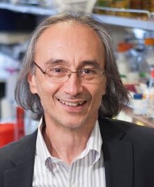 John Blenis合作论文数Sandra and Edward Meyer Cancer Center, Weill Cornell Medicine, Cornell University;Graduate School of Medical Sciences, Weill Cornell Medicine, Cornell University12
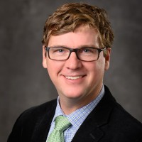 Jeffrey MacKeigan合作论文数Department of Pediatrics and Human Development, College of Human Medicine, Michigan State University12Ellen Triantafellow合作论文数10Christopher J. Wilson合作论文数9Jonathan M. Goodwin合作论文数8Liu Shanming合作论文数8Keaney Erin P合作论文数7Wang Zuncai合作论文数7Pascal Furet合作论文数6
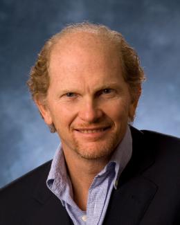 Andrea Ballabio合作论文数Department of Molecular And Human Genetics, Baylor College of Medicine;Dipartimento di Scienze Mediche Traslazionali, Universita Degli Studi di Napoli Federico II;Telethon Institute of Genetics and Medicine6
 William E Dowdle合作论文数Department of Biochemistry and Biophysics and Cardiovascular Research Institute, University of California6John A. Tallarico合作论文数Eugene F. Merkert Chemistry Center, Boston College6Rowena DeJesus合作论文数5Lawrence G Hamann合作论文数5
 William Sellers合作论文数Broad Institute;Division of Medical Sciences, Harvard Medical School5
 David J. Glass合作论文数Regeneron Pharmaceuticals, Inc.;Harvard Medical School, Harvard University5
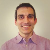 Gregory R Hoffman合作论文数Department of Chemistry and Chemical Biology, Cornell University5Suchithra Menon合作论文数5Jeffrey A. Porter合作论文数5Joan Mannick合作论文数4Philip Bergman合作论文数4Mark Knapp合作论文数4Phil Bergman合作论文数4Guiqing Liang合作论文数4
 Diane Fingar合作论文数Department of Cell & Developmental Biology, Medical School, University of Michigan4
 Sharon A. Tooze合作论文数London Research Institute;The Francis Crick Institute4
 Fulvio Reggiori合作论文数Department of Biomedicine, Aarhus University4Brachmann Saskia M合作论文数4Alicia Lindeman合作论文数4
 Daniel Klionsky合作论文数Life Sciences Institute, University of Michigan;Department of Molecular, Cell & Developmental Biology, College of Literature, Science, and the Arts, University of Michigan4
 Ramnik Xavier合作论文数Center for Computational and Integrative Biology, Massachusetts General Hospital;Department of Molecular Biology, Massachusetts General Hospital;Harvard Medical School;Division of Medical Sciences, Harvard University;Broad Institute4McAllister Gregory合作论文数4Francesco Cecconi合作论文数4John S. Reece-Hoyes合作论文数4Carleton Goold合作论文数3
 Timothy Lehmberg合作论文数3Xiaohong Mao合作论文数3
 Maria Isabel Colombo合作论文数3
 Jennifer L Brogdon合作论文数3
 David Rubinsztein合作论文数Department of Medical Genetics, Cambridge Institute for Medical Research, University of Cambridge;UK Dementia Research Institute, University of Cambridge3Maira Sauveur-Michel合作论文数3
 Nicholas Ktistakis合作论文数Babraham Institute3Cornella-Taracido Ivan合作论文数3Harrington Edmund合作论文数3
 Noboru Mizushima (水島昇)合作论文数Department of Biochemistry and Molecular Biology, Graduate School and Faculty of Medicine, The University of Tokyo3
 Kobylarz Marek J合作论文数Life Sciences Institute, University of British Columbia3Biag Jonathan合作论文数3




