Cardiovascular diseases continue to exhibit high global morbidity and mortality rates, driving urgent demand for the development of small-diameter (diameter < 6 mm) vascular grafts. Current small-diameter vascular grafts exhibit low patency rates in vascular reconstruction due to incomplete endothelial coverage. The design of vascular grafts with spontaneous endothelialization is critically needed. Herein, we developed an on-demand ROS-responsive polycaprolactone (PCL) graft modified with puerarin (PUE) and hyaluronic acid (HA) by electrospinning. In this study, PCL mimicked the structure of native blood vessels, and PUE-HA encouraged the antioxidation and endothelialization of vascular grafts with improved biocompatibility. The PUE-HA-PCL graft could promote the adhesion and proliferation of endothelial cells (ECs) and facilitate the process of tissue regeneration. Moreover, the grafts remained completely unobstructed with well-organized elastin fibers for 3 months after implantation. Taken together, the results effectively demonstrate that PUE-HA-PCL will be a promising functional small-diameter vascular graft with spontaneous endothelialization.
The failure of small-diameter vascular grafts (SDVGs) is primarily attributed to thrombosis and pathological inflammation, leading to incomplete endothelialization and ultimately poor long-term patency. Current antithrombotic strategies are limited by insufficient durability and the risk of systemic coagulation disturbances associated with continuous anticoagulation. Herein, we developed a thrombin-responsive vascular graft enabling on-demand thrombolysis and modulation of the pathological microenvironment. Curcumin was encapsulated into liposomes to overcome its poor bioavailability, endowing the graft with reactive oxygen species (ROS) scavenging and immunomodulatory effects. Thrombin-responsive curcumin-loaded liposomes (uPA-Cur-Lip) were then constructed by sequentially conjugating a thrombin-cleavable peptide and urokinase-type plasminogen activator (uPA) to the liposomes. Subsequently, uPA-Cur-Lip was immobilized onto electrospun PCL/PLGA vascular grafts via polydopamine (PDA) coating, yielding uPA-Cur-PDA@PCL/PLGA. In vitro studies confirmed sustained curcumin release from the liposomes and thrombin-responsive "on-demand" thrombolytic activity. Furthermore, uPA-Cur-PDA@PCL/PLGA enhanced endothelial cell migration, alleviated oxidative stress, and promoted M2 macrophage polarization. The results of in vivo rat abdominal aorta implantation showed that uPA-Cur-PDA@PCL/PLGA exhibited sustained patency for 3 months without thrombosis, along with improved endothelialization and reduced inflammation. Overall, we developed a thrombin-responsive curcumin liposome-modified SDVG that enables on-demand thrombolysis and modulates the local microenvironment, thereby promoting vascular regeneration and improving long-term graft performance.
OBJECTIVE:To evaluate the effects of beyond cold light whitening and desensitization on shear bond strength of orthodontic metal brackets. MATERIALS AND METHODS:Ninety-eight extracted human premolars were randomly divided into seven groups (n = 14). Group 1 was the control (no treatment). Group 2 underwent bleaching, followed by bonding after 24 h. Group 3 received desensitization, then bonding after 24 h. Group 4 combined bleaching and desensitization, with bonding after 24 h. Groups 5, 6, and 7 followed the same procedure as Group 4, but bonding occurred 1, 2, and 3 weeks later, respectively. All samples were stored in 37°C artificial saliva after treatment and bonded with 3 M light-cure composite resin using halogen light. Shear bond strength was tested with a universal testing machine (INSTRON 5848). Adhesive remnant index (ARI) scores were assessed post-debonding. Statistical analysis was conducted using Analysis of Variance (ANOVA), Tukey's test, and chi-square tests, with significance set at p < 0.05. RESULT:The shear bond strength 24 h after bleaching (7.5 ± 1.77 MPa) was significantly lower than the control (12.24 ± 3.71 MPa, p < 0.05). Desensitization alone (11.68 ± 3.49 MPa) showed no significant difference compared with the control (p > 0.05). Shear bond strength significantly decreased 24 h after bleaching and desensitization (p < 0.05) but recovered to control levels after 1 week (p > 0.05). ARI scores showed no significant differences. CONCLUSIONS:(1) Brackets bonded 24 h after bleaching or combined treatment showed reduced shear bond strength but were clinically acceptable; (2) The use of nano-biomaterial desensitizers slightly reduces the shear strength of brackets, but it will repair the damage of teeth caused by bleaching; (3) The impacts of bleaching and desensitization gradually reduce and return to normal levels after 1 week.
Small-diameter vascular grafts (SDVGs) often struggle to maintain long-term patency due to thrombus formation, intimal hyperplasia, and inflammation. Endothelialization emerges as a pivotal strategy for addressing these concerns. As a representative activator of the hypoxia-inducible factor (HIF) pathway, ML228 can stimulate the expression of downstream target genes like vascular endothelial growth factor (VEGF) to induce angiogenesis, yet it requires encapsulation by nanoparticles for optimal delivery and efficacy. However, the immune system often recognizes nanoparticles as foreign entities, posing a significant risk of clearance. In this study, we developed ML228-loaded poly (lactic-co-glycolic acid) (PLGA) nanoparticles and coated them with platelet membranes, thereby enhancing their biocompatibility and enabling immune escape. The ML228-loaded PLGA nanoparticles coated with platelet membranes (MPNP) were immobilized onto electrospinning SDVGs made of silk fibroin (SF) and polycaprolactone (PCL) to obtain MPNP-coated grafts (SF/PCL@MPNP) with the ability to promote endothelialization. In vitro biological activity studies demonstrated that SF/PCL@MPNP activated the HIF pathway, upregulating the downstream target gene VEGF, which facilitated endothelial cells migration and angiogenesis. In vivo implantation in a rat abdominal aorta model revealed that SF/PCL@MPNP promoted endothelialization, supported the regeneration of contractile smooth muscle cells, and modulated inflammatory responses. Overall, this study presents a strategy for constructing SDVGs using ML228-loaded nanoparticles with platelet membrane coating, highlighting the promises of using ML228 to activate the HIF pathway and membrane-coated nanoparticles to improve endothelialization in vascular graft applications.
Lung cancer remains the leading cause of cancer-related mortality worldwide, largely due to late-stage diagnosis. Early detection is critical for improving patient outcomes, yet current screening methods, such as low-dose computed tomography (CT), often lack the sensitivity and specificity required for early-stage detection. Here, we present a multimodal early screening platform that integrates a multiplexed laser-induced graphene (LIG) immunosensor with machine learning to enhance the accuracy of lung cancer diagnosis. Our platform enables the rapid, cost-effective, and simultaneous detection of four tumor markers─neuron-specific enolase (NSE), carcinoembryonic antigen (CEA), p53, and SOX2─with limits of detection (LOD) as low as 1.62 pg/mL. By combining proteomic data from the immunosensor with deep learning-based CT imaging features and clinical data, we developed a multimodal predictive model that achieves an area under the curve (AUC) of 0.936, significantly outperforming single-modality approaches. This platform offers a transformative solution for early lung cancer screening, particularly in resource-limited settings, and provides potential technical support for precision medicine in oncology.
Endothelial cell (EC) dysfunction within the aorta has long been recognized as a prominent contributor to the progression of atherosclerosis and the subsequent failure of vascular graft transplantation. However, the direct relationship between EC dysfunction and vascular remodeling remains to be investigated. In this study, we sought to address this knowledge gap by employing a strategy involving the release of glutamine synthetase (GS), which effectively activated endothelial metabolism and mitigates EC dysfunction. To achieve this, we developed GS-loaded small-diameter vascular grafts (GSVG) through the electrospinning technique, utilizing dual-component solutions consisting of photo-crosslinkable hyaluronic acid and polycaprolactone. Through an in vitro model of oxidized low-density lipoprotein-induced injury in human umbilical vein endothelial cells (HUVECs), we provided compelling evidence that the GSVG promoted the restoration of motility, angiogenic sprouting, and proliferation in dysfunctional HUVECs by enhancing cellular metabolism. Furthermore, the sequencing results indicated that these effects were mediated by miR-122-5p-related signaling pathways. Remarkably, the GSVG also exhibited regulatory capabilities in shifting vascular smooth muscle cells towards a contractile phenotype, mitigating inflammatory responses and thereby preventing vascular calcification. Finally, our data demonstrated that GS incorporation significantly enhanced re-endothelialization of vascular grafts in a ferric chloride-injured rat model. Collectively, our results offer insights into the promotion of re-endothelialization in vascular grafts by restoring dysfunctional ECs through the augmentation of cellular metabolism.
Hydrogel scaffolds represent an attractive tool for tissue repair. However, targeted tissue repair requires a specific shape and biological function design, and most natural-protein-based hydrogel scaffolds are predominantly confined to specific tissue repair applications. Here, we developed a versatile structural biomimetic natural protein platform through synergistic electrospinning, photopolymerization, and metal-coordination strategies. By integrating methacrylated silk fibroin (SFMA) with acrylated bisphosphonates (AcBP), we developed a dynamically functionalizable matrix that enables (1) customizable shape control via tunable electrospinning collectors and (2) on-demand biological function customization through metal-ion chelation. As a proof of concept, we demonstrate this platform's scenario-specific therapeutic efficacy: (i) Mg2+-functionalized membranes (S-LB-Mg) that orchestrate angiogenic-osteogenic coupling in critical-sized calvarial defects, (ii) Ag+-integrated dressing (S-LB-Ag) enabling bacterial eradication via a nonantibiotic mechanism and accelerating infected wound closure, and (iii) Zn2+-loaded conduits (S-LB-Zn) that drive macrophage M2 polarization to enhance peripheral nerve regeneration. This naturally derived protein-based platform overcomes the potential side effects associated with clinical bioactive factor/antibiotic composite scaffolds, offering a simple and customizable solution for the repair and regeneration of diverse tissues in a cost-effective yet highly effective manner. Overall, our strategy provides an alternative perspective for constructing protein-derived hydrogel microfibers with customizable functions and shapes for tissue repair applications.
Stem cell-derived extracellular vesicles (EVs) show great potential for promoting bone tissue regeneration. However, normal EVs (Nor-EVs) have a limited ability to direct tissue-specific regeneration. Therefore, it is necessary to optimize the osteogenic capacity of EV-based systems for repairing extensive bone defects. Herein, we show that hydrogels loaded with osteoinductive dental pulp stem cell-derived EVs (Ost-EVs) enhanced bone tissue remodeling, resulting in a 2.23 ± 0.25-fold increase in the expression of bone morphogenetic protein 2 (BMP2) compared to the hydrogel control group. Moreover, Ost-EVs led to a higher expression of alkaline phosphatase (ALP) (1.88 ± 0.16 of Ost-EVs relative to Nor-EVs) and the formation of orange-red calcium nodules (1.38 ± 0.10 of Ost-EVs relative to Nor-EVs) in vitro. RNA sequencing revealed that Ost-EVs showed significantly high miR-1246 expression. An ideal hydrogel implant should also adhere to surrounding moist tissues. In this study, we were drawn to mussel-inspired adhesive modification, where the hydrogel carrier was crafted from hyaluronic acid (HA) and polyethylene glycol derivatives, showcasing impressive tissue adhesion, self-healing capabilities, and the ability to promote bone growth. The modified HA (mHA) hydrogel was also responsive to environmental stimuli, making it an effective carrier for delivering EVs. In an ectopic osteogenesis animal model, the Ost-EV/hydrogel system effectively alleviated inflammation, accelerated revascularization, and promoted tissue mineralization. We further used a rat femoral condyle defect model to evaluate the in situ osteogenic ability of the Ost-EVs/hydrogel system. Collectively, our results suggest that Ost-EVs combined with biomaterial-based hydrogels hold promising potential for treating bone defects.
Rapid hemostasis and effective healing for the non-compressible liver wounds which are not able to be sewn, especially for those large-area wounds, remain great clinical challenges. In this study, we fabricated epidermal growth factor (EGF)-loaded chitosan microspheres (CM) and then incorporated them into a photo-crosslinking gelatin methacryloyl (GelMA) hydrogel. The results showed that the EGF-loaded CM/GelMA precursor solution could transform into a hydrogel and cease bleeding at laceration sites without external stress. Subsequently, the sustained release of EGF accelerated wound closure and promoted liver regeneration. The in vitro experiments demonstrated that the microsphere/hydrogel composite could promote the proliferation and migration of L02 cells. Moreover, the histological and immunohistological analyses indicated that EGF-CM/GelMA composite could alleviate inflammation in the mouse liver and promote liver remodeling. Overall, this multi-functional microsphere/hydrogel composite will inspire the development of clinical applications for noncompressible hemostasis and successive wound closure.
Implantable tissue-engineered vascular grafts (TEVGs) usually trigger the host reaction which is inextricably linked with the immune system, including blood–material interaction, protein absorption, inflammation, foreign body reaction, and so on. With remarkable progress, the immune response is no longer considered to be entirely harmful to TEVGs, but its therapeutic and impaired effects on angiogenesis and tissue regeneration are parallel. Although the implicated immune mechanisms remain elusive, it is certainly worthwhile to gain detailed knowledge about the function of the individual immune components during angiogenesis and vascular remodeling. This review provides a general overview of immune cells with an emphasis on macrophages in light of the current literature. To the extent possible, we summarize state-of-the-art approaches to immune cell regulation of the vasculature and suggest that future studies are needed to better define the timing of the activity of each cell subpopulation and to further reveal key regulatory switches.
Cell sheet engineering has been proven to be a promising strategy for cardiac remodeling post-myocardial infarction. However, insufficient mechanical strength and low cell retention lead to limited therapeutic efficiency. The thickness and area of artificial cardiac patches also affect their therapeutic efficiency. Cardiac patches prepared by combining cell sheets with electrospun nanofibers, which can be transplanted and sutured to the surface of the infarcted heart, promise to solve this problem. Here, we fabricated a novel cardiac patch by stacking brown adipose-derived stem cells (BADSCs) sheet layer by layer, and then they were combined with multi-walled carbon nanotubes (CNTs)-containing electrospun polycaprolactone/silk fibroin nanofibers (CPSN). The results demonstrated that BADSCs tended to generate myocardium-like structures seeded on CPSN. Compared with BADSCs suspension-containing electrospun nanofibers, the transplantation of the CPSN-BADSCs sheets (CNBS) cardiac patches exhibited accelerated angiogenesis and decreased inflammation in a rat myocardial infarction model. In addition, the CNBS cardiac patches could regulate macrophage polarization and promote gap junction remodeling, thus restoring cardiac functions. Overall, the hybrid cardiac patches made of electrospun nanofibers and cell sheets provide a novel solution to cardiac remodeling after ischemic myocardial infarction.
The periosteum plays a crucial role in both bone development and the healing process of bone fractures. However, few studies have focused on developing artificial periosteum due to the complexity of its construction and the biological risks for clinical practice. To address this issue, we proposed a strategy for the fabrication of a photocrosslinkable natural hydrogel nanofibrous membrane (SFMA-Lap@AcBP) based on methacrylated silk fibroin (SFMA), photoinitiator (Lap), acrylate bisphosphonate (AcBP) through combining electrospinning technology with hydrogel, mimicking the structure and function of periosteum to promote bone regeneration. AcBP were covalently attached to the SFMA hydrogel nanofibrous membrane via conjugation. With the degradation of SFMA-Lap@AcBP hydrogel nanofibrous membrane, the released BP-grafted derivates could promote bone regeneration. Moreover, SFMA-Lap@AcBP hydrogel membranes were found to be able to trap metal ions such as Mg2+ and Ca2+ in vivo, which might play a synergistic role in inducing complete bone regeneration. Furthermore, the hydrogel membranes had excellent cytocompatibility. They could imitate the microenvironment of the extracellular matrix, thereby offering structural and biochemical cues for the adhesion, proliferation, and differentiation of cells, without the need of exogenous cells or inductive growth factors, resulting in a facilitated osteogenesis. After implanting membranes into a cranial periosteal and bone defect in rats, both the amount and quality of new bone in the SFMA-Lap@AcBP group were found to be higher than those in the other groups. Therefore, the hydrogel membrane bionic periosteum was efficient and versatile in inducing bone regeneration, providing a potential strategy to address clinical issues.
Drug-loaded liposomes have been shown to be effective in the treatment of hepatocellular carcinoma (HCC). However, the systemic non-specific distribution of drug-loaded liposomes in tumor patients is a critical therapeutic challenge. To address this issue, we developed galactosylated chitosan-modified liposomes (GC@Lipo) that could selectively bind to the asialoglycoprotein receptor (ASGPR), which is highly expressed on the membrane surface of HCC cells. Our study demonstrated that the GC@Lipo significantly enhanced the anti-tumor efficacy of oleanolic acid (OA) by enabling targeted drug delivery to hepatocytes. Remarkably, treatment with OA-loaded GC@Lipo inhibited the migration and proliferation of mouse Hepa1-6 cells by upregulating E-cadherin expression and downregulating N-cadherin, vimentin, and AXL expressions, compared to a free OA solution and OA-loaded liposomes. Furthermore, using an axillary tumor xenograft mouse model, we observed that OA-loaded GC@Lipo led to a significant reduction in tumor progression, accompanied by concentrated enrichment in hepatocytes. These findings strongly support the clinical translation of ASGPR-targeted liposomes for the treatment of HCC.
The multi-bacterial environment of the oral cavity makes it hard for periodontal regeneration. As a class of antimicrobial peptide, beta defensin has been found to show broad-spectrum antibacterial ability. In addition, connective tissue growth factor (CTGF) is demonstrated to play a great role in multi-physiological events such as angiogenesis, wound healing and, more importantly, fibrogenesis. In this study, human β defensin 3 (hBD3) and CTGF were co-transfected into bone marrow derived mesenchymal stem cells (BMSCs) for preparing cell sheets. The transfection efficiency was detected through fluorescence of eGFP and western blot assay. Our results showed that the hBD3 and CTGF proteins were highly and stably expressed in the BMSCs after transfection. The results of RT-PCR and induced differentiation indicated that hBD3 promoted osteogenic differentiation of BMSCs, while CTGF significantly increased fibrogenic differentiation even in the presence of hBD3. The BMSCs acquired stronger capacity in terms of promoting M2 polarization of RAW 264.7 macrophages fulfilled by the transfection and secretion of hBD3 and CTGF. To further evaluate the periodontal remodeling performance of cell sheets, a coralline hydroxyapatite (CHA)-chitosan based hydrogel-human tooth system was designed to simulate the natural periodontal environment. The results showed that dense extracellular matrix, oriented fiber arrangement, and abundant collagen deposition appeared in the area of BMSCs sheets after subcutaneous transplantation. Altogether, our data showed that the lentivirus transfected BMSCs sheets had a promising application prospect for periodontal repair.
Skeletal muscle tissue cannot repair itself after volumetric muscle loss (VML) injury, which results in chronic inflammation and secondary injuries. Quercetin (QCN) possesses natural antioxidant properties and the ability of suppressing inflammation. Herein, an in situ photocurable composite hydrogel was developed by employing silk fibroin methacryloyl (SilMA) and decellularized extracellular matrix (ECM) derived from skeletal muscle. QCN loaded liposomes were introduced to create a local microenvironment which was conducive to skeletal muscle regeneration by regulating M2 polarization of macrophages and reducing oxidative stress. The SilMA-ECM-QCN composite hydrogel was characterized with suitable porosity, mechanical properties and degradation properties, as well as adhesion property for injury wound sealing via in situ polymerization. Moreover, the therapeutic effects of SilMA-ECM-QCN for promoting muscle regeneration and decreasing fibrosis were demonstrated by using a VML rat model. The results showed that the photocurable SilMA-ECM-QCN hydrogel had a huge advantage in the rapid filling of complex skeletal muscle tissue defects. Altogether, the present work could provide a promising strategy to create a pro-regenerative microenvironment for muscle regeneration by utilizing natural biomaterials.
Translocation of extrinsic molecules into living cells is becoming increasingly crucial in biological studies ranging from cell engineering to biomedical applications. The concerns regarding biosafety and immunogenicity for conventional vectors and physical methods yet challenge the effective intracellular delivery. Here, we begin with an overview of approaches for trans-membrane delivery up to now. These methods are featured with a relatively mature application but usually encounter low cell survival. Our review then proposes an advanced application for nanomaterial-sensitized photoporation triggered with a laser. We cover the mechanisms, procedures, and outcomes of photoporation-induced intracellular delivery with a highlight on its versatility to different living cells. We hope the review discussed here encourages researchers with further improvement and applications for photoporation-induced intracellular drug delivery.
This study was to explore the mechanism by how exosomes (exo) derived from BMSCs affects cardiomyocyte apoptosis. BMSCs were isolated and incubated with cardiomyocytes while the cardiomyocytes were exposed to sevoflurane or DMSO treatment. Apoptotic cells were calculated and level of apoptosis related proteins was detected by Western blot. Through transfection with microRNA-(miRNA)-312 inhibitor, we evaluated the effect of BMSC-exo on the sevoflurane-induced apoptosis. Sevoflurane significantly inhibited the viability of cardiomyocytes and induced cardiomyocyte apoptosis. Besides, sevoflurane decreased the expression of miR-312 and enhanced Bax expression in cardiomyocytes through restraining the phosphorylation of MAPK/ERK. Treatment with BMSC-exo, however, activated MAPK/ERK signaling by up-regulating miR-312, thereby inhibiting cardiomyocyte apoptosis, promoting cardiomyocyte proliferation, and elevating the level of Bcl-2. In conclusion, BMSC-exo-derived miR-312 inhibits sevoflurane-induced cardiomyocyte apoptosis by activating PI3K/AKT signaling pathway.
Quercetin (Que) has been proved to have various biological activities, including anti-oxidation, anti-inflammation and anti-virus, showing great potential in liver protection. However, its water insolubility leads to low bioavailability. Therefore, the development of a suitable drug delivery fashion is imminent. In recent years, liposomes have been widely used in the fields of drug delivery and gene transfer thanks to the cell membrane like structure, easy surface-modification and high encapsulation efficiency. Herein, we fabricated Que loaded anionic liposomes. Galactosylated chitosan (GC) was simply attached to the surfaces of liposomes through electrostatic adsorption to achieve targeted delivery by binding to asialoglycoprotein receptor (ASGPR). The results showed that Que loaded liposomes modified with GC (GC-Que-Lipo) could enrich the liver in mice through tail vein injection. Liposomes could achieve sustained drug release and GC-Que-Lipo promoted M2 polarization of macrophages. More importantly, it could maintain low content of AST, ALT, ALP and high level of GSH while reducing lipid oxidation, thereby protecting the liver from damage in acute liver injury model. In general, we expect to be able to acquire targeted and efficient delivery of quercetin through a facile approach, thus fulfill the prevention and treatment of liver diseases.
The aim of this study was to optimize the preparation method of polymethyl methacrylate (PMMA) denture base loaded with nano silver (NAg), to more effectively and safely impart sustainable antibacterial functions. NAg solution was synthetized and mixed with acrylic acid and methyl methyacrylate (MMA) monomer in order to prepare a new type of NAg solution (NS)/polymer methyl methacrylate denture base specimens (NS/PMMA). The surface morphology, mechanical strength, antimicrobial activity, anti-aging performance, cytotoxicity and biocompatibility of NS/PMMA denture base were evaluated in comparison with specimens fabricated using traditional NAg adding methods and NAg-free denture base. The aesthetic characteristics and mechanical strength of NS/PMMA denture base met the clinical application requirements. Meanwhile, NS/PMMA denture base showed better antibacterial activity, anti-aging properties, no cytotoxicity and displayed exceptional biocompatibility. NS/PMMA denture base thus has great potential for clinical application.
Background: Fractures are a medical disease with a high incidence, and about 5-10% of patients need bone transplantation to fill the defect. In this study, we aimed to synthesize a new type of coralline hydroxyapatite (CHA)/silk fibroin (SF)/glycol chitosan (GCS)/difunctionalized polyethylene glycol (DF-PEG) self-healing hydrogel and to evaluate the therapeutic effects of this novel self-healing hydrogel as a human umbilical cord mesenchymal stem cells (hucMSC)-derived exosome carrier on bone defects in SD rat. Methods: HucMSCs were isolated from fetal umbilical cord tissue and characterized by surface antigen analysis and pluripotent differentiation in vitro. The cell supernatant after ultracentrifugation was collected to isolate exosomes, which were characterized by transmission electron microscopy and western blot analysis. In vitro cell induction experiments were performed to observe the effects of hucMSC-derived exosomes on the biological behavior of mouse osteoblast progenitor cells (mOPCs) and human umbilical vein endothelial cells (HUVECs). The CHA/SF/GCS/DF-PEG hydrogels were prepared using DF-PEG as the gel factor and then structural and physical properties were characterized. HucMSCs-derived exosomes were added to the hydrogel and their effects were evaluated in SD rats with induced femoral condyle defect. These effects were analyzed by X-ray and micro-CT imaging and H&E, Masson and immunohistochemistry staining. Results: HucMSC-derived exosomes can promote osteogenic differentiation of mOPCs and promote the proliferation and migration of HUVECs. The CHA/SF/GCS/DF-PEG hydrogel has a high self-healing capacity, perfect surface morphology and the precipitated CHA crystals have a small size and low crystallinity similar to natural bone minerals. The MTT results showed that the hydrogel was non-toxic and have a good biocompatibility. The in vivo studies have shown that the hydrogel containing exosomes could effectively promote healing of rat bone defect. The histological analysis revealed more new bone tissue and morphogenetic protein 2 (BMP-2) in the hydrogel-exosome group. In addition, the hydrogel-exosome group had the highest microvessel density. Conclusion: A self-healing CHA/SF/GCS/DF-PEG hydrogel was successfully prepared. The hydrogel has excellent comprehensive properties and is expected to become a new type of bone graft material. This hydrogel has the effect of promoting bone repair, which is more significant after the addition of hucMSC-derived exosomes.
 Haifeng Liu (刘海峰)合作论文数School of Biological Science and Medical Engineering, Beihang University12
 Xinbo Wei合作论文数11
 Yubo Fan (樊瑜波)合作论文数School of Biological Science and Medical Engineering, Beihang University;Key Laboratory for Biomechanics and Mechanobiology of Ministry of Education, Beihang University;Beijing Key Lab for Design & Evaluation Technology of Implantable & Interventional Medical Devices, Beihang University9
 Zheng Xing合作论文数4Ximin Guo (郭希民)合作论文数4Yuqing Wang合作论文数3
 Xi He合作论文数3Jie Sun合作论文数3Cuimi Duan合作论文数3Shengzhao Xiao合作论文数2Rui-Deng Wang合作论文数2Depeng Yang合作论文数2XiangBin Zhou合作论文数2
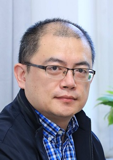 Huan Nie (聂桓)合作论文数School of Life Science and Technology, Harbin Institute of Technology;Faculty of Life Sciences and Medicine, Harbin Institute of Technology2
 Zhao Chen合作论文数2
 Gang Zhou (周钢)合作论文数Key Laboratory for Biomechanics and Mechanobiology of Ministry of Education, School of Biological Science and Medical Engineering, Beihang University2
 Fang Zhou (周方)合作论文数Peking University Third Hospital2
 HongYan Guo (郭红延)合作论文数Chinese People's Liberation Army General Hospital2Renpeng Peng合作论文数1Xinran Wei合作论文数1Jinjin Yang (杨紧紧)合作论文数1Jesse Lee合作论文数1Meiyi Jiang合作论文数1Handi Sun合作论文数1Sheng Guo合作论文数1Yabin Li合作论文数1Rui Ding合作论文数1Wang, Jian合作论文数1
 Jie Chen合作论文数Nanyang Environment and Water Research Institute, Nanyang Technological University1Qiusheng Shi合作论文数1Jialing Cai合作论文数1Bo Wei合作论文数1Peng Chen合作论文数1Jinwu Bai合作论文数1Haoran Su (苏浩然)合作论文数1
 Xiao-Xiao Yang合作论文数1Yuying Gao合作论文数1Jian Wang合作论文数1
 Lu Zhou合作论文数1Li Xia合作论文数1
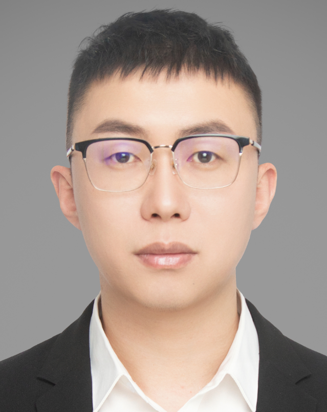 Yang Wang (王杨)合作论文数School of Medical Science and Engineering, Beihang University1Yu Zhou合作论文数1Biao Zhu合作论文数1XiaoYe Tuo (庹晓晔)合作论文数1Zhang Tong合作论文数1
 Qian Feng (冯茜)合作论文数Bioengineering College, Chongqing University1
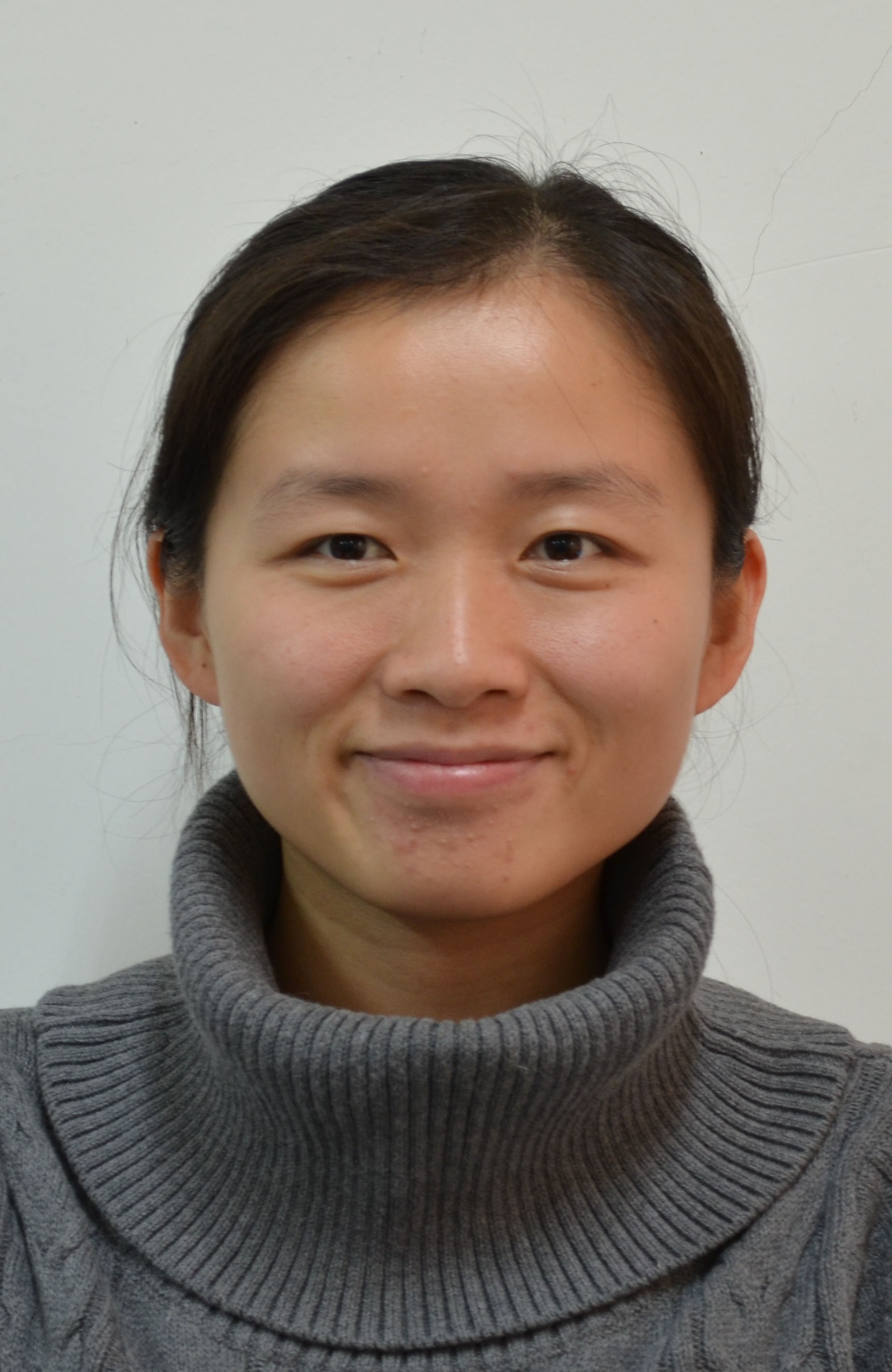 Chen Peng (彭琛)合作论文数School of Medicine, Tongji University;Tenth People's Hospital, Tongji University1




