Although recognized as a key regulator of gastrointestinal tissues, Wnt signaling pathway function in the stomach is poorly understood. This study aimed to define Wnt functions and identify Wnt-regulated genes in the stomach. Reporter mouse analysis localized Wnt signaling to the base and proliferative region in both the corpus and the antrum. Canonical Wnt inhibition in vivo using Sox2-CreERT2; Ctnnb1fl/fl mice reduced epithelial cell proliferation with loss of gastric stem cells. Wnt-regulated genes and potential effector pathways were studied by bulk RNA sequencing (RNA-Seq) analysis of corpus and antral organoids 24 h after Wnt inhibition in vitro. Cell signature analysis revealed that gastric organoids adopt a surface cell transcriptional profile following Wnt inhibition instead of a basal cell profile. Furthermore, retinoid metabolism terms were differentially expressed after Wnt inhibition, with decreased expression of retinoic acid target genes. Inhibition of retinoic acid signaling in corpus and antral organoids showed a marked increase in surface cell marker expression, consistent with the effects of Wnt inhibition. In the mouse, immunostaining showed differential localization of retinoid metabolic components in luminal pit cells (ALDH3A1) and basal chief/deep mucous cells (STRA6), with expression changes after β-catenin deletion in Sox2-CreERT2; Ctnnb1fl/fl mice, consistent with the Wnt-regulated cell fate changes observed in organoids. Together, these studies showed that Wnt signaling is required for gastric stem cell survival and promotes differentiation of cell types at the gland base. We identified retinoid metabolism as a candidate Wnt-regulated pathway, with cell-specific expression of key components, and regulation of surface cell marker expression by retinoic acid signaling.NEW & NOTEWORTHY Using mouse genetic and organoid models, we show that canonical Wnt signaling promotes gastric epithelial cell proliferation and regulates differentiation along the base-lumen gland axis in the stomach. Transcription profiling of Wnt-inhibited gastric organoids identified retinoic acid signaling as a potential Wnt-regulated effector pathway in the stomach. Accordingly, manipulation of retinoic acid signaling in organoids altered differentiated marker expression consistent with Wnt inhibition, supporting the presence of a Wnt-retinoic acid signaling axis in the stomach.
Microvillus inclusion disease (MVID) is a congenital diarrheal disorder, caused by inactivating mutations in myosin Vb (MYO5B). MYO5B-deficient mice and cell lines have demonstrated the importance of MYO5B in brush border development; however, the previous models lacked specificity to test intestinal stem cell functions. In the present study, we investigated the effects of progressive MYO5B deficiency originating in intestinal crypt cells utilizing mouse models. Our transcriptomic and multiplex immunostaining datasets demonstrate that MYO5B is critical for intestinal stem cell function. MYO5B-deficient crypts acquire a hyperproliferative phenotype with incomplete cell differentiation in vivo and an elevated organoid formation rate compared to control crypts. An evident disruption in mitochondrial structure and fatty acid metabolism likely underlies these crypt phenotypes. Consistent with mouse models, MVID patient biopsies demonstrate abnormal expansion of the proliferative zone along with villus blunting. These data reveal the direct role of MYO5B in intestinal epithelial progenitor cell functions.
Gastrin is secreted following a rise in gastric pH, leading to gastric acid secretion. Sleeve gastrectomy (SG), a bariatric surgery where 80% of the gastric corpus is excised, presents a challenge for gastric pH homeostasis. Using histology, and single-cell RNA sequencing of the gastric epithelium in 12 women, we observed that SG is associated with an increase in a sub-population of acid-secreting parietal cells that overexpress respiratory enzymes and an increase in histamine-secreting enterochromaffin-like cells (ECLs). ECLs of SG-operated patients overexpressed genes coding for biosynthesis of neuropeptides and serotonin. Mathematical modeling showed that pH homeostasis by gastrin is analogous to non-linear proportional and integral control, that drives adaptation of the epithelium to acid-secretion demand. Quantitative model predictions were validated in patients. The results demonstrate human gastric epithelium remodeling following SG at the molecular and cellular levels, and more generally how trophic hormones enable robust adaptation of tissue function to meet physiological demand.
There is an urgent need to find targeted agents for T cell acute lymphoblastic leukemia (T-ALL). NOTCH1 is the most frequently mutated oncogene in T-ALL, but clinical trials showed that pan-Notch inhibitors caused dose-limiting toxicities. Thus, we shifted our focus to ETS1, which is one of the transcription factors that most frequently co-bind Notch-occupied regulatory elements in the T-ALL context. To identify the most essential enhancers, we performed a genome-wide CRISPRi screen of the strongest ETS1-dependent regulatory elements. The top-ranked element is located in an intron of AHI1 that interacts with the MYB promoter and is amplified with MYB in approximately 8.5% of patients with T-ALL. Using mouse models, we showed that this enhancer promoted self-renewal of hematopoietic stem cells and T cell leukemogenesis, maintained early T cell precursors, and restrained myeloid expansion with aging. We named this enhancer the hematopoietic stem cell MYB enhancer (H-Me). The H-Me showed limited activity and function in committed T cell progenitors but was accessed during leukemogenesis. In one T-ALL context, ETS1 bound the ETS motif in the H-Me to recruit cBAF to promote chromatin accessibility and activation. ETS1 or cBAF degraders impaired H-Me function. Thus, we identified a targetable stem cell element that was co-opted for T cell transformation.
INTRODUCTION: Notch signaling is a principal niche pathway crucial for intestinal stem cells (ISCs), directing ISC self-renewal and lineage commitment to absorptive enterocytes. Because Notch signaling requires cell-cell contact, Paneth cells are considered to be the Notch niche cells as they neighbor each ISC and express the key Notch ligands Dll1 and Dll4. However, no functional studies have investigated the Paneth cell-specific role in the Notch niche. Additionally, ISCs function normally after Paneth cell ablation, yet how they maintain Notch signaling without Paneth cells remains unknown. We aimed to define the ISC response to Paneth cell-specific Notch ligand depletion. We hypothesized that loss of DLL1 and DLL4 in Paneth cells inhibits Notch signaling in ISCs, leading to loss of ISCs as well as impaired proliferation and differentiation. METHODS: To deplete Dll1 and Dll4 in Paneth cells, we crossed Dll1 f/f ; Dll4 f/f mice with a constitutive Paneth cell-specific Cre reporter mouse ( Defensin4-Cre; Rosa-LSL-tdTomato ). To deplete these Notch ligands after intestinal maturation, we crossed Dll1 f/f ; Dll4 f/f mice to a tamoxifen-inducible Paneth cell Cre strain ( Lysozyme-CreER T2 ; Rosa-LSL-tdTomato ), and induced ligand gene deletion in adult mice by treatment with 100mg/kg tamoxifen for 5 days, harvesting intestines 2 days later. Crypt cells were characterized by histological analysis of proliferation (EdU), and ISC (OLFM4), Paneth cell (tdTomato, LYZ, MMP7, UEA1), goblet cell (MUC2, UEA1), and endocrine cell (CHGA) marker expression. RESULTS: Surprisingly, constitutive Paneth cell-specific deletion of Dll1 and Dll4 did not affect overall morphology or proliferation of the intestinal epithelium, suggesting that normal ISC function was maintained throughout development. However, analysis of the ISC marker and Notch target OLFM4, showed a loss of ISC Notch signaling at the crypt base. Instead, cells expressing OLFM4 were shifted higher into the transit-amplifying region of the crypt, indicating that ISCs migrated away from defective Paneth cells and next to Notch ligand-expressing progenitors in the upper crypt. Paneth cells remained clustered at the crypt base, exhibiting hyperplasia with increased numbers of marker-expressing cells. Furthermore, the villus epithelium harbored mislocated tdTomato-positive cells that co-expressed Paneth and goblet cell markers, a feature of Paneth cell progenitors. These changes imply a drive to generate new Paneth cells to replace the ones lacking Notch ligands. In addition, increased numbers of endocrine cells were observed at the crypt base; although their numbers were not sufficient to replace the Paneth cell Notch niche. Like the constitutive model, tamoxifen-inducible deletion of Dll1 and Dll4 in Paneth cells promoted OLFM4 mislocation to the upper crypt, indicating ISC movement out of the crypt base. Interestingly, cleaved caspase 3 staining revealed increased apoptotic cells at the crypt base after inducible Dll1 and Dll4 deletion, suggesting that acute cell death plays a role in the crypt cell remodeling. CONCLUSION: Paneth cell-specific Dll1 and Dll4 deletion induces crypt remodeling to rebuild the ISC Notch niche. This project was funded by NIH R01 DK118023 awarded to L.C. Samuelson. This abstract was presented at the American Physiology Summit 2025 and is only available in HTML format. There is no downloadable file or PDF version. The Physiology editorial board was not involved in the peer review process.
Intestinal stem cells replenish the epithelium throughout life by continuously generating intestinal epithelial cell types, including absorptive enterocytes, and secretory goblet, endocrine, and Paneth cells. This process is orchestrated by a symphony of niche factors required to maintain intestinal stem cells and to direct their proliferation and differentiation. Among the various mature intestinal epithelial cell types, Paneth cells are unique in their location in the stem cell zone, directly adjacent to intestinal stem cells. Although Paneth cells were first described as an epithelial cell component of the innate immune system due to their expression of anti-microbial peptides, they have been proposed to be niche cells due to their close proximity to intestinal stem cells and expression of niche factors. However, function as a niche cell has been debated since mice lacking Paneth cells retain functional stem cells that continue to replenish the intestinal epithelium. In this review, we summarize the intestinal stem cell niche, including the Notch, Wnt, growth factor, mechanical, and metabolic niche, and discuss how Paneth cells might contribute to these various components. We also present a nuanced view of the Paneth cell as a niche cell. Although not required, Paneth cells enhance stem cell function, particularly during intestinal development and regeneration. Furthermore, we suggest that Paneth cell loss induces intestinal stem cell remodeling to adjust their niche demands.
Tissue architecture and function are influenced by mechanical cues. Yet, how cell nuclei sense forces within 3D tissues and dictate differentiation remains unknown as prior studies focused on isolated mesenchymal cells, which fail to fully predict tissue-level mechanical properties. We fill this knowledge gap utilizing live reporters and material-based organoid models. We posit the nucleus as an active mechanosensor of tissue shape, with levels of the nuclear scaffolding protein lamin-A varying across intestinal stem cell differentiation trajectory. Elevated forces on differentiated Paneth cell nuclei, in both organoids and tissue explants, increase lamin-A and nuclear wrinkling. Enhancing nuclear mechanotransduction primes cell differentiation, in otherwise stem promoting conditions, revealing that nuclear mechanics can direct stem cell fate. By engineering spatiotemporally controlled de novo tissue curvature with photo-degradable hydrogels, we direct spatially patterned lamin-A levels across mouse and human organoids of healthy and diseased origin, uncovering conserved nuclear mechanosensing pathway in epithelial tissues.
Microvillus inclusion disease (MVID) is a congenital disorder characterized by severe diarrhea, caused by inactivating mutations in myosin Vb (MYO5B). Mice lacking MYO5B in their intestinal epithelium ( VilCre ERT2 ; MYO5B fl/fl ) demonstrate the importance of MYO5B in enterocyte brush border development and crypt function, including differentiation and proliferation. However, previous MYO5B knockout models lacked specificity to the crypt, therefore, the observed crypt deficits could be secondary effects of nutrient malabsorption. This study aims to better understand the crypt-specific effects caused by MYO5B loss. Tamoxifen-inducible Lrig1-Cre ERT2 ;R26R-YFP;MYO5B fl/fl (Lrig1DMYO5B) mice were generated by crossbreeding of established strains. Adult Lrig1DMYO5B mice and control littermates received tamoxifen at Day 0, and GI tissues were collected up to Day 5. Single cell RNA-sequencing (scRNA-seq) was conducted on the jejunal epithelium and lamina propria of Day 3 and 5. To validate the small intestinal stem cell-specific effects of MYO5B loss, we generated Olfm4-Cre ERT2 ;R26R-TdTOM;MYO5B fl/fl (Olfm4DMYO5B) mice. Lrig1DMYO5B mice began exhibiting weight loss at Day 4 and lost 20% of their starting body weight on Day 5. The proliferative crypt region was expanded in Lrig1DMYO5B mice beginning at Day 3. Correspondingly, MYO5B expression was diminished in the crypts at Day 3 and nearly absent from the entire epithelium by Day 5. Differentiation of epithelial cells derived from MYO5B-lacking progenitors was altered: decreased tuft cells, abnormal goblet cell shape, and mislocalized Paneth cells. Electron Microscopy of the Day 3 jejunum revealed a disruption in progenitor cell mitochondrial morphology. Day 3 Lrig1DMYO5B progenitor cells, but not villus cells, demonstrated significantly less Fatty Acid Oxidation (FAO) activity compared to control. scRNA-seq data revealed a lack of stem cell identity in progenitor cells of the Day 3 and 5 Lrig1DMYO5B jejunum, marked by a decrease in stem cell gene expression and numbers. TA, absorptive and secretory progenitor cell populations were expanded at both timepoints. Lrig1DMYO5B fibroblasts had decreased expression of Wnt s, Bmp s, and R-spondin s, which are critical for epithelial progenitor cell function. Mitochondrial oxidation gene expression was decreased in Day 3 Lrig1DMYO5B progenitor cells. In the Olfm4DMYO5B mouse small intestine, MYO5B – /TdTOM+ cells were present across the crypt-villus axis at Day 7. Mirroring the observations in Lrig1DMYO5B, the Olfm4DMYO5B small intestine demonstrated a progressive crypt expansion and cell differentiation deficits. Lrig1DMYO5B and Olfm4DMYO5B strains revealed the negative impact of MYO5B loss on intestinal epithelial cell differentiation and interaction between progenitor cell and mesenchymal niche. Funding: NIH R01DK128190 This abstract was presented at the American Physiology Summit 2025 and is only available in HTML format. There is no downloadable file or PDF version. The Physiology editorial board was not involved in the peer review process.
Background and aim: Microvillus inclusion disease (MVID) is a congenital disorder characterized by severe diarrhea and is caused by inactivating mutations in motor protein myosin Vb (MYO5B). Mice lacking MYO5B in intestinal and colonic epithelia ( Vil-CreERT2;Myo5bfl/fl) display an MVID phenotype characterized by significant weight loss, lack of solid feces, and brush border defects. However, previous models have not discerned if this diarrheal phenotype is due to MYO5B loss in the small intestine specifically or both in the small intestine and colon. Using the small intestinal stem cell specific Cre driver under Olfm4, we phenotyped the mice lacking MYO5B only in the small intestine, while normal MYO5B expression is maintained in the colon. Methods: Olfm4-GFP-CreERT2;R26R-LSL-tdTom and Myo5bfl/fl mice were crossbred to generate Olfm4-GFP-CreERT2;R26R-LSL-tdTom;Myo5bfl/fl (Olfm4DMYO5B) mice. Adult Olfm4DMYO5B mice and control littermates received tamoxifen injection at day 0. GI tissues were collected 5 and 7 days post tamoxifen. Results: Following Cre induction, Olfm4DMYO5B mice exhibited progressive weight loss starting at day 5 and on average lost 20.6% on day 7. At day 7, the small intestine contained clear luminal contents, similar to that of the previous MVID model mice. However, the colon of day 7 Olfm4DMYO5B mice contained small, solid feces. An elongation of PCNA+ crypts was identified on day 5 throughout the small intestine. Villus blunting and microvillus inclusions were prominent in the small intestinal enterocytes on day 7, but not on day 5. The day 7 Olfm4DMYO5B small intestine demonstrated a decrease in tuft cell numbers, suggesting differentiation deficits in the MYO5B-lacking stem cells. However, the colon of Olfm4DMYO5B mice showed intact morphology. Colonic crypt depth and tuft cell numbers at both days 5 and 7 were similar to those of control mice. Immunostaining confirmed the maintenance of MYO5B expression in the colon and progressive loss of MYO5B in the small intestine of Olfm4DMYO5B mice. At day 5, MYO5B was specifically lost in small intestinal crypts, and the loss of MYO5B expanded to the villus tips on day 7. These MYO5B-lacking cells were tdTom+, including enterocytes, goblet, and rare tuft cells. Following the loss of MYO5B, small intestinal enterocytes lacked proper apical localization of numerous transporters and enzymes, including Na+-glucose cotransporter 1 and dipeptidyl peptidase 4. Conclusion: Recently established Olfm4DMYO5B mice demonstrated that the specific MYO5B deletion in the small intestinal stem cells causes hyperproliferation and differentiation deficits. Colonic MYO5B function is suffcient for the formation of solid feces, however, these mice experience prominent weight loss, likely due to the MVID-like malabsorption in the small intestine. NIH R01DK128190, RC2DK118640. This is the full abstract presented at the American Physiology Summit 2024 meeting and is only available in HTML format. There are no additional versions or additional content available for this abstract. Physiology was not involved in the peer review process.
BACKGROUND:Tissue repair and regeneration in the gastrointestinal system are crucial for maintaining homeostasis, with the process relying on intricate cellular interactions and affected by micro- and macro-nutrients. Iron, essential for various biological functions, plays a dual role in tissue healing by potentially causing oxidative damage and participating in anti-inflammatory mechanisms, underscoring its complex relationship with inflammation and tissue repair. OBJECTIVE:The study aimed to elucidate the role of low dietary iron in gastrointestinal tissue repair. METHODS:We utilized quantitative iron measurements to assess iron levels in inflamed regions of patients with ulcerative colitis and Crohn's disease. In addition, 3 mouse models of gastrointestinal injury/repair (dextran sulfate sodium-induced colitis, radiation injury, and wound biopsy) were used to assess the effects of low dietary iron on tissue repair. RESULTS:We found that levels of iron in inflamed regions of both patients with ulcerative colitis and Crohn's disease are elevated. Similarly, during gastrointestinal repair, iron levels were found to be heightened, specifically in intestinal epithelial cells across the 3 injury/repair models. Mice on a low-iron diet showed compromised tissue repair with reduced proliferation. In standard diet, epithelial cells and the stem cell compartment maintain adequate iron stores. However, during a period of iron deficiency, epithelial cells exhaust their iron reserves, whereas the stem cell compartments maintain their iron pools. During injury, when the stem compartment is disrupted, low iron levels impair proliferation and compromise repair mechanisms. CONCLUSIONS:Low dietary iron impairs intestinal repair through compromising the ability of epithelial cells to aid in intestinal proliferation.
Biliary obstruction and cholangiocyte hyperproliferation are important features of cholangiopathies affecting the large extrahepatic bile duct (EHBD). The mechanisms underlying obstruction-induced cholangiocyte proliferation in the EHBD remain poorly understood. Developmental pathways, including WNT signaling, are implicated in regulating injury responses in many tissues, including the liver. To investigate the contribution of WNT signaling to obstruction-induced cholangiocyte proliferation in the EHBD, we used complementary in vivo and in vitro models with pharmacologic interventions and transcriptomic analyses. To model obstruction, we used bile duct ligation (BDL) in mice. Human and mouse biliary organoids and mouse biliary explants were used to investigate the effects of WNT activation and inhibition in vitro. We observed an upregulation of WNT ligand expression associated with increased biliary proliferation following obstruction. Cholangiocytes were identified as both WNT ligand-expressing and WNT-responsive cells. Inhibition of WNT signaling decreased cholangiocyte proliferation in vivo and in vitro, while activation increased proliferation. WNT effects on cholangiocyte proliferation were β-catenin dependent, and we showed a direct effect of WNT7B on cholangiocyte growth. Our studies suggested that cholangiocyte-derived WNT ligands can activate WNT signaling to induce proliferation after obstructive injury. These findings implicate the WNT pathway in injury-induced cholangiocyte proliferation within the EHBD.
The metal ion transporter SLC39A8 is associated with physiological traits and diseases, including blood manganese (Mn) levels and inflammatory bowel diseases (IBD). The mechanisms by which SLC39A8 controls Mn homeostasis and epithelial integrity remain elusive. Here, we generate Slc39a8 intestinal epithelial cell-specific-knockout (Slc39a8-IEC KO) mice, which display markedly decreased Mn levels in blood and most organs. Radiotracer studies reveal impaired intestinal absorption of dietary Mn in Slc39a8-IEC KO mice. SLC39A8 is localized to the apical membrane and mediates 54Mn uptake in intestinal organoid monolayer cultures. Unbiased transcriptomic analysis identifies alkaline ceramidase 1 (ACER1), a key enzyme in sphingolipid metabolism, as a potential therapeutic target for SLC39A8-associated IBDs. Importantly, treatment with an ACER1 inhibitor attenuates colitis in Slc39a8-IEC KO mice by remedying barrier dysfunction. Our results highlight the essential roles of SLC39A8 in intestinal Mn absorption and epithelial integrity and offer a therapeutic target for IBD associated with impaired Mn homeostasis.
INTRODUCTION: Intestinal epithelial cells are rapidly renewed throughout life by intestinal stem cells (ISCs). ISC function is regulated by niche factors that orchestrate cellular proliferation and differentiation. Of these, Notch signaling is essential for ISC self-renewal and regulates the cell fate choice between absorptive cells (enterocyte) vs secretory (Paneth, goblet, enteroendocrine, and tuft) cells. Yet, the Notch niche is not fully defined. In the mature small intestine, Paneth cells neighbor each ISC and act as Notch niche cells, providing the Notch signal by expressing the Notch ligands DLL1 and DLL4 on their surface. However, studies in mice have shown that ISCs can function in the immature intestine before Paneth cells are formed, and when Paneth cells are depleted in the adult intestine. We previously observed that acute Notch inhibition via the inhibitor dibenzazepine (DBZ) resulted in Paneth cell apoptosis with intact ISCs displaying transient dysfunction. Here, we aimed to define the Notch niche after Paneth cell apoptosis in adult mice and in the developing intestine. We hypothesized that during acute Notch inhibition, ISCs remodel to compensate for Paneth cell loss by activating Notch ligand expression, and that this remodeling mimics the state of immature ISCs prior to Paneth cell emergence. METHODS: We isolated Lgr5+ ISCs by FACS from Lgr5-GFP-CreERT2 mice 24 hours after DBZ or vehicle treatment and performed bulk RNAseq analysis. To define the Notch niche during development, we analyzed postnatal intestines from Notch ligand reporter mice (Dll1-mCherry and Dll4-mCherry) crossed with the ISC reporter mice (Lgr5-GFP-CreERT2). RESULTS: ISCs isolated from Paneth cell-depleted intestines upregulated expression of Notch ligands Dll4 and Dll4 and various secretory cell markers, with expected downregulation of Notch target genes, but no alteration of other stem cell genes. This suggests that after acute Notch inhibition, Lgr5+ ISCs maintain partial stem cell identity while acquiring a signature resembling that of secretory progenitors. In the immature intestine, we showed by immunohistology that sporadic cells expressing secretory cell markers (LYZ, MUC2, and CHGA) localized to the intervillus region and co-stained with Dll1- or Dll4-mCherry+. This suggests that multiple differentiated cell types could provide the Notch signal to immature ISCs. We also observed occasional Lgr5-GFP+ ISCs that were co-positive for Dll1-mCherry, suggesting that immature ISCs can express Notch ligands at homeostasis. Further experimentation will determine whether these Dll1+ ISCs are similar to the remodeled adult ISCs after Paneth cell depletion. CONCLUSIONS: Acute Notch inhibition directly remodels ISCs to express the Notch ligands, Dll1 and Dll4 in the adult intestine. In the immature intestine, multiple secretory cell types appear to comprise the immature Notch niche. This project was funded by NIH R01 DK118023 awarded to Dr. Linda Samuelson. This is the full abstract presented at the American Physiology Summit 2024 meeting and is only available in HTML format. There are no additional versions or additional content available for this abstract. Physiology was not involved in the peer review process.
Germline adenomatous polyposis coli (APC) mutation in patients with familial adenomatous polyposis (FAP) promotes gastrointestinal polyposis, including the formation of frequent gastric fundic gland polyps (FGPs). In this study, we investigated how dysregulated Wnt signaling promotes FGPs and why they localize to the corpus region of the stomach. We developed a biobank of FGP and surrounding nonpolyp corpus biopsies and organoids from patients with FAP for comparative studies. Polyp biopsies and polyp-derived organoids exhibited enhanced Wnt target gene expression. Polyp-derived organoids with intrinsically upregulated Wnt signaling showed poor tolerance to further induction, suggesting that high Wnt restricts growth. Targeted genomic sequencing revealed that most gastric polyps did not arise via APC loss of heterozygosity. Studies in genetic mouse models demonstrated that heterozygous Apc loss increased epithelial cell proliferation in the corpus but not the antrum, while homozygous Apc loss was not maintained in the corpus yet induced hyperproliferation in the antrum. Our findings suggest that heterozygous APC mutation in patients with FAP may be sufficient to drive polyp formation in the corpus region while subsequent loss of heterozygosity to further enhance Wnt signaling is not tolerated. This finding contextualizes the abundant yet benign nature of gastric polyps in FAP patient corpus compared with the rare, yet adenomatous polyps in the antrum.
This study demonstrates that human gastric corpus organoids have a lower Wnt signaling threshold to drive optimal growth relative to patient-matched antral organoids. Paradoxically, supramaximal Wnt levels suppress corpus organoid proliferation, yet promote differentiation toward deep glandular cell types while simultaneously enhancing progenitor cell function. These findings provide novel insights into how Wnt signaling differentially regulates homeostasis in the human gastric corpus and antrum and contextualizes patterns of Wnt activation diseases.
Notch signaling regulates gastrointestinal stem cell proliferation and differentiation yet Notch-regulated transcriptional effectors of gastric epithelial cell differentiation are poorly understood. Here we tested the role of the bHLH transcription factor Achaete-Scute homolog 1 (ASCL1) in gastric epithelial cell differentiation, and its regulation by Notch. Newborn Ascl1 null mice showed a loss of expression of markers of neurogenin-3-dependent enteroendocrine cells, with normal expression of enterochromaffin-like cells, mucous cells, chief cells, and parietal cells. In adult mice, Ascl1 gene expression was observed in the stomach, but not the intestine, with higher expression in antral than corpus epithelium. Lineage tracing in Ascl1-CreER(T2); Rosa26-LSL-tdTomato mice revealed single, scattered ASCL1(+) cells in the gastric epithelium, demonstrating expression in antral gastrin- and serotonin-producing endocrine cells. ASCL1-expressing endocrine cells persisted for several weeks posttamoxifen labeling with a half-life of approximately 2 months. Lineage tracing in Gastrin-CreER(T2) mice demonstrated a similar lifespan for gastrin-producing cells, confirming that gastric endocrine cells are long-lived. Finally, treatment of Ascl1-CreER(T2); Rosa26-LSL-tdTomato mice with the pan-Notch inhibitor dibenzazepine increased the number of lineage-labeled cells in the gastric antrum, suggesting that Notch signaling normally inhibits Ascl1 expression. Notch regulation of Ascl1 was also demonstrated in a genetic mouse model of Notch activation, as well as Notch-manipulated antral organoid cultures, thus suggesting that ASCL1 is a key downstream Notch pathway effector promoting endocrine cell differentiation in the gastric epithelium. NEW & NOTEWORTHY Although Notch signaling is known to regulate cellular differentiation in the stomach, downstream effectors are poorly described. Here we demonstrate that the bHLH transcription factor ASCL1 is expressed in endocrine cells in the stomach and is required for formation of neurogenin-3-dependent enteroendocrine cells but not enterochromaffin-like cells. We also demonstrate that Ascl1 expression is inhibited by Notch signaling, suggesting that ASCL1 is a Notch-regulated transcriptional effector directing enteroendocrine cell fate in the mouse stomach.
Introduction: The Wnt signaling pathway regulates gastrointestinal stem cell identity and function. Throughout the stomach, a gradient of Wnt signaling is established across the epithelium with highest levels at the gland base. However, the proximal (corpus) and distal (antrum) regions of the stomach have strikingly different epithelial architectures, with antral stem cells and proliferation localized to the gland base while corpus proliferation is localized higher-up in the glands where Wnt levels are diminished. It is unknown how Wnt may differentially regulate cell function and identity within these regions. Furthermore, activation of Wnt signaling through genetic diseases, such as familial adenomatous polyposis (FAP), results in abundant polyposis of the corpus while the antrum is mostly spared. This study investigated how activation of Wnt signaling in human gastric corpus- and antral-derived organoids regulates stem cell function, with the hypothesis that corpus stem/progenitor cells have a lower threshold for optimal Wnt signaling. Methods: Human patient-matched corpus and antral organoids from two individuals were grown in the presence of the Wnt agonist CHIR99021 to assess Wnt sensitivity in vitro. Corpus organoids were then grown in high levels of CHIR99021 to assess how Wnt activation affected growth, proliferation, differentiation, and stem cell maintenance. Results: Treatment with CHIR99021 in media free of exogenous Wnt/R-spondin stimulated a maximal growth response in corpus organoids at lower concentrations than their respective patient-matched antral organoids. Increased Wnt signaling beyond the maximal growth concentration also suppressed proliferation at a rapid rate in corpus organoids. Chronic treatment with increasing levels of CHIR99021 reduced organoid size, suppressed surface cell differentiation, and promoted expression of markers for differentiated deep glandular cells, including neck and chief cells. Surprisingly, organoids retained in high levels of CHIR99021 had enhanced organoid formation ability when passaged, indicating that progenitor cell status was maintained in these non-proliferative, deep glandular cell-enriched organoids. Passaging these quiescent organoids into low Wnt conditions led to a rapid rescue of normal growth, organoid morphology, and surface cell differentiation. Conclusion: Our findings suggest that progenitor cells in human corpus organoids have a lower threshold for optimal Wnt signaling than progenitor cells in the antrum. We demonstrate that Wnt signaling in the corpus plays a central role in defining a bimodal axis of differentiation, and that high activation of Wnt signaling promotes stemness while simultaneously suppressing proliferation. Funding support for this research was provided through T32DK094775 (KPM), R25GM086262 (ED), and R01DK126451 (LCS). This is the full abstract presented at the American Physiology Summit 2023 meeting and is only available in HTML format. There are no additional versions or additional content available for this abstract. Physiology was not involved in the peer review process.
EditorialInnovating and Building Momentum for Physiology’s FutureJennifer S. Pollock, , Linda Samuelson, , and Dee Silverthorn, Jennifer S. PollockUniversity of Alabama at Birmingham, Birmingham, Alabama, APS President, Linda SamuelsonUniversity of Michigan, Ann Arbor, Michigan, APS Past President, and Dee SilverthornUniversity of Texas at Austin, Austin, Texas, APS President-ElectPublished Online:29 Dec 2021https://doi.org/10.1152/physiol.00042.2021This is the final version - click for previous versionMoreSectionsPDF (158 KB)Download PDFDownload PDFPlus ToolsExport citationAdd to favoritesGet permissionsTrack citations ShareShare onFacebookTwitterLinkedInEmail Let me start by congratulating all colleagues and friends in the physiology community in making it through another year of the tumultuous pandemic. Although we began to see signs of a “return to normal—many of us getting back to more regular hours in the laboratory, restarting important work that had been put on hold, and occasionally getting to see each other in-person—life was still nowhere close to what it had been in 2019. However, despite it all, our connections as a community remained and physiologists found a way to continue building on and growing scientific knowledge. So, too, has the American Physiological Society (APS) continued on its path to support, publish, and promote the physiology community and provide resources, funding, knowledge, and connection opportunities.In late 2020, the APS Council approved a new strategic plan focused on prioritizing scientific excellence; elevating the discipline; providing more value to our members; championing diversity, equity and inclusion (DEI) in our membership and the greater science, technology, engineering, and mathematics (STEM) community; and expanding engagement among all our members and, especially, our international member community. As part of meeting these goals in 2021, we continued to expand our digital offerings; launch new products and resources for physiology researchers, educators, and trainees; and plan an exciting annual meeting for 2023 that will educate and energize.I am pleased to share some of our biggest accomplishments of the last year and several exciting new programs and member benefits that will launch in the near future.A Centennial Celebration and New Resources from APS PublicationsA major highlight in our publishing portfolio in 2021 is the 100th anniversary of Physiological Reviews. Founded in 1921, the journal is an authoritative voice in the discipline, long-ranked the No. 1 among physiology journals. As Editor-in-Chief Sadis Matalon, PhD, FAPS, wrote in a recent issue of The Physiologist Magazine (https://bit.ly/100YearsofPRV), “A century later, Physiological Reviews is the most respected and cited physiological journal in the world. We publish highly authoritative, non-biased and informative reviews that are highly cited by basic, translational and clinical scientists engaged in biomedical research and patient care. It is a mark of excellence for an author to be asked to contribute an article. Authors labor for years to complete the final versions.” The Society joins the Physiological Reviews editorial team in celebrating the centennial of this important publication.The APS Publications Committee, journal editors-in-chief, and publishing staff have also continued to innovate, finding new ways to promote research and engage and educate authors and reviewers. In 2021, they have rolled out several new initiatives, including the following:APS Open Access: Authors will find enhanced guidance and information on the benefits of publishing open access in APS journals (https://bit.ly/APSOpenAccess). The Society now also offers a discount for APS members who publish open access.Targeting the Peer Review Pipeline: In addition to building new resources for reviewers (https://journals.physiology.org/reviewers), we have introduced several new programs to provide reviewer training and resources including a new reviewer training webinar for the American Journal of Physiology-Regulatory, Comparative and Integrative Physiology.Improving Rigor and Reproducibility: APS journals implemented new rigor and reproducibility requirements for all original research submissions beginning in July 2021. Author compliance with the new requirements are being assessed using an Artificial Intelligence tool (SciScore), through a pilot program lasting into 2022. The SciScore report is included with the first decision letter, and authors are asked to revise their manuscript to meet the rigor and reproducibility requirements. After the first quarter of implementation, the Publishing team has noted an improvement in scores on revisions from 4.48 to 4.92, nearly a 0.5 increase between initial submission (n = 652) and revision (n = 229).Empowering Researchers to Promote Their Work: The Society has created a guide (https://bit.ly/PromoteYourWork) to help our published authors promote their articles, gain readership, increase their scientific reputation and, ultimately, their article’s citations. The new pages also detail the many ways APS amplifies article reach to a variety of audiences—media, researchers, educators, students, librarians, and the general public—to drive the impact of physiological research.APS Spotlight Cover Program: The Publishing team launched the new Spotlight Cover Program (https://journals.physiology.org/spotlightcovers) as another tool to help authors to increase visibility of their work. Selected images are highlighted on journal websites and social media pages. Authors also receive an oversized printed poster of their covers and high-resolution digital files ready for sharing to their networks.Diversity, Equity, and InclusionOne of the key initiatives outlined in the APS 2020 strategic plan was to champion DEI within the APS member community and the discipline, creating an environment in which all individuals are encouraged to join, thrive and lead. The Society continued to make progress toward this goal in 2021 by instituting several programs and initiatives. These included the following:A bylaw change, approved by APS members, that appoints the chair of the DEI Committee as an ex-officio member of APS Council, ensuring greater consideration of DEI in the strategic direction of the Society.A comprehensive DEI survey, managed and distributed by Impact Consulting LLC to APS members and staff. The survey measured how DEI is embedded in APS culture, the member and staff experience of DEI at APS, and a review of policies and procedures, among other areas of interest. The initial survey results have been delivered to APS leadership, including DEI Committee Chair Karla Haack, PhD, and Women in Physiology Committee Chair Kedra Wallace, PhD. I thank these leaders and all of those involved as they work to make actionable recommendations that drive us to be a more inclusive Society.A well-attended series of 10 webinars focused on DEI, including celebrations of the heritage and experience of underrepresented researchers and educational content on topics such as implicit bias, creating inclusive work environments, and cluster hiring.Thousands Attend the APS Scientific Webinar SeriesWe have seen continued success with our scientific webinar series in 2021. I am pleased to report that through November, we have hosted 13 webinars on a range of topics including obesity, aging, and inflammation and immunophysiology. Leading researchers, both APS members and nonmembers alike, are among the faculty who have presented these sessions to some 4,300 attendees. The sessions in the series have received an average satisfaction score of 9.2 on a 10-point scale. We are thrilled at the reception that these webinars have received and look forward to bringing you more top-tier content with our webinars in 2022. The theme for the 2022 scientific webinar series will be cardiovascular physiology.Experimental Biology Kicks off a Successful 2021 Conference SeasonThough our scientific conference program remained virtual in 2021, we planned and executed several highly attended meetings and conferences this year.The Society welcomed nearly 3,100 physiology attendees to Experimental Biology (EB) in April. These researchers presented 167 oral sessions and 1,201 poster presentations. Overall, EB attendees had access to 13 session channels that served to accommodate 195 h of programming over the course of the meeting. Many thanks to Kirsteen Browning, PhD (chair), the APS Joint Program Committee and the APS Scientific Meetings staff for planning an engaging 4 days of content.We are hard at work planning the final Experimental Biology meeting, to be held April 2–5, 2022, in Philadelphia. We hope you join us as we reflect back on the history of EB and look ahead to our new annual meeting—the American Physiology Summit 2023—with more details below.APS also hosted two scientific conferences in 2021. Organizers Michelle Gumz, PhD, and Kelly Hyndman, PhD, planned the Seventeenth International Conference on Endothelin, which APS hosted virtually October 4–7, 2021. This conference is held biennially for the community of researchers studying developments in endothelin (ET) and clinical applications of blocking the ET system and using ET as a biomarker for disease. This year’s meeting convened more than 120 researchers representing 14 countries. The program featured presentations from leading ET authorities along with professional development opportunities for young investigators.APS also held the New Trends in Sex and Gender Medicine conference online from October 19–22, 2021. The conference convened more than 260 scientists from 19 countries who specialize in research on sex and gender differences in diseases of the cardiovascular, renal, endocrine, and immune systems. Co-organizers Licy Yanes Cardozo, MD, and Vesna Garovic, MD, PhD, secured a speaker lineup of internationally recognized researchers. The conference attracted hundreds of submitted abstracts.The Launch of the APS Career GatewayResearchers today are expected to do much more than science. The demands of our jobs include managing and motivating teams, mentoring young researchers, communicating to nonscientific audiences about our work, and so many more things that we may not have learned as part of the traditional academic research training.Over the summer, we asked our members to tell us the professional areas where they needed the most help. Themes surrounding planning an intentional career path, management, rigor and reproducibility, publishing their work, and making a name in science began to emerge. APS has responded by creating the new Career Gateway, an extensive set of more than 60 curated resources—with more on the way—on these and other topics aimed at helping researchers round out their professional skill set. As next steps, we will be rolling out a more robust jobs board, career coaching and mentoring services, too.I hope you visit the website and take a look around. I guarantee you that you will learn something you did not already know and hope that you will pass on the information to your colleagues.American Physiology Summit 2023APS leaders and staff are deep into the planning of our inaugural annual meeting, and, let me tell you, it will be all about and for PHYSIOLOGISTS, all the time. We will continue to post new information about the meeting at www.physiology.org/APS2023. Importantly, save the dates April 20–23, 2023, for APS 2023 in your calendar and plan to join us in Long Beach, California!We will also need your help. We want this meeting to provide the top tier science, speakers, professional development and community that you need to support your work and career. We would love to hear from you about what would make this meeting a “must attend” experience. We are regularly posting new questions and are working hard to incorporate your ideas into the new meeting. Please tell us what you would like to see by visiting the website (www.physiology.org/APS2023) or emailing [email protected]org.Looking Ahead at What Is to ComeTherefore, dear colleagues, from my view, I believe we have many things to celebrate as members of the APS community. I hope that in the days, months, and years ahead, you find even more ways to engage with the Society and discover new offerings and opportunities that enrich your experience as a scientist.As I look forward to the final months of my presidency, I thank you for the opportunity to lead and represent your interests as we move forward to the future. I look forward to hearing from you about how APS can serve you better ([email protected]edu).No conflicts of interest, financial or otherwise, are declared by the author.This article has no references to display. Back to Top Next FiguresReferencesRelatedInformation More from this issue > Volume 37Issue 1January 2022Pages 2-3 Crossmark Copyright & PermissionsCopyright © 2022 Int. Union Physiol. Sci./Am. Physiol. Soc.https://doi.org/10.1152/physiol.00042.2021PubMed34806419History Received 17 November 2021 Accepted 17 November 2021 Published online 29 December 2021 Published in print 1 January 2022 PDF download Metrics Downloaded 562 times
BACKGROUND AND AIMS:In extrahepatic bile duct (EHBD) cholangiopathies, including primary sclerosing cholangitis, a reactive cholangiocyte phenotype is associated with inflammation and epithelial hyperproliferation. The signaling pathways involved in EHBD injury response are poorly understood. In this study, we investigated the role of Hedgehog (HH) signaling and its downstream effectors in controlling biliary proliferation and inflammation after EHBD injury. APPROACH AND RESULTS:Using mouse bile duct ligation as an acute EHBD injury model, we used inhibitory paradigms to uncover mechanisms promoting the proliferative response. HH signaling was inhibited genetically in Gli1-/- mice or by treating wild-type mice with LDE225. The role of neutrophils was tested using chemical (SB225002) and biological (lymphocyte antigen 6 complex locus G6D [Ly6G] antibodies) inhibitors of neutrophil recruitment. The cellular response was defined through morphometric quantification of proliferating cells and CD45+ and Ly6G+ immune cell populations. Key signaling component expression was measured and localized to specific EHBD cellular compartments by in situ hybridization, reporter strain analysis, and immunohistochemistry. Epithelial cell proliferation peaked 24 h after EHBD injury, preceded stromal cell proliferation, and was associated with neutrophil influx. Indian HH ligand expression in the biliary epithelium rapidly increased after injury. HH-responding cells and neutrophil chemoattractant C-X-C motif chemokine ligand 1 (CXCL1) expression mapped to EHBD stromal cells. Inhibition of HH signaling blocked CXCL1 induction, diminishing neutrophil recruitment and the biliary proliferative response to injury. Directly targeting neutrophils by inhibition of the CXCL1/C-X-C motif chemokine receptor 2/Ly6G signaling axis also decreased biliary proliferation. CONCLUSIONS:HH-regulated CXCL1 orchestrates the early inflammatory response and biliary proliferation after EHBD injury through complex cellular crosstalk.
 Theresa M. Keeley合作论文数50
 Juanita L. Merchant合作论文数University of Michigan Medical School31
 Deborah Gumucio合作论文数Department of Cell & Developmental Biology, University of Michigan Medical School21Yana Zavros合作论文数Department of Molecular and Cellular Physiology, University of Cincinnati20
 Elise S Demitrack合作论文数19
 Kelli L Vandussen合作论文数Molecular and Integrative Physiology, University of Michigan17
 Andrea Todisco合作论文数Division of Gastroenterology and Hepatology, Department of Internal Medicine, Medical School, University of Michigan-Ann Arbor16
 Miriam Meisler合作论文数Michigan Neuroscience Institute, University of Michigan;Department of Human Genetics, Medical School, University of Michigan15
 Nataliya Razumilava合作论文数Division of Gastroenterology and Hepatology, Department of Internal Medicine, Medical School, University of Michigan-Ann Arbor14Renu N Jain合作论文数13
 Kathryn A. Eaton合作论文数Department of Microbiology and Immunology, Medical School, University of Michigan12Peter J. Dempsey合作论文数12Karen L. Hinkle合作论文数12Elise S. Hibdon合作论文数10Jm Lay合作论文数10
 Junya Shiota合作论文数10Joseph M. Metzger合作论文数10Cs Brunkan合作论文数9
 Nureen Mohamad Zaki合作论文数9Gail B Gifford合作论文数8Ka Lacourse合作论文数8L Friishansen合作论文数8
 Alexis J Carulli合作论文数8
 Patrick Gillespie合作论文数8
 Thomas L. Saunders合作论文数Department of Internal Medicine, Medical School, University of Michigan7
 Jens Rehfeld合作论文数Department of Clinical Medicine, Faculty of Health and Medical Sciences, University of Copenhagen7Chew Catherine S合作论文数7Yu-Hwai Tsai合作论文数7Maria Y. Mao合作论文数7Lymari Lopez-Diaz合作论文数7Gc Bane合作论文数6Howard Crawford合作论文数6Elizabeth Delgado合作论文数6Lj Swanberg合作论文数6K Wiebauer合作论文数6
 Joel K. Greenson合作论文数Department of Pathology, Medical School, University of Michigan6
 Ivan P. Maillard合作论文数Division of Hematology and Oncology, Department of Medicine, Perelman School of Medicine, University of Pennsylvania6
 Mohamad El-Zaatari合作论文数Department of Internal Medicine, University of Michigan6
 Kevin Mcgowan合作论文数6WI LIN合作论文数5
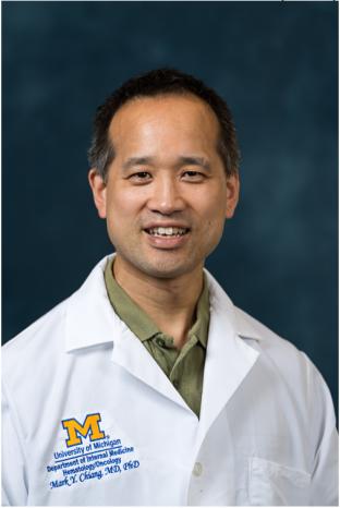 Mark Chiang合作论文数University of Michigan Cancer Center;Hematology/Oncology Division, Medical School, University of Michigan5
 Yatrik Shah合作论文数Department of Molecular & Integrative Physiology, Medical School, University of Michigan5Margaret Westfall合作论文数Department of Biomedical Engineering, College of Engineering, University of Michigan-Ann Arbor5
 Jason Spence合作论文数Department of Biomedical Engineering, University of Michigan;Department of Internal Medicine, University of Michigan;Department of Cell & Developmental Biology, Medical School, University of Michigan;The Spence Laboratory, University of Michigan5
 Patrick Tso合作论文数Department of Pathology, University of Cincinnati5
 Natacha Bohin合作论文数5
 Christian W Siebel合作论文数Dept Discovery Oncol, Genentech Inc5Horita Nobukatsu合作论文数5Elena M. Stoffel合作论文数5




