Aims:All-cause mortality ranges between 33% and 42% for individuals with untreated moderate to severe aortic stenosis (AS). Transcatheter aortic valve replacement makes this a treatable condition, if identified early. Machine learning-based tools show great promise to predict cardiovascular outcomes. Methods and results:We developed and validated a machine learning model for 3-year prediction of AS risk (ASrisk) using serum biomarkers and vital sign measurements. We then evaluated the tool's capacity to identify diagnoses of AS sequelae, echocardiographic outcomes in individuals not diagnosed with AS, as well as enrichment and 3-year aortic valve area reduction in individuals with high ASrisk. Among 919 954 participants, 429 996 were from the Mount Sinai Data Warehouse (MSDW) [2179 (0.5%) AS cases] and 489 958 were from the UK Biobank [5066 (1%) AS cases]. Odds ratio (OR) of AS sequelae increased quantitatively with ascending deciles of ASrisk [OR 1.63 (95% CI 1.60-1.67) in MSDW]. Increasing ASrisk by 1 SD resulted in higher odds of echocardiographic findings in undiagnosed individuals [OR 1.88 (95% CI 1.71-2.06) for Doppler velocity index, OR 2.50 (95% CI 2.36-2.64) for aortic valve area, and OR 2.61 (95% CI 1.89-2.71) for mean gradient]. Three years after risk assessment, individuals with ASrisk > 0.95 show an 11-fold enrichment for AS diagnosis in both cohorts and an average reduction in aortic valve area of 0.42 cm2. Conclusion:ASrisk can predict risk of AS 3 years ahead of diagnosis in the general population.
Objectives The study developed framework that leverages an open-source Large Language Model (LLM) to enable clinicians to ask plain-language questions about a patient's entire echocardiogram report history. This approach is intended to streamline the extraction of clinical insights from multiple echocardiogram reports, particularly in patients with complex cardiac diseases, thereby enhancing both patient care and research efficiency.Materials and Methods Data from over 10 years were collected, comprising echocardiogram reports from patients with more than 10 echocardiograms on file at the Mount Sinai Health System. These reports were converted into a single document per patient for analysis, broken down into snippets and relevant snippets were retrieved using text similarity measures. The LLaMA-2 70B model was employed for analyzing the text using a specially crafted prompt. The model's performance was evaluated against ground-truth answers created by faculty cardiologists.Results The study analyzed 432 reports from 37 patients for a total of 100 question-answer pairs. The LLM correctly answered 90% questions, with accuracies of 83% for temporality, 93% for severity assessment, 84% for intervention identification, and 100% for diagnosis retrieval. Errors mainly stemmed from the LLM's inherent limitations, such as misinterpreting numbers or hallucinations.Conclusion The study demonstrates the feasibility and effectiveness of using a local, open-source LLM for querying and interpreting echocardiogram report data. This approach offers a significant improvement over traditional keyword-based searches, enabling more contextually relevant and semantically accurate responses; in turn showing promise in enhancing clinical decision-making and research by facilitating more efficient access to complex patient data.
HomeCirculation: Cardiovascular ImagingVol. 16, No. 11Beware Before Mitral Balloon Valvuloplasty: Parachute Mitral Valve Can Mimic Rheumatic Mitral Stenosis No AccessCase ReportRequest AccessFull TextAboutView Full TextView PDFView EPUBSections ToolsAdd to favoritesDownload citationsTrack citationsPermissions ShareShare onFacebookTwitterLinked InMendeleyReddit Jump toNo AccessCase ReportRequest AccessFull TextBeware Before Mitral Balloon Valvuloplasty: Parachute Mitral Valve Can Mimic Rheumatic Mitral Stenosis Sarah Goldman, Hartzell Schaff, Kimberly Capustin, Annapoorna Kini, Samin Sharma, David Power, Waqas Malick, Jose Meller, Steve Liao, Lori Croft and Martin Goldman Sarah GoldmanSarah Goldman Correspondence to: Sarah Goldman, MD, Zucker School of Medicine at Hofstra Northwell, Department of Internal Medicine, Lenox Hill Hospital New York, NY 917-446-4860. Email E-mail Address: [email protected] https://orcid.org/0009-0009-1227-2074 Zucker School of Medicine at Hofstra Northwell, Department of Internal Medicine, Lenox Hill Hospital New York (S.G.). , Hartzell SchaffHartzell Schaff https://orcid.org/0000-0003-0994-027X Department of Cardiovascular Surgery, Mayo Clinic, Rochester, MN (H.S.). , Kimberly CapustinKimberly Capustin The Zena and Michael A. Wiener Cardiovascular Institute, Icahn School of Medicine at Mount Sinai, New York (K.C., A.K., S.S., D.P., W.M., J.M., S.L., L.C., M.G.). , Annapoorna KiniAnnapoorna Kini https://orcid.org/0000-0002-7189-3307 The Zena and Michael A. Wiener Cardiovascular Institute, Icahn School of Medicine at Mount Sinai, New York (K.C., A.K., S.S., D.P., W.M., J.M., S.L., L.C., M.G.). , Samin SharmaSamin Sharma https://orcid.org/0000-0002-1888-0793 The Zena and Michael A. Wiener Cardiovascular Institute, Icahn School of Medicine at Mount Sinai, New York (K.C., A.K., S.S., D.P., W.M., J.M., S.L., L.C., M.G.). , David PowerDavid Power https://orcid.org/0000-0002-5409-1943 The Zena and Michael A. Wiener Cardiovascular Institute, Icahn School of Medicine at Mount Sinai, New York (K.C., A.K., S.S., D.P., W.M., J.M., S.L., L.C., M.G.). , Waqas MalickWaqas Malick The Zena and Michael A. Wiener Cardiovascular Institute, Icahn School of Medicine at Mount Sinai, New York (K.C., A.K., S.S., D.P., W.M., J.M., S.L., L.C., M.G.). , Jose MellerJose Meller The Zena and Michael A. Wiener Cardiovascular Institute, Icahn School of Medicine at Mount Sinai, New York (K.C., A.K., S.S., D.P., W.M., J.M., S.L., L.C., M.G.). , Steve LiaoSteve Liao The Zena and Michael A. Wiener Cardiovascular Institute, Icahn School of Medicine at Mount Sinai, New York (K.C., A.K., S.S., D.P., W.M., J.M., S.L., L.C., M.G.). , Lori CroftLori Croft The Zena and Michael A. Wiener Cardiovascular Institute, Icahn School of Medicine at Mount Sinai, New York (K.C., A.K., S.S., D.P., W.M., J.M., S.L., L.C., M.G.). and Martin GoldmanMartin Goldman The Zena and Michael A. Wiener Cardiovascular Institute, Icahn School of Medicine at Mount Sinai, New York (K.C., A.K., S.S., D.P., W.M., J.M., S.L., L.C., M.G.). Originally published10 Oct 2023https://doi.org/10.1161/CIRCIMAGING.123.015483Circulation: Cardiovascular Imaging. 2023;16FootnotesFor Sources of Funding and Disclosures, see page 928.Correspondence to: Sarah Goldman, MD, Zucker School of Medicine at Hofstra Northwell, Department of Internal Medicine, Lenox Hill Hospital New York, NY 917-446-4860. Email sgoldman7@northwell.eduREFERENCES1. Hakim FA, Kendall CB, Alharthi M, Mancina JC, Tajik JA, Mookadam FSO. Parachute mitral valve in adults - a systematic overview.Echocardiogr. 2010; 27:581–586. doi: 10.1111/j.1540-8175.2009.01143.xCrossrefMedlineGoogle Scholar2. Suraci N, Goldman H, Baruqui D, Santana O. Parachute mitral valve.Ann Card Anesthesia. 2021; 24:75–76. doi: 10.4103/aca.ACA_82_19CrossrefMedlineGoogle Scholar3. Casavecchia G, Gravina M, Zicchino S, Capalbo S, Di Biase M, Brunetti ND. Parachute mitral valve assessed by cardiac magnetic resonance.Interv Med Appl Sci. 2019; 11:65–67. doi: 10.1556/1646.11.2019.03CrossrefMedlineGoogle Scholar4. Yuan SM. Parachute mitral valve: morphology and surgical management.Turk Gogus Kalp Damar Cerrahisi Derg. 2020; 28:219–226. doi: 10.5606/tgkdc.dergisi.2020.18041CrossrefMedlineGoogle Scholar Previous Back to top Next FiguresReferencesRelatedDetails November 2023Vol 16, Issue 11 Advertisement Article InformationMetrics © 2023 American Heart Association, Inc.https://doi.org/10.1161/CIRCIMAGING.123.015483PMID: 37814885 Originally publishedOctober 10, 2023 Keywordschildhoodechocardiogramfeverheart ratepalpitationsPDF download Advertisement SubjectsEchocardiographyMagnetic Resonance Imaging (MRI)Nuclear Cardiology and PETRheumatic Heart DiseaseValvular Heart Disease
Background: Right ventricular (RV) function is important in the evaluation of cardiac function, but its assessment using standard transthoracic echocardiography (TTE) remains challenging. Cardiac magnetic resonance imaging (CMR) is considered the gold standard. The American Society of Echocardiography recommends surrogate measures of RV function and RV ejection fraction (RVEF) by TTE, including fractional area change (FAC), free wall strain (FWS), and tricuspid annular planar systolic excursion (TAPSE), but they require technical expertise in acquisition and quantification.Methods: The aim of this study was to evaluate the sensitivity, specificity, and positive and negative predictive values of FAC, FWS, and TAPSE derived using a rapid, novel artificial intelligence (AI) software (LVivoRV) from a single-plane transthoracic echocardiographic apical four-chamber, RV-focused view without ultrasound enhancing agents for detecting abnormal RV function compared with CMR-derived RVEF. RV dysfunction was defined as RVEF < 50% and RVEF < 40% on CMR.Results: TTE and CMR were performed within a median of 10 days (interquartile range, 2-32 days) of each other in 225 consecutive patients without interval procedural or pharmacologic intervention. The sensitivity and negative predictive value to detect CMR-defined RV dysfunction when all three AI-derived parameters (FAC, FWS, and TAPSE) were abnormal were 91% and 96%, while those of expert physician reads were 91% and 97%. Specificity and positive predictive value were lower (50% and 32%) compared with expert physician-read echocardiograms (82% and 56%).Conclusions: AI-derived measurements of FAC, FWS, and TAPSE had excellent sensitivity and negative predictive value for ruling out significant RV dysfunction (CMR RVEF < 40%), comparable with that of expert physician readers, but lower specificity. Thus AI, using American Society of Echocardiography guidelines, may serve as a useful screening tool for rapid bedside assessment to exclude significant RV dysfunction.
Introduction: Patients (pts) with pulmonary embolism (PE) and right ventricular (RV) dysfunction have a worse prognosis. We previously validated a real-time artificial intelligence software (AI, LVivoRV®) which calculates RV fractional area changes (FAC), free wall strain (FWS), and tricuspid annular planar systolic excursion (TAPSE) from a single unedited, non-ultrasound agent-enhanced apical 4-chamber (A4C) TTE view. Hypothesis: AI-calculated TTE parameters of RV function will accurately identify pts with intermediate-high and high-risk PE that would otherwise have been identified by comprehensive physician assessment of all TTE parameters, physical exam and biomarkers (“physician assessment”). Methods: We retrospectively identified pts in whom both a TTE (60.7% with ultrasound contrast) and chest CTA were performed for PE evaluation (median 1 day between studies). Based on comprehensive physician assessments, pts were stratified by the 2019 ESC guidelines from low-risk to high-risk for PE mortality. The accuracy of AI-TTE thresholds for RV dysfunction (previously defined) to identify physician-assessed high-risk PE was examined. Results: Of the 107 pts, 66 (61.7%) had confirmed PE on CTA. By physician assessment, 28 of these 66 cases were classified as intermediate-high/high-risk PE. Across all AI-TTE parameters, the sensitivities and negative predictive values for intermediate-high/high-risk PE (n=28) ranged from 79-86% and 83-87% respectively (Table). In contrast, the specificities and positive predictive values ranged from 30-56% and 29-38%. Conclusions: A simple to use, fully automated, AI-based TTE assessment of RV dysfunction at the bedside identified ~85% of all cases that would otherwise have been identified as intermediate-high and high-risk PE by comprehensive physician assessment (although the false positive rate was high). Further studies are warranted to examine how best to integrate this AI into clinical care pathways.
ABSTRACT Importance SARS-CoV-2 infection directly causes severe acute respiratory illness, leading to systemic tissue hypoxia and ischemia including the heart. Myocardial cytopathy associated with hypoxic response has been largely overlooked in COVID-19 patients. Additionally, histology analysis and cardiac function of COVID-19 cases are often reported separately, rendering an incomplete understanding of COVID-19 cardiac symptoms. Objective To examine the relationship between myocardial cellular responses to hypoxic stress versus cardiac functional alterations within the same COVID-19 patients. Design, Setting, and Participants Cellular hypoxia Inducible Factor 1 alpha (HIF1α) expression was analyzed by immunohistochemistry using post-mortem COVID-19 heart and lung tissues with known cardiac echocardiography records from a total of 8 patients. Clinical echocardiography data were obtained from Mount Sinai Heart between March to December, 2020. All gender and age groups were considered as long as cardiac involvement meets the preserved (EF > 50%) or moderate to severe (EF < 45%) criteria with confirmed SARS-CoV-2 infection. Cell-type specific subcellular localization of HIF1α expression and nuclear stability was examined by immunohistochemistry and transmission electronic microscopy (TEM). Terminal deoxynucleotidyl transferase dUTP nick end labeling (TUNEL) was used to quantify apoptosis. Main Outcomes and Measures No planned outcomes of this study as this is a retrospective analysis based on post-mortem specimens exclusively. Results Cardiac HIF1α expression was found to be significantly higher in patients with preserved EF levels than it was in the low EF group. In the preserved EF group, HIF1α is protective against apoptosis predominantly in endothelial cells and cardiac fibroblasts. In the low EF group, HIF1α protects cardiomyocyte nuclear integrity as evident by its nuclear accumulation with nuclear envelope preservation. Conclusions and Relevance This study establishes a direct link of cardiac cellular responses to hypoxic stress with matching functional and histological data, serving as one of the first studies to bridge previous stand-alone clinical data and cellular data. The protective role of HIF1α in hearts may help predict cardiac involvement in not only COVID-19 patients, but also decipher the underlying mechanisms in other forms of viral cardiomyopathy. KEY POINTS Question Are hypoxic signaling pathways associated with cardiac functional alterations in COVID-19 patients? Findings Cardiac HIF1α expression of COVID-19 patients with EF>50% or EF<45% was analyzed and quantified. Increased cardiac HIF1α + cells were found in patients with higher EF. HIF1α + endothelial cells are resistant to apoptosis, and HIF1α + cardiomyocytes are able to retain nuclear envelope under hypoxic stress. Meaning HIF1α is cardioprotective in hearts of COVID-19 patients.
Objectives Obstructive sleep apnoea (OSA) is often linked to cardiovascular disease. A limited number of studies have reported an association between OSA and left ventricular diastolic dysfunction (LVDD). However, prior studies were performed on small patient populations. Studies have shown a high prevalence of OSA among first responders to the 9/11 World Trade Center (WTC) terrorist attack. We investigated the relationship between OSA and LVDD in a large population of WTC responders. Design Cross-sectional study. Setting One-time screening programme as part of the WTC-CHEST Study (NCT10466218), performed at a quaternary medical centre in New York City, from November 2011 to June 2014. Participants A total of 1007 participants with mean age of 51 years of mostly non-Hispanic white men were evaluated. Patients from the WTC Health Program-Clinical Center of Excellence, who were over the age of 39 years, were eligible to participate. Results Evaluation of those without OSA diagnosis showed no significant association with LVDD when comparing those screened (Berlin Questionnaire) as OSA high risk versus OSA low risk (p=0.101). Among those diagnosed with LVDD, there was a significant association when comparing those with and without patient-reported OSA (OR 1.50, 95% CI 1.13 to 2.00, p=0.005), but the significance was not maintained after adjusting for pertinent variables (OR 1.3, 0.94 to 1.75, p=0.119). Notably, comparing those with OSA diagnosis and those low risk of OSA, the OR for LVDD was significant (1.69, 1.24 to 2.31, p=0.001), and after adjusting for waist-hip ratio, diabetes and coronary artery calcium score percentile, the relationship remained significant (OR 1.45, 1.03 to 2.04, p=0.032). Conclusion The strong association of OSA with LVDD in this population may inform future guidelines to recommend screening for LVDD in high-risk asymptomatic patients with OSA.
Background: There are currently no clear guidelines regarding the use of ultrasound enhancing agents (UEAs) with transthoracic echocardiography (TTE) for patients hospitalized with Covid-19. We investigated whether the performance of TTE with UEAs provides more diagnostic information and allows for shorter acquisition time compared to unenhanced TTE imaging in this patient population. Methods: We analyzed the TTEs of 107 hospitalized Covid-19 patients between April and June 2020 who were administered UEAs (Definity (R), Lantheus). The time to acquire images with and without UEAs was calculated. A level III echocardiographer determined if new, clinically significant findings were visualized with the addition of UEAs. Results: There was a mean of 11.84 +/- 3.59 UEA cineloops/study vs 20.74 +/- 8.10 non-UEA cineloops/study (p < 0.0001). Mean time to acquire UEA cineloop images was 72.28 +/- 28.18 s/study compared to 188.07 +/- 86.04 s/ study for non-UEA cineloop images (p < 0.0001). Forty-eight patients (45%) had at least one new finding on UEA imaging, with a total of 62 new findings seen. New information gained with UEAs was more likely to be found in patients with acute respiratory distress syndrome (21 vs 9, p < 0.001) and in those on mechanical ventilation (21 vs 15, p = 0.046). Conclusions: TTE with UEAs required less time and fewer cineloop images compared to non-UEA imaging in patients hospitalized with Covid-19. Additionally, Covid-19 patients with severe respiratory disease benefited most with regard to new diagnostic information. Health care personnel should consider early use of UEAs in select hospitalized Covid-19 patients in order to reduce exposure and optimize diagnostic yield.
BACKGROUND:Quantification of left ventricular ejection fraction (LVEF) by transthoracic echocardiography (TTE) is operator-dependent, time-consuming, and error-prone. LVivoEF by DIA is a new artificial intelligence (AI) software, which displays the tracking of endocardial borders and rapidly quantifies LVEF. We sought to assess the accuracy of LVivoEF compared to cardiac magnetic resonance imaging (cMRI) as the reference standard and to compare LVivoEF to the standard-of-care physician-measured LVEF (MD-EF) including studies with ultrasound enhancing agents (UEAs).METHODS:In 273 consecutive patients, we compared MD-EF and AI-derived LVEF to cMRI. AI-derived LVEF was obtained from a non-UEA four-chamber view without manual correction. Thirty-one patients were excluded: 25 had interval interventions or incomplete TTE or cMRI studies and six had uninterpretable non-UEA apical views.RESULTS:In the 242 subjects, the correlation between AI and cMRI was r = .890, similar to MD-EF and cMRI with r = .891 (p = 0.48). Of the 126 studies performed with UEAs, the correlation of AI using the unenhanced four-chamber view was r = .89, similar to MD-EF with r = .90. In the 116 unenhanced studies, AI correlation was r = .87, similar to MD-EF with r = .84. From Bland-Altman analysis, LVivoEF underreported the LVEF with a bias of 3.63 ± 7.40% EF points compared to cMRI while MD-EF to cMRI had a bias of .33 ± 7.52% (p = 0.80).CONCLUSIONS:Compared to cMRI, LVivoEF can accurately quantify LVEF from a standard apical four-chamber view without manual correction. Thus, LVivoEF has the ability to improve and expedite LVEF quantification.
BackgroundCOVID-19 has been associated with an increased incidence of ischemic stroke. The use echocardiography to characterize the risk of ischemic stroke in patients hospitalized with COVID-19 has not been explored.MethodsWe conducted a retrospective study of 368 patients hospitalized between 3/1/2020 and 5/31/2020 who had laboratory-confirmed infection with SARS-CoV-2 and underwent transthoracic echocardiography during hospitalization. Patients were categorized according to the presence of ischemic stroke on cerebrovascular imaging following echocardiography. Ischemic stroke was identified in 49 patients (13.3%). We characterized the risk of ischemic stroke using a novel composite risk score of clinical and echocardiographic variables: age <55, systolic blood pressure >140 mmHg, anticoagulation prior to admission, left atrial dilation and left ventricular thrombus.ResultsPatients with ischemic stroke had no difference in biomarkers of inflammation and hypercoagulability compared to those without ischemic stroke. Patients with ischemic stroke had significantly more left atrial dilation and left ventricular thrombus (48.3% vs 27.9%, p = 0.04; 4.2% vs 0.7%, p = 0.03). The unadjusted odds ratio of the composite novel COVID-19 Ischemic Stroke Risk Score for the likelihood of ischemic stroke was 4.1 (95% confidence interval 1.4-16.1). The AUC for the risk score was 0.70.ConclusionsThe COVID-19 Ischemic Stroke Risk Score utilizes clinical and echocardiographic parameters to robustly estimate the risk of ischemic stroke in patients hospitalized with COVID-19 and supports the use of echocardiography to characterize the risk of ischemic stroke in patients hospitalized with COVID-19.
Background Atrial myxomas are the most common benign cardiac tumours. This case highlights an unusual presentation and complex management of a patient who was incidentally found to have a left atrial tumour concerning for a myxoma. Case summary A 54-year-old-woman presented with symptoms of nausea and vomiting and was found to have a left atrial mass incidentally in addition to a renal infarct. She was also found to have COVID-19 and the mass was initially thought to be a thrombus. With the help of multimodality imaging, it was determined that the mass was an atrial myxoma and she was started on short-term anticoagulation to prevent recurrent embolization. After 6 weeks of anticoagulation, she successfully underwent elective resection of the mass which was confirmed to be myxoma with superimposed thrombus. Discussion It is difficult to differentiate cardiac tumours from intracardiac thrombus and multimodality cardiac imaging is crucial to make an accurate diagnosis. While the treatment of atrial myxomas involves early surgical resection, it becomes more complicated with concurrent COVID-19 infection.
Introduction: Cardiac MRI (CMR) is the gold standard for right ventricular function (RVF) because echo assessment is limited. Potential echo parameters to assess RVF include RV fractional area change (FAC), RV free wall strain (FWS), and tricuspid annular plane systolic excursion (TAPSE) on apical 4-chamber (A4C) view. We compared a new Artificial Intelligence method that tracks the RV almost instantaneously in a single 4-chamber view (AI, LVivo RV®, DiA Imaging, Figure) to quantify RVF vs CMR. Methods: We compared AI RVF against CMR in 125 pts. Abnormal LVEF and RVEF were defined as <57% and <49% respectively. Echo closest to CMR date was analyzed with AI RV to obtain FAC, FWS, and TAPSE. We defined abnormal RVF by DiA Imaging’s predetermined thresholds. Sensitivities and specificities for abnormal RVF and chi-square (χ 2 ) tests were calculated for each variable against CMR RVEF. Results: Of the 125 pts, 55 (44%) were female with median age 55 [Q1 44, Q3 67] years. Thirty pts (24%) had abnormal RVEF and 78 (62.4%) had abnormal LVEF by CMR. All pts with abnormal RVEF had abnormal LVEF. Compared to CMR, AI RV sensitivities and specificities for abnormal RVEF were: FAC 87% and 60%, FWS 80% and 61%, TAPSE 77% and 54%, any 2 criteria 83% and 61%, and all 3 criteria 63% and 69%. AI RV χ 2 values were: FAC 19.9 (p<0.001), FWS 15.4 (p<0.001), TAPSE 8.4 (p=0.004), any 2 criteria 18.0 (p<0.001), and all 3 criteria 10.4 (p=0.001). Conclusions: This is the first validation of a novel AI method (LVivo RV®) that can detect RV dysfunction using 3 standard RV measurements from a single A4C view with good sensitivity and specificity compared to volumetric CMR as the gold standard.
COVID-19 affects multiple organs. Clinical data from the Mount Sinai Health System show that substantial numbers of COVID-19 patients without prior heart disease develop cardiac dysfunction. How COVID-19 patients develop cardiac disease is not known. We integrated cell biological and physiological analyses of human cardiomyocytes differentiated from human induced pluripotent stem cells (hiPSCs) infected with severe acute respiratory syndrome coronavirus 2 (SARS-CoV-2) in the presence of interleukins (ILs) with clinical findings related to laboratory values in COVID-19 patients to identify plausible mechanisms of cardiac disease in COVID-19 patients. We infected hiPSC-derived cardiomyocytes from healthy human subjects with SARS-CoV-2 in the absence and presence of IL-6 and IL-1β. Infection resulted in increased numbers of multinucleated cells. Interleukin treatment and infection resulted in disorganization of myofibrils, extracellular release of troponin I, and reduced and erratic beating. Infection resulted in decreased expression of mRNA encoding key proteins of the cardiomyocyte contractile apparatus. Although interleukins did not increase the extent of infection, they increased the contractile dysfunction associated with viral infection of cardiomyocytes, resulting in cessation of beating. Clinical data from hospitalized patients from the Mount Sinai Health System show that a significant portion of COVID-19 patients without history of heart disease have elevated troponin and interleukin levels. A substantial subset of these patients showed reduced left ventricular function by echocardiography. Our laboratory observations, combined with the clinical data, indicate that direct effects on cardiomyocytes by interleukins and SARS-CoV-2 infection might underlie heart disease in COVID-19 patients. IMPORTANCE SARS-CoV-2 infects multiple organs, including the heart. Analyses of hospitalized patients show that a substantial number without prior indication of heart disease or comorbidities show significant injury to heart tissue, assessed by increased levels of troponin in blood. We studied the cell biological and physiological effects of virus infection of healthy human iPSC-derived cardiomyocytes in culture. Virus infection with interleukins disorganizes myofibrils, increases cell size and the numbers of multinucleated cells, and suppresses the expression of proteins of the contractile apparatus. Viral infection of cardiomyocytes in culture triggers release of troponin similar to elevation in levels of COVID-19 patients with heart disease. Viral infection in the presence of interleukins slows down and desynchronizes the beating of cardiomyocytes in culture. The cell-level physiological changes are similar to decreases in left ventricular ejection seen in imaging of patients' hearts. These observations suggest that direct injury to heart tissue by virus can be one underlying cause of heart disease in COVID-19.
 Martin E. Goldman合作论文数36Milena J. Henzlova合作论文数23
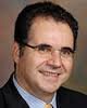 Stamatios Lerakis合作论文数Department of Medicine, School of Medicine, Emory University21Duvall W Lane合作论文数18
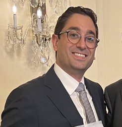 Solomon Bienstock合作论文数Icahn School of Medicine at Mount Sinai, Zena and Michael A. Wiener Cardiovascular Institute13
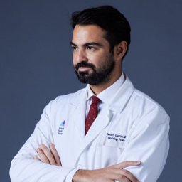 Giustino Gennaro合作论文数Interventional Cardiology Unit, San Raffaele Scientific Hospital13
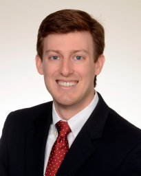 Connor Patrick Oates合作论文数Icahn School of Medicine at Mount Sinai, Zena and Michael A. Wiener Cardiovascular Institute10Ashton Lai合作论文数Icahn School of Medicine at Mount Sinai, Zena and Michael A. Wiener Cardiovascular Institute9Annapoorna Kini合作论文数8Eric Stern合作论文数8Steve Liao合作论文数8Rajeev Samtani合作论文数8
 Vivek Reddy合作论文数Icahn School of Medicine at Mount Sinai8
 Samin K. Sharma合作论文数Cardiovascular Clinical Institute, Mount Sinai Hospital;Mount Sinai Heart Network, Mount Sinai Hospital;Eternal Heart Care Center, Eternal Hospital;Department of Cardiology, Eternal Hospital8Samantha Buckley合作论文数8Nina Kukar合作论文数7Guma Krista A合作论文数7George Titus合作论文数6
 Usman Baber合作论文数5Elliot J. Levine合作论文数5Supreet Singh合作论文数5
 Gina Larocca合作论文数5William L. Duvall合作论文数5Frans Beerkens合作论文数Icahn Sch Med Mt Sinai, Dept Med, New York, NY 10017 USA5
 Karishma Rahman合作论文数Icahn Sch Med Mt Sinai, Cardiovasc Res Inst5Argulian Edgar合作论文数College of Physicians &, Columbia University;Surgeons, New York, NY 10025, USA. eargulian@chpnet.org;St. Luke's-Roosevelt Hospital Center, Columbia University College of Physicians & Surgeons5
 Tatyana A Danilov合作论文数5Cynara Maceda合作论文数5
 Anton Camaj合作论文数5Marco Vicenzi合作论文数4Nada Shaban合作论文数4eric ginsberg合作论文数4Javier Sanz合作论文数4Nikola Kocovic合作论文数4
 Gregg Stone合作论文数The Zena and Michael A. Wiener Cardiovascular Institute, Icahn School of Medicine at Mount Sinai4Simonette T. Sawit合作论文数4
 Rupa Iyengar合作论文数4
 Mary McLaughlin合作论文数4Andrew J. Einstein合作论文数4
 Luke Hermann合作论文数4
 Jeffrey Silbiger合作论文数4
 Giulio Stefanini合作论文数Humanitas University4Renato Bragato合作论文数4Joseph Sweeny合作论文数4
 Richard Ro合作论文数4Brian C. Hsia合作论文数3Gregory W Serrao合作论文数3Waqas Malick合作论文数3Irene Rota合作论文数3




