7032 Background: The impact of immune cell counts pre LD and during immune reconstitution on CAR T therapy outcomes is poorly understood. We investigated the association between CD4, CD8 and NK cell counts and CAR T therapy outcomes in patients with LBCL. Methods: Retrospective study of R/R LBCL patients who received CAR T cells between 2016-24 at Mayo Clinic, Rochester, were included in this analysis. Peripheral blood CD4, CD8 and NK counts were measured pre LD and at M1 post CAR T infusion. The receiver operating characteristic (ROC) curve was used to determine the optimal cutoff for pre LD and M1 immune cells to predict patients who were alive and in remission at 6 months (M6) post CAR T infusion. Patients who progressed or were lost to follow up prior to day 30 were excluded from M1 ROC analysis. Results: Of 140 patients, 81 were alive and in remission at M6 (Group A), while 59 had relapse or death (Group B). Axicabtagene ciloleucel was the CAR T product given in 81% of patients (114/140). Median pre LD CD4 counts were significantly lower in Group B compared to group A (143 vs 280 cells/µL, p = 0.001). No significant difference was observed in median pre LD CD8 counts (205 vs 221 cells/µL, p = 0.8). ROC analysis identified an optimal pre LD CD4 count of 124.5 cells/µL for M6 alive+remission. Lower pre LD CD4 counts (<124.5) predicted worse progression free survival (PFS) in univariate (HR = 3.02, 95% confidence interval [CI]: 1.73–5.27, p< 0.01) and multivariable analysis (MVA) adjusted for IPI and the number of prior lines (aHR = 2.54, 95% CI: 1.39–4.62, p< 0.01).Lower pre LD CD4 counts also predicted inferior overall survival (OS) in MVA (aHR = 2.27, 95% CI: 1.08–4.77, p = 0.03). (Table 1) On day 30 landmark analysis, ROC identified M1 optimal CD4 count of ≥ 99.5 cells/µL to be associated with a trend toward superior PFS (P=0.09), but not OS (p=0.90) in MVA. Median pre LD NK cell counts were significantly lower in Group B (73 vs 98 cells/µL, p = 0.04). ROC analysis identified an optimal pre LD NK count of 151 cells/µL for M6 alive + remission. Lower pre LD NK counts (<151) predicted worse PFS, both on univariate (HR = 4.17, 95% CI: 1.29–13.47, p = 0.02) and MVA (aHR = 4.64, p< 0.01). The lower pre LD NK cell count group had worse OS in MVA (aHR = 5.69, 95% CI: 1.16–28.1, p = 0.01). (Table 1) On D30 landmark analysis, M1 NK cell count of ≥ 128.5 cells/µL was associated with a trend toward superior PFS (P=0.08), but not OS (p=0.30) in MVA. Conclusions: Pre LD CD4 and NK cell counts are significantly associated with PFS and OS post CAR T therapy in patients with R/R LBCL. Pre-treatment immune subset levels may identify patients at higher risk of relapse or death after CAR-T therapy. Groups Median PFS 2 Year PFS Median OS 2 Year OS CD4 ≥124.5 cells/µL (N=75) NR 58% 3.31 years 67% CD4 <124.5 cells/µL (N=30) 2.2 months 22% 1.59 years 45% NK ≥151 cells/µL (N=17) NR 77% NR 84% NK <151 cells/µL (N=82) 10.6 months 44% 3.31 years 62%
Abstract: Primary central nervous system lymphoma (PCNSL) is a rare form of non-Hodgkin lymphoma involving the brain, cerebrospinal fluid, or retina/vitreous without systemic involvement. Induction with high-dose methotrexate (HD-MTX) followed by consolidation with autologous stem cell transplant (auto-SCT) has become the standard treatment paradigm for most patients. However, limited data are available regarding the efficacy of a maintenance approach with HD-MTX. Herein, we retrospectively reviewed the characteristics and outcomes of 148 patients diagnosed with PCNSL between October 2010 and June 2022, who underwent HD-MTX–based induction therapy followed by either auto-SCT consolidation (n = 70) or HD-MTX maintenance therapy (n = 37). At a median follow-up time of 4.5 years, the progression-free survival (PFS) was 8.3 years and the overall survival (OS) was not reached. Compared to patients who underwent auto-SCT, patients who received maintenance HD-MTX had a higher median age at diagnosis of 72 vs 62 years and a trend toward higher proportion of patients being Eastern Cooperative Oncology Group 2 or higher (41% vs 29%). At 5-years postinduction treatment initiation, the PFS rates in the auto-SCT cohort and HD-MTX maintenance cohort were 74.6% and 72.6%, respectively, and the OS rates were 76.0% and 82.4%, respectively. Overall, there was no significant difference in PFS or OS based on postinduction management strategy. Our data suggest that maintenance HD-MTX may be a reasonable, time-limited treatment strategy for patients with PCNSL responding to initial induction therapy.
e18508 Background: BPDCN is a rare and aggressive hematologic malignancy. Despite the advent of targeted therapies, median overall survival (mOS) remains <1 year (y) in the absence of stem cell transplant (SCT). This study evaluates clinical and molecular predictors of outcomes in a large cohort of BPDCN patients at a tertiary center. Methods: BPDCN patients seen at Mayo Clinic (AZ, FL, MN) between 2000-2024 were identified retrospectively. Analyses considered parameters at diagnosis. Next generation sequencing (NGS) was performed using a 47-gene assay. Standard statistical methods were used; calculations used R. Results: Among 69 patients, the median age was 70y (range 19-94) with male predominance (83%). At presentation, skin (80%), bone marrow (73%), lymph nodes (52%), spleen (27%), and CNS (12%) were involved; 26% had a concurrent hematologic malignancy. Among 20 cases with NGS, the median number of mutations was 3 (range 0-5), most frequently TET2 (n=16), ASXL1 (n=13), SRSF2 (n=7), and NRAS (n=5). Frontline therapy (Table) yielded a complete response (CR) rate of 63% and overall response rate of 76%. CR rates were similar among regimens, but patients who received an ALL (hyper-CVAD based) regimen (8/13) or tagraxofusp (6/9) were more likely to be bridged to SCT (6/29, p<0.01). After median follow up of 38 months (mo), mOS for all was 15 (95% CI 12-21) mo; 44 (64%) were deceased at last follow up. Among 62 evaluable patients, 20 (32%, median age 60 vs 74y, p<0.01) underwent SCT which improved survival (mOS 51 vs 10 mo, 3y OS 52% vs 5%, p<0.01); mOS was not different between allogeneic (n=17) and autologous (n=3) SCT (54 vs 38 mo, p=0.45). When censored for SCT, the 1y OS for patients receiving an ALL regimen, tagraxofusp, or other regimen was 82%, 67%, and 47%, respectively (p=0.15). In univariate analysis, age ≥50y, lymph node involvement, bone marrow involvement, hemoglobin <10 g/dL, leukocytes (WBC) >11 x10 9 /L, abnormal karyotype, and HCT were significantly associated with OS. In multivariate analysis, WBC >11 x10 9 /L (HR 3.7, p<0.03), abnormal karyotype (HR 5.4, p<0.01), and SCT (HR 0.23, p<0.01) remained significant. Separately, SRSF2 mutation was associated with inferior OS (15 vs 51 mo, p=0.02), but significance was lost when adjusted for age, abnormal karyotype, and SCT. Conclusions: Newly-diagnosed BPDCN patients treated with an ALL regimen or tagraxofusp followed by SCT had longer OS compared to those treated with all other regimens. WBC >11 x10 9 /L and abnormal karyotype were independent predictors of inferior survival whereas SCT was beneficial. Regimen N Age (y) ANNOVA CR Chi Sq HCT Chi Sq 1y OS mOS (mo) Log-rank Tagraxofusp 9 68 (40-70) <0.001 7 (78%) 0.548 6 (67%) 0.018 88% 20.8 0.121 ALL 13 43 (20-72) 8 (67%) 8 (62%) 74% 50.7 Hyper-CVAD 11 43 (20-72) 6 (60%) 6 (60%) 67% 35.8 AML 18 70 (48-80) 13 (76%) 5 (28%) 58% 14.2 Intensive 8 66 (48-77) 7 (88%) 3 (38%) 57% 12.4 HMA+Venetoclax 8 69 (65-80) 6 (75%) 2 (25%) 50% 14.0 Lymphoma 8 74 (49-85) 3 (43%) 1 (13%) 50% 14.0 CHOP 6 74 (49-85) 3 (50%) 1 (17%) 50% 17.7 Other 3 66 (63-73) 1 (33%) 0 (0%) 33% 12.0
Introduction: Our group previously published that the infusion autograft inhibitor KIR2DL2 (CD158b+CD337-) and activating NKp30 (CD158b- CD337+) Natural killer (NK) cells receptors were predictors of clinical outcomes in lymphoma patients undergoing autologous peripheral blood hematopoietic stem cell transplantation (APBHSCT) (Porrata et al. Leukemia Research 2019; 81: 1-9). To assess if these subsets of NK cells still hold clinical relevance, we set up to investigate their prognostic ability to predict clinical outcomes at Day 100 post-APBHSCT. Methods: 107 lymphoma patients who participated in our double-blind phase III trail (Porrata et al, Biology of Blood and Marrow Transplantation 2016; 22(6); 1017-1023) and had clinical assessment at Day 100 post-APBHSCT were included in this study. Nkp30 and KIR2DL2 at Day 100 were assessed by flow cytometry analysis. Results: With a median follow-up for the entire cohort from Day 100 was 94.7 months (range: 4.83-158.1 months). Figure 1 shows the overall survival (OS) and progression-free survival (PFS) based on the absolute numbers of KIR2DL2 and Nkp30 NK cells. At Day 100, patients with a KIR2DL2 < 0.08 and Nkp30 ≥0.19 experienced superior OS and PFS compared with patients with a Day 100 KIR2DLD2 ≥0.08 and NKp30 < 0.19. In the multivariate analysis both the KIR2DL2 [OS: HR = 1.767, 95%CI, 1.302-30.31, p < 0.04; and PFS: HR = 2.663, 95%CI, 1.108-6.403, p < 0.03] and Nkp30 [OS: HR = 2.594, 95% CI, 1.239-5.432, p < 0.01; and PFS: HR = 3.676, 95% CI, 1.875-7.206, p < 0.01] were independent predictors for OS and PFS. Conclusion: Day 100 inhibitory KIR2DL2 and activating NKp30 NK cells are prognostic immune-biomarkers in lymphoma patients undergoing APBHSCT.
Chimeric antigen receptor T-cell therapy is the new standard of care in fit patients with refractory or early relapsed diffuse large B-cell lymphoma (DLBCL). However, there may still be a role for salvage chemotherapy (ST) and autologous stem cell transplant (ASCT) in certain circumstances (e.g., lack of resources for chimeric antigen receptor T-cell therapy, chemosensitive relapses). We retrospectively studied 230 patients with refractory or early relapsed DLBCL who underwent ST and ASCT. The median line of ST was one (range, 1-3). Best response before ASCT was complete response in 106 (46%) and partial response in 124 (54%) patients. The median follow-up after ASCT was 89.4 months. The median progression-free (PFS) and overall survival (OS) were 16.1 and 43.3 months, respectively. Patients relapsing between 6 to 12 months after frontline therapy had a numerically better median PFS (29.6 months) and OS (88.5 months). Patients who required one line of ST, compared to those requiring more than one line, had a better median PFS (37.9 vs. 3.9 months; P=0.0005) and OS (68.3 vs. 12.0 months; P=0.0005). Patients who achieved complete response had a better median PFS (71.1 vs. 6.3 months; P<0.0001) and OS (110.3 vs. 18.9 months; P<0.0001) than those in partial response. Patients who achieved complete response after one line of ST had the most favorable median PFS (88.5 months) and OS (117.2 months). Post-ASCT survival outcomes of patients with refractory or early relapsed DLBCL appeared reasonable and were particularly favorable in those who required only one line of ST to achieve complete response before ASCT, highlighting the role of this procedure in select patients with chemosensitive disease.
INTRODUCTION:The infusion of autograft Natural Killer Cells (NKC)/CD14+ HLA-DRDIM ratio is a predictor of survival in lymphoma patients undergoing autologous peripheral blood hematopoietic stem cell transplantation (APBHSCT). This study evaluated if the Day 100 NKC/CD14+ HLA-DRDIM ratio still functions as a prognostic immune-biomarker.METHODS:This was a retrospective, single-institution, cohort analysis including 107 patients in this study that had clinical assessment at Day 100 post-APBHSCT from our prior phase III trial. We evaluated the prognostic ability of the Day 100 NKC/CD14+ HLA-DRDIM ratio to predict overall survival (OS) and progression-free survival (PFS) using Cox regression model for outcome analysis and survival by Kaplan-Meier method.RESULTS:The median follow-up from day 100 was 94.7 months (range 4.83-158.1 months) for the entire cohort. Patients with a Day 100 NKC/CD14+ HLA-DRDIM ratio ≥1.67 experienced better OS and PFS versus those with a Day 100 NKC/CD14+ HLA-DRDIM ratio <1.67: median OS was not reached versus 49.7 months, the 5-year OS rates were 91% (95% CI, 81%-96%) versus 40% (95% CI, 27%-55%), p < .0001, respectively; and median PFS was not reached versus 23.5 months, the 5-year PFS rates were 66% (95% CI, 55%-81%) versus 21% (95% CI, 15%-40%), p < .0001, respectively. Day 100 NKC/CD14+ HLA-DRDIM ratio was an independent predictor for OS and PFS in the multivariate analysis.CONCLUSIONS:Day 100 NKC/CD14+ HLA-DRDIM ratio is a prognostic immune-biomarker in lymphoma patients post- APBHSCT.
Background: In the ZUMA2 study, a higher peak chimeric antigen receptor (CAR) T cell expansion correlated with better outcomes in patients with relapsed/refractory (R/R) mantle cell lymphoma (MCL) receiving brexucabtagene autoleucel (brexu-cel). However, in clinical practice, peak CAR T cell expansion is not routinely assessed for. Since patients receive lymphodepleting agents prior to CAR T cell infusion, it is hypothesized that peak absolute lymphocyte count (ALC), which can be easily measured, in the first two weeks after CAR T cell infusion represents peak CAR T cell expansion. Our group previously showed that peak ALC was associated with response durability and outcomes in patients with R/R large B-cell lymphoma receiving axicabtagene ciloleucel. In this retrospective study, we assessed the association of peak ALC with clinical outcomes after brexu-cel infusion in patients with R/R MCL. Methods: Patients with R/R MCL who received brexu-cel between March 2020-March 2024 at Mayo Clinic were included in this analysis. Peak ALC was defined as maximum ALC value in the first 14 days after CAR T cell infusion. Progression-free survival (PFS) was defined as time from CAR T cell infusion to lymphoma relapse, progression, or death, and overall survival (OS) was defined as time from to CAR T cell infusion to death. The optimal cutoff point for peak ALC that correlated with PFS was determined by the log-rank method using the Cutoff Finder application, a computational method which fits Cox proportional hazard models to the dichotomized variable and survival. The optimal cutoff was the point with the most significant PFS split by log-rank test. Results: A total of 43 patients were identified. The median age at time of CAR T cell infusion was 68 years (IQR 58, 73). 34 (79%) were male, 28 (74%) had Ki-67 >30%, 14 (33%) had blastoid or pleomorphic histology, and 19 (44%) had TP53 mutation or deletion. Median number of prior lines of therapy was 3 (IQR 2, 4) and 13 (30%) received prior autologous stem cell transplant. 32 (74%) received bridging therapy prior to CAR T cell infusion. All patients received fludarabine plus cyclophosphamide for lymphodepletion. The best overall response rate was 88%, with a complete response rate of 84%. After a median follow-up of 25.6 months (95% CI 14.4-31.4), the median PFS and OS were 16.6 (95% CI 8.6-NE) and 35.1 (95% CI 24.5-NE) months, respectively. The incidence of cytokine release syndrome (CRS) was 95% (4.7% grade ≥3). The median time to initial CRS and maximum grade CRS onset was 4.5 days (IQR 1, 6.25) and 5.5 days (IQR 2.75, 7), respectively. The incidence of immune effector cell-associated neurotoxicity syndrome (ICANS) was 65% (18% grade ≥ 3). The median time to initial and maximum grade ICANS onset was 9 days (IQR 6, 11) and 10.5 days (IQR 7, 12), respectively. The median ALC at time of CAR T cell infusion was 0.03 x10⁹/L (IQR 0.02, 0.05). Among 37 patients with identifiable ALC peaks, the median ALC peak was 0.84 (IQR 0.41, 2.45) and the median time to peak ALC was 12 days (IQR 9, 12). 6 patients had no identifiable peak ALC due to no frequent CBC differentials post CAR T cell infusion. A peak ALC of ≥0.45 × 10⁹/L correlated significantly with PFS, with median PFS not reached for the group with peak ALC of ≥0.45 × 10⁹/L (n=27) compared to 9.3 months for the group with peak ALC of <0.45 × 10⁹/L (n=10) and a 1-year PFS rate of 68% vs 33% (p=0.02). OS was also significantly higher in patients with a peak ALC of ≥0.45 × 10⁹/L (median OS not reached vs 11 months, 1-year OS rate of 84% vs 44%, p=0.01). The incidence of ICANS was significantly higher (74% vs 30%, p=0.02) in patients with a peak ALC of ≥0.45 × 10⁹/L. Numerically, the group with peak ALC of ≥0.45 × 10⁹/L had a later onset of initial and maximum grade ICANS, but the difference was not statistically significant. Peak ALC was not associated with ICANS severity. There was a trend towards higher incidence of CRS (100% vs 80%, p=0.07) and earlier CRS onset (3 vs 5 days, p=0.07) in the high peak ALC group. There was no difference in CRS severity. Conclusion: Peak ALC after CAR T cell infusion was significantly associated with better PFS and OS and higher incidence of ICANS in patients with R/R MCL receiving brexu-cel. Peak ALC may serve as a surrogate measure for peak CAR T cell expansion in routine practice, but confirmatory studies are needed. Larger studies are warranted to confirm the association of peak ALC with PFS, OS and toxicities in patients with R/R MCL receiving brexu-cel.
The infusion autograft absolute number of inhibitory killer immunoglobulin-like receptor (KIR) 2DL2 and activating natural killer (NK)p30 cells are predictors of clinical outcomes in lymphoma patients undergoing autologous peripheral blood hematopoietic stem cell transplantation (APBHSCT). To assess if the long-term recovery of these NK cell subsets still holds clinical relevance, we set up to investigate their prognostic ability at day 100 post-APBHSCT. This was a retrospective single-institution study including 107 patients from our prior phase III trial who had a clinical assessment at day 100 post-APBHSCT. The median follow-up from day 100 was 168.19 months (interquartile range: 156.85-181.28 months). Patients with day 100 inhibitory KIR2DL2 < 0.08 cells/µL and activating NKp30 ≥ 0.19 cells/µL experienced superior overall survival (OS) and progression-free survival (PFS). A multivariate analysis revealed both the day 100 inhibitory KIR2DL2 [OS: HR = 1.449, 95%CI, 1.231-1.895,
Immune checkpoint inhibitors (ICIs) and brentuximab vedotin (BV) are novel agents for classic Hodgkin lymphoma, including relapse after autologous stem cell transplant (ASCT). However, their impact on survival post-ASCT relapse, in comparison with conventional therapy, is less known due to the lack of randomized controlled trials. Clinical characteristics and outcomes of 115 patients with relapse (or progression) after ASCT are studied. After a median follow-up of 8.59 years from post-ASCT relapse, the median progression-free survival (PFS) and overall survival (OS) were 0.91 and 5.07 years, respectively. Median lines of therapy after post-ASCT relapse was 2 (range, 1–12). The median PFS was not reached (NR) versus 1.11 versus 0.50 versus 0.85 versus 0.78 years ( P = 0.006) and OS was NR versus 7.60 versus 3.08 versus 3.51 versus 3.17 years ( P = 0.28) in patients first treated with ICIs versus BV versus investigational agents versus chemotherapy versus radiation therapy (RT). First-line treatment with novel agents (ie, ICIs and BV) was associated with superior outcomes compared with investigational agents and chemotherapy/RT with a median PFS of 1.65 versus 0.50 versus 0.79 years ( P = 0.003) and a median OS of 7.60 versus 3.08 versus 3.32 years ( P = 0.08). Regardless of lines of therapy, the treatment with ICIs had the most favorable outcome with a median PFS and OS of 3.98 and NR years, respectively. Allogeneic stem cell transplant (allo-SCT) was done in 23 patients (20%), and the median post-allo-SCT PFS and OS were 1.31 and 2.35 years, respectively. In conclusion, survival following post-ASCT relapse improves significantly when patients receive novel agents.
Autologous stem cell transplantation (ASCT) is an important treatment that can offer a cure for patients with lymphoma. However, advanced age is an important factor that determines eligibility and outcomes after ASCT. Over the past decade, attributed to improved supportive care, ASCT for older patients has become more feasible. In this study, we report the single-center outcomes of older patients with lymphoma undergoing ASCT at Mayo Clinic Rochester to highlight its interval improvement over time and to help redefine the implications of ASCT in the chimeric antigen receptor T cell therapy era. This single-center retrospective study evaluated the characteristics and outcomes of older patients with lymphoma who underwent ASCT between 2000 and 2021. We report various relevant transplantation-related outcomes, including progression-free survival, overall survival (OS), relapse incidence, and nonrelapse mortality (NRM) in older patients with various lymphoma histologic subtypes. The main outcome was NRM, defined as the time from ASCT to non-lymphoma-related death, with relapse as a competing event. Of 492 patients age ≥65 years were analyzed. The median age at ASCT was 68.8 years. The most common indication for ASCT was diffuse large B cell lymphoma, accounting for 59.3% of cases. In multivariate analyses, patients undergoing ASCT in 2009 to 2021, an Eastern Cooperative Oncology Group Performance Status of 0, and low Hematopoietic Cell Transplantation Comorbidity Index (HCT-CI) (0 to 3) had a significantly lower NRM. Factors associated with OS included age, lactate dehydrogenase level, and HCT-CI. The 1-year NRM in older patients was low at 6.0%, in concordance with previous reports. Age should not be the sole factor determining a patient's ASCT eligibility. With the proper patient selection, ASCT remains a reasonable option for older patients with lymphoma.
Background: Primary central nervous system lymphoma (PCNSL) is a rare form of non-Hodgkin lymphoma with historically poor outcomes. Induction therapy with high-dose methotrexate (HD-MTX)-based regimens followed by consolidation with autologous stem cell transplant (auto-SCT) has become the mainstay of treatment with overall response rates (ORR) of 69-87% and two-year overall survival (OS) of 66-70% reported in prospective clinical trials. Limited data are available regarding a maintenance approach with single-agent HD-MTX, particularly in patients who are not auto-SCT candidates. Herein, we describe a comparison of outcomes in patients with PCNSL who underwent HD-MTX-based induction followed by either HD-MTX maintenance or auto-SCT consolidation at Mayo Clinic, Rochester. Methods: Patients with a diagnosis of PCNSL who received HD-MTX as part of induction therapy at Mayo Clinic between October 2010 and June 2022 were identified. Patients with prior or concurrent diagnosis of systemic lymphoma were excluded. Primary endpoints were progression-free survival (PFS) defined as time from post-induction treatment initiation to relapse, progression, or death due to any cause; and OS defined as time from post-induction treatment initiation to death due to any cause. We compared baseline characteristics by post-induction therapy using descriptive statistics. The primary endpoints PFS and OS were evaluated using Kaplan-Meier curves and compared by risk scores using a log-rank test. Results: A total of 148 patients were identified (51% female, 95% white) with median age at diagnosis of 66 years (range 29-85), with 48 patients (32%) age > 70. Most patients had multifocal disease (n=89, 60%) and deep brain involvement (n=100, 70%) at diagnosis. Few had vitreoretinal (n=15, 10%) or CSF involvement (n=9, 6%) at diagnosis. Using the Memorial Sloan Kettering (MSKCC) prognostic score for PCNSL, 35 patients (26%) scored class 3 (poor prognosis), 85 (62%) scored class 2, and 17 (12%) scored class 1. In total, 117 patients (79%) received methotrexate, rituximab, and temozolomide (MRT) induction therapy, and the remaining 31 patients (21%) received methotrexate and rituximab (MR) induction therapy (Table 1). A total of 70 patients (47%) underwent consolidation with auto-SCT and 37 patients (25%) received maintenance methotrexate. The patients who received maintenance methotrexate had higher median age of 72 versus 62 years, higher proportion of patients older than 70 (54% versus 16%, p<0.001), higher proportion of MSKCC prognostic score class 3 patients (33% versus 17%, p=0.026), and trend towards higher proportion of patients ECOG 2 or higher (41% versus 29%, p=0.109). A higher proportion of patients who underwent auto-SCT received MRT (vs MR) induction (n=64, 91%) compared to those who received maintenance methotrexate (n=24, 65%, p=0.004) (Table 1). The median follow-up for all patients was 4.5 years. At 1-, 3-, and 5-years post-induction treatment initiation, the PFS in the auto-SCT cohort was 85.8% (95% CI 77.7-94.9), 81.0% (95% CI 72.0-91.0), and 74.6% (95% CI 63.3-87.9), respectively; and the PFS in the HD-MTX maintenance cohort was 88.6% (95% CI 78.7-99.8), 72.6% (95% CI 58.8-89.8), and 72.6% (95% CI 58.8-89.8), respectively. At the same time points, the OS in the auto-SCT cohort was 94.0% (95% CI 88.4-99.9), 81.3% (95% CI 71.8-92.0), and 76.0% (95% CI 65.0-89.0), respectively; and the OS in the HD-MTX maintenance cohort was 94.3% (95% CI 86.9-100.0), 87.5% (76.8-99.8%), and 82.4% (69.0-98.4), respectively. Overall, there was no significant difference in PFS (p=0.64) or OS (p=0.95) based on post-induction treatment (Figure 1). Conclusions: Our study demonstrates comparable outcomes in PCNSL between a post-induction treatment approach with maintenance HD-MTX and consolidation auto-SCT, even though baseline characteristics indicate that patients who went on to receive auto-SCT were younger and had overall better performance status. This suggests that maintenance HD-MTX is a reasonable, time-limited strategy for patients with PCNSL responding to initial induction therapy, particularly in patients who are not candidates for auto-SCT.
Background Central nervous system (CNS) involvement is a rare complication (<5% of cases) of mantle cell lymphoma (MCL) and is associated with poor outcomes, highlighting the need for novel treatment approaches. We report the outcomes of a large cohort of patients (pts) with CNS MCL from Mayo Clinic and Memorial Sloan Kettering Cancer Center (MSKCC). Methods MCL pts with CNS involvement seen at Mayo Clinic and MSKCC between 1/2000-1/2021 were included. CNS involvement was defined by histologically confirmed CNS MCL, CSF analysis demonstrating lymphoma cells, and/or neuroimaging findings compatible with CNS lymphoma. Medical records were reviewed for baseline characteristics, treatment, and outcomes. First-line MCL chemotherapy was categorized into higher intensity chemotherapy (i.e., anthracycline-based +/- high-dose cytarabine), moderate intensity chemotherapy (i.e., bendamustine-based regimens or single agent chemotherapy), and other. CNS treatment regimens were categorized as intrathecal (IT) therapy alone, systemic therapy (with CNS penetration such as IV cytarabine or IV methotrexate) with/without IT therapy, regimens with Bruton's tyrosine kinase inhibitors (BTKi), chimeric antigen receptor T cell (CAR-T) therapy, or radiation alone. Kaplan-Meier method was used for time to event analysis. Forest plots were developed using univariable Cox proportional hazards models. Results Seventy patients were included in the cohort, 34 from MSKCC and 36 from Mayo Clinic. Median age at MCL diagnosis was 65 years (range 56-72) and 52 were male (74%). MIPI score was available in 60 pts: low (n=10, 17%), intermediate (n=20, 33%), and high (n=30, 50%) risk. First line therapy included: higher intensity chemotherapy (n=42, 60%), moderate intensity chemotherapy (n=25, 36%) and other (n=3, 4%). Twenty-five (36%) pts underwent autologous hematopoietic cell transplantation in CR1. Median time from initial MCL diagnosis to CNS involvement was 24 months (m) (range 0-167). Four (6%) pts had CNS involvement at initial diagnosis. Abnormal CNS imaging was reported in 53 (76%) pts: leptomeningeal (n=37), leptomeningeal and parenchymal (n=4), parenchymal (n=8), orbital (n=4). The most common regimen following CNS diagnosis was systemic therapy with or without IT therapy (n=34, 54%) followed by treatment including BTKi (n=13, 21%), IT alone (n=10, 16%), radiation alone (n=3, 5%), and treatment including CAR-T (n=3, 5%). Three patients elected hospice care and four patients had missing/unknown CNS treatment. The overall CNS response to therapy was available in 54 patients: CR in 22 (41%) pts, PR in 8 (15%), SD in 11 (20%), and PD in 13 (24%) pts. Of the 70 patients, 63 died. Median follow up was 6.9 years (range: 1.6-11.2) from initial MCL diagnosis and 2.1 years from CNS diagnosis (range: 0.4-7.9). Median OS from MCL diagnosis was 3.4 years (95% CI: 2.4-4.6). In univariable Cox models, sex, stage, performance status, Ki67, or first line systemic chemotherapy type were not significantly associated with OS from MCL diagnosis. Low risk MIPI score (score < 5.7) was associated with favorable OS (HR 0.31, 94% CI: 0.12-0.77; p value 0.01) compared to high risk. First-line treatment and treatment intensity were not associated with a significant difference in OS. Median PFS for the first CNS-directed therapy was 2.4 m (95% CI: 1.7-4.3). Cause of death was disease-related in a majority (n=49, 78%) of pts and therapy-related in 10% (n=6). The remaining 8 deaths had cause of death reported as missing or other (13%). Median OS from CNS involvement was 5.1 m (95% CI: 3.3-10.0) (Figure 1). When assessing OS from time of CNS diagnosis, treatment regimens incorporating BTK inhibitors were associated with a more favorable OS compared to systemic therapy +/- IT therapy (HR=0.37, 95% CI: 0.16-0.89, p value=0.03) (Figure 2). Conclusions Our dual center series confirms patients with CNS involvement by MCL have poor outcomes. When evaluating OS from the time of CNS diagnosis, novel therapies, including BTKi and CAR-T, may be more effective than CNS-penetrant chemotherapy for treatment of CNS relapse suggesting newer therapeutic approaches are required to improve outcomes.
The autograft absolute lymphocyte count (A-ALC) >= 0.5 x 10(9) cells/kg is a survival prognostic factor for lymphoma patients undergoing autologous peripheral blood hematopoietic stem cell transplantation (APBHSCT). However, the A-ALC has not be tested as prognostic factor against double hit/triple hit lymphomas (DHL/THL). Thus, we set up to investigate if A-ALC is a prognostic factor for overall survival (OS) and progression-free survival (PFS) for DHL/THL post-APBHSCT. From January 2012 until December 2020, we identified 77 DHL/THL patients treated with APBHSCT. All patients required to have the diagnosis of DHL/THL by FISH for rearrangements of MYC, BCL2, and BCL6. With a median follow-up of 20.4 months (range, 0.4-94.5 months), DHL/THL patients infused with A-ALC >= 0.5 x 10(9) cells/kg experienced superior OS (HR = 0.251, 95%CI 0.117-0.539, p < 0.0004) and PFS (HR = 0.347, 95%CI 0.160-0.753, p < 0.007). Multivariate analysis showed that A-ALC was an independent predictor for OS (HR =0.119, 95%CI 0.030-0.473, p < 0.003) and PFS (HR = 0.400, 95%CI 0.189-0.850, p < 0.02). Our study showed that A-ALC is a prognostic factor for survival in DHL/THL. Our current practice for lymphoma patients is to collect enough stem cell but also A-ALC to improve clinical outcomes post-APBHSCT.
Autologous stem cell transplantation treatment has been viewed as a therapeutic modality to enable the infusion of higher doses of chemotherapy to eradicate tumor cells. Nevertheless, recent reports have shown that, in addition to stem cells, infusion of autograft immune effector cells produces an autologous graft-versus-tumor effect, similar to the graft-versus-tumor effect observed in allogeneic-stem cell transplantation, but without the clinical complications of graft-versus-host disease. In this review, I assess the impact on clinical outcomes following infusions of autograft-antigen presenting cells, autograft innate and adaptive immune effector cells, and autograft immunosuppressive cells during autologous stem cell transplantation. This article is intended to provide a platform to change the current paradigmatic view of autologous stem cell transplantation, from a high-dose chemotherapy-based treatment to an adoptive immunotherapeutic intervention.
Abstract Our phase III trial reported that autograft‐absolute lymphocyte count (A‐ALC) improved survival post‐autologous peripheral blood hematopoietic stem cell transplantation (APBHSCT) for a short‐term follow‐up of 2 years. We evaluated retrospectively in our phase III trial patients that the A‐ALC still confers survival benefit with a longer follow‐up. With a median follow‐up of 127.6 months, patients infused with an A‐ALC ≥ 0.5 × 10⁹ cells/kg experienced better overall survival (HR = 0.392, 95% confidence of interval [CI]: 0.224–0.687, p < 0.001) and progression‐free survival (HR = 0.413, 95% CI: 0.253–0.677), p < 0.0004). This study supports that A‐ALC provides long‐term survival benefit post APBHSCT.
7522 Background: Patients with newly diagnoseddiffuse large B-cell lymphoma (DLBCL) who achieve event-free survival at 24 months (EFS24) following immunochemotherapy (IC) have excellent overall survival (OS) similar to that of age- and sex-matched general population. The standard of care for patients with relapsed or refractory (RR) DLBCL following frontline IC is salvage therapy followed by autologous stem cell transplant (ASCT). The goal of this study is to evaluate the role of progression-free survival (PFS) at 24 months (PFS24) as a landmark after ASCT in patients with RR DLBCL. Methods: Patients with RR DLBCL after frontline R-CHOP or R-CHOP-like IC who underwent salvage therapy and ASCT at Mayo Clinic or University of Iowa between 07/2000 and 4/2020 were identified from institutional lymphoma transplant databases. Clinical characteristics, treatment information, and outcome data were abstracted. Post-ASCT PFS, OS, and post-relapse survival (PRS) were plotted by Kaplan-Meier method, and cumulative incidences of relapse vs non-relapse mortality (NRM) and different causes of death were compared accounting for competing events. Statistical analyses were performed in EZR v1.54. Results: A total of 437 patients were identified. Median age at ASCT was 61 years (range 19-78), and 280 (64%) were male. After a median post-ASCT follow up of 8.0 years (95% CI 7.2-8.7), 215 patients had a relapse (or disease progression), 180 within 2 years and 35 after 2 years. For the entire cohort, post-ASCT relapse rate was much higher than NRM rate (48.1 vs 9.1% at 5-year). Median PFS and OS after ASCT was 2.7 and 5.4 years, respectively. Lymphoma was the primary cause of death after ASCT. In contrast, for patients who had achieved PFS24 (n = 220), rates of post-PFS24 relapse and NRM were similar (14.8% and 12.3% at 5-year). Median PFS and OS after achieving PFS24 was 10.0 and 11.5 years, respectively. Lymphoma related and unrelated death rates were similar after achieving PFS24 (Table). For all patients who had a post-ASCT relapse, median PRS was 0.7 years (95% CI 0.5-0.9), and late relapse ( > 2 vs ≤2 years after ASCT) was associated with better PRS (median 2.3 [1.7-4.8] vs 0.5 [0.3-0.7] years, p < 0.001). Conclusions: Post-ASCT PFS24 is an important prognostic predictor of post-ASCT outcomes in patients with RR DLBCL following frontline IC.[Table: see text]
The infusion of autograft immune effector cells directly impacts the clinical outcomes of patients treated with autologous peripheral blood hematopoietic stem cell transplantation, suggesting the possibility of an autologous graft-versus tumor cells. Furthermore, the early recovery of immune effector cells also affects survival post-autologous peripheral blood hematopoietic stem cell transplantation. Natural killer cells are among the immune effector cells reported to be collected, infused, and recovered early post-autologous peripheral blood hematopoietic stem cell transplantation. In this review, I attempt to give an update on the role of natural killer cells regarding improving survival outcomes on patients treated with autologous peripheral blood hematopoietic stem cell transplantation.
PURPOSE: High-dose methotrexate (HDMTX; > 500 mg/m2) is an important component of lymphoma therapy. Serum MTX monitoring at 48 hours is the standard approach to identify those at increased risk of developing MTX toxicity. Our aim was to characterize the incidence of complications and their association with MTX levels. METHODS: A retrospective review of our institutional electronic medical record was conducted to identify patients with lymphoma who received HDMTX between January 1, 2002, and December 31, 2018. We characterized the incidence of acute kidney injury (AKI), intensive care unit (ICU) admission, length of hospital stay (LOS), and 30-day mortality across 48-hour MTX levels. To establish an association between 48-hour MTX levels and the complications listed, we performed chi-square analysis for dichotomous variables and Kruskal-Wallis for nonparametric data. Receiver operator characteristic curve analysis was performed to identify the MTX level where AKI grade ≥ 2 was more likely. Multivariate logistic regression analysis was performed to identify risk factors for this MTX level. RESULTS: We identified 642 patients with 2,804 cycles of HDMTX. The incidence of AKI was 19.1% with AKI grade ≥ 2 making up 21% of cases. Rates of AKI, ICU admission, and 30-day mortality are associated with elevated 48-hour MTX levels. There was a significant increase in median LOS with elevated MTX levels (P < .001). Receiver operator characteristic curve analysis for AKI grade ≥ 2 demonstrated a 48-hour MTX level threshold of 1.28 μmol/L. Multivariate logistic regression analysis revealed age, male sex, elevated body surface area, higher MTX dose, monotherapy, and first cycle as independent factors. CONCLUSION: Elevated MTX levels are associated with a significant increased rate of AKI, ICU admission, prolonged LOS, and 30-day mortality. Elevated 48-hour MTX levels, particularly > 1.28 μmol/L, should alert clinicians for complications and to initiate measures to reduce MTX levels.
 Stephen M. Ansell合作论文数Division of Hematology, Department of Internal Medicine, Mayo Clinic200
 Patrick B. Johnston合作论文数Department of Hematology, Mayo Clinic;Mayo Clinic Comprehensive Cancer Center;Transplant Center, Mayo Clinic160
 Svetomir N. Markovic合作论文数134
 Ivana N Micallef合作论文数127
 Thomas E. Witzig合作论文数Division of Hematology, Mayo Clinic College of Medicine121
 David J. Inwards合作论文数Mayo Clinic116
 Thomas M. Habermann合作论文数Mayo Clinic Comprehensive Cancer Center;Departments of Hematology, Mayo Clinic101
 Mark R. Litzow合作论文数Division of Hematology, Department of Internal Medicine, Mayo Clinic77
 Grzegorz S. Nowakowski合作论文数Division of Hematology, Department of Internal Medicine, Mayo Clinic;Mayo Clinic Comprehensive Cancer Center75David J Inwards合作论文数75
 Martha Q. Lacy合作论文数Mayo Clinic-Rochester64Joseph P. Colgan合作论文数62
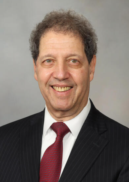 Morie A. Gertz合作论文数Mayo Clinic Comprehensive Cancer Center;Division of Hematology, Department of Internal Medicine, Mayo Clinic61
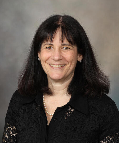 Angela Dispenzieri合作论文数Comprehensive Cancer Center, Mayo Clinic;Transplant Center, Mayo Clinic58
 Ivana N. Micallef合作论文数Division of Hematology, Mayo Clinic53
 Dennis A Gastineau合作论文数49
 Carrie A. Thompson合作论文数Mayo Clinic43
 Shaji Kumar合作论文数Division of Hematology, Department of Internal Medicine, Mayo Clinic40Kay Ristow合作论文数40Suzanne R. Hayman合作论文数37
 Paludo Jonas合作论文数Department of Internal Medicine, Mayo Clinic35
 Ayalew Tefferi合作论文数Comprehensive Cancer Center, Mayo Clinic35
 William Macon合作论文数Department of Pathology, Vanderbilt University Medical Center34
 Michelle A. Elliott合作论文数Departments of Hematology, Mayo Clinic;Comprehensive Cancer Center, Mayo Clinic31Francis K. Buadi合作论文数31
 Villasboas José C合作论文数29
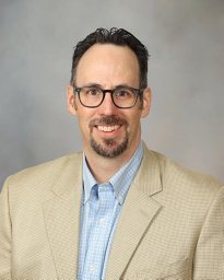 Matthew J. Maurer合作论文数Mayo Clinic29
 Hogan William合作论文数Department of Health Outcomes and Policy,University of Florida26Betsy Laplant合作论文数26Hogan WJ合作论文数25
 James R. Cerhan合作论文数College of Medicine and Science, Mayo Clinic;Mayo Clinic Comprehensive Cancer Center;Mayo Clinic Center for Individualized Medicine23D. Gastineau合作论文数22
 Craig B. Reeder合作论文数Division of Hematology/Oncology, Department of Internal Medicine, Mayo Clinic21
 Yucai Wang (王育才)合作论文数School of Life Sciences, University of Science and Technology of China;The CAS Key Laboratory of Innate Immunity and chronic Disease21
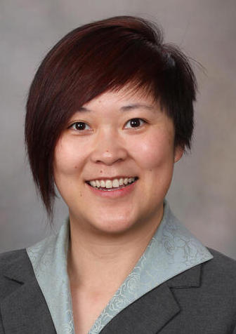 Yi Lin合作论文数Division of Hematology, Department of Internal Medicine, Mayo Clinic;Mayo Clinic Comprehensive Cancer Center20
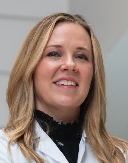 Shernan Holtan合作论文数Department of Medicine, Roswell Park Comprehensive Cancer Center;Blood and Marrow Transplantation Section, Roswell Park Comprehensive Cancer Center;Jacobs School of Medicine and Biomedical Sciences, State University of New York at Buffalo18Brian K Link合作论文数Univ Iowa, Holden Comprehens Canc Ctr, Iowa City, IA 52242 USA17
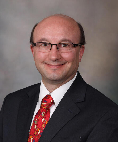 David Dingli合作论文数Department of Hematology, Mayo Clinic;Mayo Clinic Comprehensive Cancer Center, Mayo Clinic;Transplant Center, Mayo Clinic17N. Nora Bennani合作论文数14
 Jeffrey L. Winters合作论文数Mayo Clinic14Gita Thanarajasingam合作论文数13
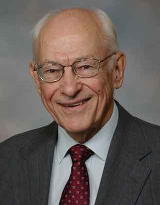 Robert Kyle合作论文数College of Medicine, Mayo Clinic12
 Arushi Khurana合作论文数Division of Hematology, Mayo Clinic11
 S. Vincent Rajkumar合作论文数Division of Hematology, Mayo Clinic;Mayo Clinic Comprehensive Cancer Center11
 Nelson Leung合作论文数Department of Hematology, Mayo Clinic11
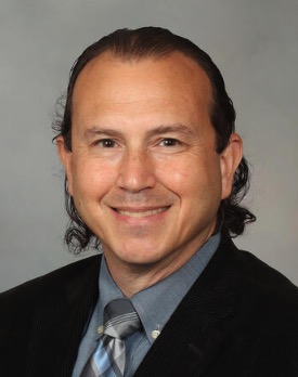 Andrew L. Feldman合作论文数Division of Hematopathology, Department of Laboratory Medicine and Pathology, Mayo Clinic;Division of Experimental Pathology and Laboratory Medicine, Department of Laboratory Medicine and Pathology, Mayo Clinic11Umar Farooq合作论文数10Rosenthal Allison合作论文数10
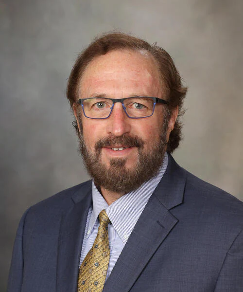 Gregory A. Wiseman合作论文数Mayo Clinic Comprehensive Cancer Center10




