PURPOSE:To determine the maximum-tolerated dose (MTD) of dose-escalated hypofractionated adaptive radiation therapy with atezolizumab in patients with head and neck squamous cell carcinomas (HNSCCs) in a phase 1 trial. METHODS AND MATERIALS:Dose-escalated hypofractionated adaptive radiation therapy was a single-center phase 1 trial. Eligible patients were aged ≥18 years with de novo metastatic HNSCC or localized American Joint Committee on Cancer 8th edition T3-T4 N0-N3, T0-T4 N1-N3 HNSCC meeting one of the following criteria: (1) not candidates for concurrent cisplatin mirroring eligibility for NRG HN-004; (2) refused concurrent cisplatin-based chemoradiation; (3) had unresectable oral cavity cancer; or (4) had recurrent disease after definitive surgical resection alone. Patients received 15 fractions of radiation to escalating total doses of 50, 55, or 60 Gy to gross disease based on a time-to-event continual reassessment methodology. Atezolizumab (1680 mg) was delivered every 4 weeks for up to 1 year after treatment. The primary endpoint of the study was to determine the MTD of radiation. RESULTS:Eighteen patients were enrolled (11 men and 7 women, median age of 74 years [range, 52-89 years]). Five patients were enrolled in radiation dose level 1 (50 Gy in 15 fractions) and received atezolizumab on the first day of radiation. Three of these patients developed Herpes Simplex Virus Type 1 reactivation with excess toxicity. As such, the study was amended to remove the concurrent atezolizumab dose. Thirteen additional patients enrolled and began receiving atezolizumab after radiation. No dose-limiting toxicities were seen after the amendment, and the MTD of radiation was 60 Gy. No loco-regional failures were seen among 7 patients treated to 60 Gy with a 1-year progression-free survival of 71.4%. CONCLUSIONS:Radiation dose may be safely escalated to 60 Gy in 15 fractions with adjuvant atezolizumab with no new safety signals. However, concurrent atezolizumab with hypofractionated radiation promoted Herpes Simplex Virus Type 1 reactivation in a previously unreported manner, resulting in excess dose-limiting toxicities.
Abstract Standard-of-care non-operative management for locally advanced head & neck squamous cell carcinoma (HNSCC) is definitive radiation with concurrent cisplatin. NRG-HN004 demonstrated the benefit of concurrent cetuximab in patients with a medical contraindication to cisplatin including those older than 70 with a Charleson-Deyo (CD) score ≥ 1 and those under 70 with a CD score ≥ 2. In these populations, the survival benefits of concurrent systemic therapies may be limited due to medical co-morbidities. We evaluated overall survival (OS) of these populations in the National Cancer Database (NCDB) comparing radiation-treated patients who received concurrent chemotherapy against concurrent immunotherapy (most likely cetuximab given it is the only FDA-approved immunotherapy for this indication). The NCDB was queried for data from 2015-2023 to include stage III, IVa, and IVb non-metastatic HNSCC patients that met the HN-004 criteria. Histological subtypes of squamous cell carcinoma (SCC) were identified using ICD-O-3 codes 8070-8078. Patients with clinical T3-T4 and/or clinically node-positive disease with a known primary were included. Patients who received less than 60 Gy were excluded. Groups were stratified based on radiation with chemotherapy (RT-C) or radiation with immunotherapy (RT-I) without overlap. Concurrent systemic therapy was defined as having started within 30 days of the initiation of radiation. OS was evaluated using Kaplan-Meier (KM) statistics and differences were evaluated using log-rank testing. A p < 0.05 was considered statistically significant. To minimize the effect of informative censoring, patients without documented follow-up after January 1, 2023 were counted as deaths unless they had greater than 5 years of observed follow-up. Additionally, KM estimates were capped at 5 years. A total of 6299 patients were identified: 5657 treated with RT-C and 642 treated with RT-I. 2-year OS was 65.0% (95% CI, 63.8-66.3) and 52.9% (95% CI, 49.2-57.0) for RT-C and RT-I, respectively. 5-year OS was 20.7% (95% CI, 19.6-21.9) and 21.8% (95% CI, 18.7-25.5) for RT-C and RT-I, respectively. KM analysis ultimately showed no significant difference between the two treatment cohorts (log-rank p=0.059). In a subgroup of patients with medical contraindications to cisplatin, there was no statistically significant improvement in OS with concurrent chemotherapy over concurrent immunotherapy (likely cetuximab) for locally advanced HNSCC in this real-world dataset. Critically, early survival benefits seen with concurrent chemotherapy dissipate with long-term follow-up possibly due to deaths from other causes in this population with extensive co-morbidity. Further prospective study is needed to evaluate the benefit of concurrent chemotherapy over cetuximab in this frail population. Citation Format: Deniz C. Demircioglu, Sohil Singh, Johnny Belcher, Joseph Zenga, Stuart J. Wong, Musaddiq Awan. Survival outcomes with concurrent systemic therapy in patients with a medical contraindication to cisplatin in the non-operative management of locally advanced HNSCC, an NCDB Study [abstract]. In: Proceedings of the American Association for Cancer Research Annual Meeting 2026; Part 1 (Regular Abstracts); 2026 Apr 17-22; San Diego, CA. Philadelphia (PA): AACR; Cancer Res 2026;86(7 Suppl):Abstract nr 7366.
Abstract Background: Although immunotherapy with B cell maturation antigen (BCMA)-targeted chimeric antigen receptor (CAR) T cells, or BCMA CARTs for short, has yielded unprecedented treatment responses in patients with relapsed and/or refractory multiple myeloma (RRMM), efficacy is limited by lack of durability in most cases. This has been attributed, in large part, to the immunosuppressive bone marrow environment (BME) of RRMM, leading to inactivation, exhaustion and attrition of BCMA CARTs. Next-generation CARTs, capable of overcoming BME-dependent inhibitory constraints, may lead to more durable treatment options for RRMM. Methods: To inform the design of BCMA CARTs that can maintain long-term activity in the myeloma-laden bone marrow, we leveraged a recently published single-cell RNA sequencing dataset, GSE210079, of the BME from 9 patients with RRMM. Based on clinical outcomes, patients were stratified into early relapse (<3 months) and late relapse (>12 months) groups. Biocomputational analyses revealed that activation of TGF-β signaling and T cell exhaustion pathways, along with suppression of IL-18-driven immune responses, were implicated in the mechanisms driving early-relapse post-BCMA CAR-T treatment. Inspired by these findings, we employed standard molecular biology tools to manufacture BCMA CARTs that feature both TGF-β resistance and simultaneously disrupt PD-1 expression and enabling inducible IL-18 secretion. Result: Compared to T cells from late-relapse myeloma, T cells from early-relapse myeloma demonstrated upregulated oncogenic signaling circuits (p53, FoxO, AMPK), elevated expression of exhaustion-related genes (PDCD1, HAVCR2, LAG3, CTLA4, TIGIT; p < 0.01), and increased mRNA levels of key TGF-β pathway genes (TGFB1, SMAD2/3/4, TGFBR2). Early-relapse myeloma exhibits reduced TCR signaling and Th1/Th2 differentiation, which are regulated by IL-18. BCMA CARTs were rendered resistant to TGF-β by enforced expression of a dominant-negative ligand-binding but non-signaling transforming growth factor beta receptor 2 (dnTFGBR2). Monoallelic “knock-in” of an IL-18 encoding gene in the PDCD1 locus relied on CRISPR/Cas9-mediated editing. The genetically modified BCMA CAR-T cells demonstrated potent cytotoxic activity against myeloma cells in vitro, increased resistance to TGF-β-mediated suppression, and inducible secretion of IL-18 driven by the endogenous PDCD1 promoter. IL-18 levels increased in correlation with T cell activation, accompanied by a concomitant silencing of PD-1 expression. Conclusion: TGF-β-resistant BCMA CARTs that produce IL-18 when the cells are challenged by exhaustion in the BME, and thus disrupt PD-1, may be viable way forward to a more potent and durable therapy for patients with RRMM than available today. This hypothesis will be evaluated in greater depth in preclinical in vivo studies ongoing in our laboratory. Citation Format: Sandhya Rai, Shijing Wang, Bin Sun, Tyce Kearl, Peiman Hematti, Joseph Zenga, Heather Himburg, Musaddiq Awan, Deepak Parashar, Binod Dhakal, Siegfried Janz, Fumou Sun, . Improving BCMA-targeted CAR-T cell therapy of myeloma using TGF-β resistance and inducible production of IL-18 [abstract]. In: Proceedings of the American Association for Cancer Research Annual Meeting 2026; Part 1 (Regular Abstracts); 2026 Apr 17-22; San Diego, CA. Philadelphia (PA): AACR; Cancer Res 2026;86(7 Suppl):Abstract nr 1520.
BACKGROUND:Cisplatin remains the cornerstone radiosensitizer for definitive and adjuvant chemoradiation in patients with locally advanced squamous cell carcinoma of the head and neck. However, despite decades of clinical use, many practical aspects of cisplatin administration remain unstandardized in clinical practice and trials. METHODS:Drawing on the collective experience of the NRG Oncology Head and Neck Working Group, this consensus article provides practical guidance on the administration of cisplatin during radiation therapy. We review and propose recommendations regarding the timing of chemotherapy and radiation initiation, premedication and hydration regimens by dose, monitoring and grading of cisplatin-induced ototoxicity, and management strategies during cisplatin shortages. Our guidance is informed by clinical trial protocols, retrospective and prospective data, and multidisciplinary expert consensus. We emphasize the importance of protocol flexibility to support equitable trial accrual, minimize treatment delays, and improve patient-centered outcomes. CONCLUSIONS:This article offers a unified framework to optimize the use of cisplatin in chemoradiation protocols, improve adherence, reduce toxicity, and preserve oncologic efficacy. Our recommendations are particularly timely in the context of evolving clinical practices and recent cisplatin shortages. Insights from the NRG Oncology collaborative group aim to inform future trial designs and clinical practice guidelines, ensuring consistent and equitable care for patients with head and neck cancer.
BACKGROUND:Patients with locally advanced head and neck squamous cell carcinoma who are medically ineligible for cisplatin lack a standard concurrent systemic therapy. METHODS:We used the National Cancer Database to identify patients diagnosed from 2015 onward with non-metastatic, locally advanced squamous HNSCC treated with definitive radiation therapy and concurrent systemic therapy, receiving either chemotherapy or cetuximab (using immunotherapy as a surrogate for cetuximab in the NCDB). Cisplatin-ineligibility was defined using the age and comorbidity criteria from NRG-HN004. Overall survival was compared using Kaplan-Meier analysis and an inverse probability weighted Cox proportional hazards model. RESULTS:The final cohort included 6045 patients: 5441 receiving chemotherapy and 604 receiving cetuximab. In the weighted Cox model, chemotherapy was not significantly associated with improved overall survival compared to cetuximab (HR, 1.08; 95% CI, 0.94-1.23; p = 0.281). CONCLUSIONS:In this retrospective analysis, no survival advantage was found with chemotherapy over cetuximab in cisplatin-ineligible patients. Future, randomized trials are necessary to define optimal systemic therapy in this population.
Abstract Introduction: HPV-negative head and neck squamous cell carcinoma (HNSCC) remains highly lethal, and even the recent KEYNOTE-689 trial showed limited response to PD-1 blockade. To clarify mechanisms of immunoresistance, we performed multi-omic single-cell profiling of HPV-negative HNSCC to define the states, clonotypes, and spatial niches of tumor-reactive T cells. Methods: Two patients with untreated HPV-negative HNSCC underwent surgical tumor sampling. Biopsies were allocated for dissociative single-cell RNA and TCR sequencing, for single-cell spatial whole transcriptome imaging with TCR add-in on the Bruker CosMx platform, and for functional co-culture of autologous tumor cells and tumor-infiltrating lymphocytes (TILs) on the Bruker Beacon system with robotic retrieval of reactive clones followed by single-cell TCR sequencing. Results: Dissociative and spatial datasets were high quality, and dissociative clustering guided spatial annotation. Two tumor-reactive TIL populations emerged: a transcriptionally reactive population marked by terminal exhaustion and clonal hyper-expansion, and a functionally reactive population identified on the Beacon platform through tumor-killing and IFNγ secretion. Neither appeared in adjacent normal mucosa. Spatial projection showed functionally reactive clonotypes progressing along a pseudotime continuum from minimally exhausted stromal states toward a deeply exhausted yet proliferative state within malignant epithelial islands. Transcriptionally reactive TILs shared this exhausted-proliferative phenotype and primarily localized within tumor islands. Conversely, Tregs and immunosuppressive macrophages were confined to stromal regions and showed minimal spatial overlap with tumor-reactive T cells. Malignant epithelium strongly enriched CXCL14 (> ten-fold over stroma), yet tumor-reactive TILs remained rare, with a tumor-to-reactive T cell ratio greater than 20 to 1. Conclusions: This is the first study to integrate dissociative, spatial, and functional single-cell analytics to define tumor-reactive TIL biology in solid tumors. We find that tumor-reactive T cells can infiltrate malignant epithelial islands, and that their deepest infiltration corresponds to the most exhausted states, indicating that chronic antigen stimulation is the primary driver of dysfunction. The confinement of suppressive cells to stromal niches suggests that their influence is exerted through maintenance of a restrictive barrier stroma rather than through proximity-based inhibition. Together, these findings highlight insufficient chemotactic recruitment of tumor-reactive T cells as a central barrier to effective antitumor immunity. The strong epithelial enrichment of CXCL14 positions it as a promising candidate for enhancing tumor-directed T cell migration. Citation Format: Joseph Zenga, Musaddiq Awan, Fumou Sun, Stuart J. Wong, Abdullah Memon, Tyce Kearl, Ean Norenberg, Margaret L. Hoang, Joseph M. Beechem, Eric Hobbs, Ashley Heck, Rachel Liu, Daniel McGuire, Erin Piazza, Peiman Hematti, Heather Himburg. Dissociative, spatial, and functional single-cell profiling reveals antigen-driven exhaustion and chemotactic failure of tumor-reactive T cells in HPV-negative HNSCC [abstract]. In: Proceedings of the American Association for Cancer Research Annual Meeting 2026; Part 1 (Regular Abstracts); 2026 Apr 17-22; San Diego, CA. Philadelphia (PA): AACR; Cancer Res 2026;86(7 Suppl):Abstract nr 4984.
Background and purpose: Quantitative MRI (qMRI) has been explored for detecting tumor changes during radiation therapy (RT) in head and neck squamous cell cancer (HNSCC). Clinical trials show prolonged survival with PD-1 targeted immune checkpoint inhibition. Hypofractionated radiation regimens are being studied to counteract radioresistant clonogen formation. This study aims to use daily qMRI monitoring in these therapies. The objective of this exploratory study was to investigate if qMRI can detect tumor microenvironment changes during hypofractionated RT in a phase I trial of Dose-Escalated Hypofractionated Adaptive Radiotherapy (DEHART). Materials and methods: Seventeen subjects with advanced HNSCC underwent MR-guided RT with daily qMRI using a 15-fraction regimen to a cumulative dose of 50, 55, or 60 Gy. A 1.5 T MRI-Linac collected daily intravoxel incoherent motion (IVIM), T1, and T2 mappings. Median primary tumor ADC, D, D*, f, T1, and T2 were calculated, using paraspinal muscle as a control. qMRI parameters were analyzed by treatment condition and length using linear mixed effect models and nonparametric tests. Results: Significant (p < 0.05) increases in ADC, D, f, and T2 were observed over treatment duration for multiple conditions. Daily monitoring enhanced result significance compared to weekly collection. Conclusions: Daily qMRI effectively monitors tumor response over short periods and varying treatment conditions. Further studies on radiation and systemic therapy combinations in HNSCC could benefit from daily qMRI data collection.
11066 Background: This study aimed to determine to what extent area-level social determinants of health (SDOH) interact with individual, institutional, and biological factors to predict outcomes in head and neck cancer (HNC) trials. Methods: Five NRG Oncology HNC trials (2635 patients receiving chemoradiation) were analyzed. Area-level SDOH coded by patient ZIP codes included rurality (rural-urban commuting area code), neighborhood socioeconomic deprivation (Area Deprivation Index [ADI] categorized as upper vs. lower quartile), and travel burden (distance and time to treatment site). Individual (demographic, cancer and treatment-related factors), institutional (accrual volume), biological (HPV+/-) factors, and outcomes (overall survival [OS], progression free survival [PFS], quality of life [QOL], and symptoms) were analyzed. Multivariable Cox proportional hazards regression and mediation analysis using logistic regression assessed associations using hazard ratio (HR) or odds ratios (OR) and 95% confidence intervals (CI). Results: Most patients were White (88%), non-Hispanic (92.7%), of mean age of 57 years, HPV+ (64.6%), and received intensity-modulated radiotherapy (95.9%) and cisplatin (94.2%). ADI and rurality were not associated with OS and PFS. OS and PFS were higher in patients with travel time <1 hour (HR=0.85, 95% CI [0.75, 0.98]; HR=0.85, 95% CI [0.73, 0.98]) and travel distance <50 miles (HR=0.84, 95% CI [0.72, 0.96]; HR=0.85, 95% CI [0.73, 0.98]). ADI, travel time, and travel distance were not associated with QOL decline. Patients treated at institutions with high rural accrual volume had worse QOL decline from baseline (OR=0.36, 95% CI [0.15, 0.85]). The impact of travel distance but not time varied by race to influence QOL decline (OR=0.38, 95% CI [0.16, 0.93]). ADI was not associated with symptoms, but patients from institutions with high rural accrual volume had worse symptoms (OR=7.83, 95% CI [1.98, 31.01]). HPV status had a significant indirect effect on the relationship between travel distance and survival at 1 year (estimate [β]=0.03, 95% CI [0.01, 0.05]) and 5 years (β=0.03, 95% CI [0.004, 0.05]), as well as a direct and total mediation effect of travel distance on QOL decline at 1 year (direct β=-0.08, 95% CI [-0.16, -0.004]; total β=-0.89, 95% CI [-0.17, -0.01]). Conclusions: This study showed the impact of area-level SDOH and their interactions with race and institutional accrual volume, which are associated with survival, QOL, and symptom changes. HPV status potentially mediated the effects of travel distance on outcomes. Our findings provide novel approaches to identify patients at risk for poor outcomes, such as those with travel burden at institutions with high rural accrual, to design community-based interventions to improve cancer outcomes.
Background and Purpose:Quantitative MRI (qMRI) is an emerging technique for characterizing tissue microenvironments. Prior work has shown that daily qMRI during MR-guided radiotherapy (MRgRT) detects differential responses to hypofractionated radiotherapy (RT) in head and neck squamous cell carcinoma (HNSCC) within the primary gross tumor volume (GTVp). This exploratory study investigates whether subregions of HNSCC tumors show distinct qMRI responses to hypofractionated RT using contoured GTVp subregions and voxelwise data. Materials and Methods:Eighteen subjects with advanced HNSCC underwent MRgRT with daily qMRI over 15 fractions (total dose 50, 55, or 60 Gy). Daily intravoxel incoherent motion (IVIM) and relaxometry sequences were acquired on a 1.5 T MR-Linac. Median ADC, D, D*, f, T1, and T2 were calculated for concentric GTVp subregions and an intra-subject control contour in paraspinal muscle. Mixed effect models were fit for each parameter. Voxelwise analysis included quantile tracking, histograms, and paired tests over the length of RT. Results:Significant changes (p < 0.05) were observed in ADC, D, f, T2, and T1 within GTVp subregions. ADC, D, f, and T2 changes were uniform, while T1 varied spatially. Spatial patterns were detected in ADC and T1, with marginal effects in D, f, and T2. Voxelwise analysis revealed significant changes in all parameters except D* after correction. ADC, D, and f increased consistently; T1 and T2 changes were quantile dependent. These trends may reflect biological processes such as declining cellularity, reperfusion, or edema. Conclusions:In this exploratory study, qMRI detected spatial heterogeneity in HNSCC. These findings support its potential role in guiding biologically adaptive RT strategies.
Purpose/Objective(s) One commercially available MR Linac imposes a 22 cm field length restriction along the superior/inferior (S/I) direction. This limitation denies patients with long planning target volumes (PTVs) the benefits of receiving MR-guided radiation therapy (MRgRT). We investigate here the feasibility of a multi-isocenter technique using an alternative planning system to enable MRgRT in patients with S/I PTV lengths exceeding 22cm. Materials/Methods A research version of a treatment planning system, with prototype Monte Carlo photon beam model incorporating a 1.5T transverse magnetic field, was installed on the local machine network of a precision radiation medicine company. Dual isocenter plans were created for one head and neck (HNC) patient and one prostate bed and nodes (PBN) patient. The distance between isocenters along the S/I direction was 6 and 10 cm for the HNC and PBN plans, respectively. For each plan, two separate identical beam sets were simultaneously optimized to generate uniform dose distributions throughout the target volume. The online workflow was then simulated in phantom: Two Rad-Rx groups containing longitudinal couch coordinates at the two isocenters were created in an electronic patient information management system. A treatment session was initiated at the first isocenter (Iso1). After simultaneous optimization, the Iso1 plan was downloaded to the sequencer and delivered to a patient-specific quality assurance. A second treatment session was then initiated at the second isocenter (Iso2), the couch was moved to Iso2, and the Iso2 plan was immediately downloaded to the sequencer and delivered to a patient-specific quality assurance. Additional patient-specific quality assurance measurements were performed by shifting the couch by 1mm increments to increase the effective “detector resolution” in the S/I direction. Couch accuracy and reproducibility was evaluated by imaging the MV alignment phantom in 0.1mm increments and registering with reference image with ball bearings. The treatment plans were tested for robustness by recalculating with the lower isocenter shifted relative to the upper, by +/- 0.2, 0.5 and 1mm; and the DVHs of important structures were compared over whole points. Results Over a one-year period, 25 HNC patients and 4 rectal cancer and nodes patients in our clinic were denied MRgRT due to PTV lengths exceeding 22cm. The treatment planning system dual isocenter plans were calculated, uploaded to an electronic patient information management system, and delivered successfully. Repeated image registration with an MV alignment phantom revealed couch accuracy and reproducibility of within 0.2mm. Patient-specific quality assurance comparison plans passed at >99% using TG218 criteria. Plans with shifted lower isocenter by +/-0.2mm, 0.5mm and 1mm had 99% of all DVH points of important OARs within 1%, 3% and 5% in Dose and Volume axis of each other, except small OARs such as optic nerves and inner ears. The target volume DVHs were all within 1% both dose and volume wise of the unshifted plans. Conclusion Efficient multi-isocenter treatments on the precision radiation medicine company are feasible, enabling the benefits of MRgRT for patients with long target volumes.
Purpose/Objective(s) Accurate representation of patient anatomy is essential for radiation therapy. Surface changes arising from weight gain/loss or tissue edema occurring during a treatment course can currently only be detected by on-treatment imaging modalities during a clinical visit. However, delayed detection of surface changes may result in postponed treatments due to the need to generate a new treatment plan. We investigate here the feasibility of a new paradigm: patient-driven radiation therapy, in which patients can self-perform 3D surface scans using affordable smartphone photogrammetry. The surface-scanned models can then be compared to reference surfaces obtained from planning images to detect surface changes, triggering the need for an adaptive plan. Materials/Methods An initial feasibility study was performed using an anthropomorphic head phantom and two healthy volunteers. The combination of an iPhone 15 Pro Max (Apple Inc., USA) and smartphone application Polycam were used to create 3D surface models using photogrammetry. Ground truth surface contours of the phantom and volunteers were obtained with CT and MRI, respectively. Image processing and analysis was performed in 3D Slicer. The CT and MRI defined surface contours were saved as standard tessellation language (STL) files and registered with smartphone-generated 3D surface models. The accuracy of the smartphone generated models relative to reference models was evaluated using Hausdorff distances. To evaluate the ability of photogrammetry to detect surface changes, smartphone surface models were also measured after placing a 1cm thick 3D printed bolus on the phantom and volunteers. Results Overall, good agreement was obtained between surface models generated from smartphone photogrammetry and ground truth CT or MRI scans. As shown in Table 1, mean and standard deviation Hausdorff distances were 1.25 ± 0.70mm, 1.04 ± 0.74mm, and 0.64 ± 0.66 mm for the phantom and two human studies, respectively. Smartphone photogrammetry was able to detect the changes in surface anatomy, induced by the 3D printed bolus. The maximum Hausdorff distance in the surface region where 3D printed bolus was placed was 10.9 mm. Conclusion Accurate 3D surface model generation using smartphone photogrammetry is feasible. The portable and affordable surface scanning with smartphone presents a promising solution for early detection of patient surface changes using home-based monitoring. Early detection can prompt the need for plan adaptation outside the clinic, preventing further treatment delays.
While much of the focus has shifted toward optimizing outcomes for the more favorable human papillomavirus (HPV)–associated head and neck squamous cell carcinomas (HNSCC), outcomes for HPV-negative HNSCCs have remained static for more than 2 decades. Concurrent systemic therapies beyond cisplatin have failed to deliver improved disease control, although the door remains open for emerging agents and adjuvant therapies. Riding a wave of noninferiority trials, radiation oncologists have adopted hypofractionation in a variety of other cancers.
Purpose HPV-associated oropharyngeal squamous cell carcinoma (OPSCC) is a distinct disease from other head and neck tumors. This guideline provides evidence-based recommendations on the critical decisions in its curative treatment, including both definitive and postoperative radiation therapy (RT) management. Methods The American Society for Radiation Oncology convened a task force to address 5 key questions on the use of RT for management of HPV-associated OPSCC. These questions included indications for definitive and postoperative RT and chemoradiation; dose-fractionation regimens and treatment volumes; preferred RT techniques and normal tissue considerations; and post-treatment management decisions. The task force did not address indications for primary surgery versus RT. Recommendations were based on a systematic literature review and created using a predefined consensus-building methodology and system for grading evidence quality and recommendation strength. Results Concurrent cisplatin is recommended for patients receiving definitive RT with T3-4 disease and/or one node >3 cm, or multiple nodes. For similar patients who are ineligible for cisplatin, concurrent cetuximab, carboplatin/5-fluorouracil, or taxane-based systemic therapy are conditionally recommended. In the postoperative setting, RT with concurrent cisplatin (either schedule) is recommended for positive surgical margins (PSM) or extranodal extension (ENE). Postoperative RT alone is recommended for pT3-4 disease, >2 nodes, or a single node >3 cm. Observation is conditionally recommended for pT1-2 disease and a single node ≤3 cm without other risk factors. For patients treated with definitive RT with concurrent systemic therapy, 7000 cGy in 33 to 35 fractions is recommended, and for patients receiving postoperative RT without PSM and ENE, 5600-6000 cGy is recommended. For all patients receiving RT, intensity modulated RT over 3-D techniques with reduction in dose to critical organs-at-risk (including salivary and swallowing structures) is recommended. Reassessment with positron emission tomography-computed tomography (PET-CT) is recommended approximately 3 months following definitive RT/chemoradiation, and neck dissection is recommended for convincing evidence of residual disease; for equivocal PET-CT findings, either neck dissection or repeat imaging is recommended. Conclusions The role and practice of RT continues to evolve for HPV-associated OPSCC, and these guidelines inform best clinical practice based on the available evidence.
Purpose/Objective(s) To determine the immunologic effects of hypofractionated radiation (RT) pre-operatively on patients with oral squamous cell carcinomas (OSCC) slated to undergo definitive resection. Materials/Methods HyPR-HN (NCT05538533) is a Phase I trial to determine the safety of three isoequivalent (EQD2 70 Gy at a/b = 3) but increasingly condensed preoperative RT regimens (46 Gy in 10 fractions, 40 Gy in 7 fractions and 35 Gy in 5 fractions) for patients with OSCC using a Time-to-event Continuous Reassessment Methodology (TiTE-CRM) for dose finding. Three patients enrolled on the HyPR-HN trial and assigned to the first dose level (46 Gy in 10 fractions) underwent biopsy prior to RT (3 patients), immediately following RT on the day of the last radiation fraction (3 patients) and at the time of surgery (2 patients) six weeks after completion of RT. Flow cytometry of immune cell populations and single cell RNA sequencing was performed at each time point for all three patients. T-cell receptor sequencing was performed on one patient at all three time points. Single cell RNA data was imported into R (v.4.3.2) and analyzed using the following packages: Seurat (v5.0) and scRepertoire (v2.0.0). Results On flow cytometry, CD8 T cell populations were diminished on the last fraction of RT compared to before RT and failed to recover at the time of surgery in all three patients. This suggests that post-RT immune checkpoint inhibition may be less effective due to T cell depletion. Single cell analysis showed the presence of canonical exhausted and regulatory T cell populations prior to RT, however after radiation these populations were diminished. Additionally, a proliferative CD8+ T cell population (TProlif_Tox) was readily identified in all three patients prior to RT, but unlike the canonical exhausted and regulatory populations, TProlif_Tox persisted after RT. TProlif_Tox co-expressed several cytotoxic markers (GZMB, IFNG), exhaustion markers (TIGIT, TIM3, LAG3, CTLA4, PD1) and cell proliferation markers (STMN, MKI47, CENPF, TOP2A, TOX), suggestive of persistent T cell stimulation in vivo. Analysis of a benign oral mucosa dataset did not demonstrate the presence of a TProlif_Tox population, suggesting tumor specificity. Further, clonal network analysis both pre- and post-RT demonstrated cycling between the TProlif_Tox cluster and canonical pre-exhausted clusters suggesting repopulation of the immune microenvironment with this TProlif_Tox cluster. Conclusion RT rapidly and persistently depletes T cells. A proliferative T cell cluster may contain tumor specific T cells and repopulates the tumor microenvironment after RT.
PurposeChanges in quantitative magnetic resonance imaging (qMRI) are frequently observed during chemotherapy or radiation therapy (RT). It is hypothesized that qMRI features are reflective of underlying tissue responses. It's unknown what underlying genomic characteristics underly qMRI changes. We hypothesized that qMRI changes may correlate with DNA damage response (DDR) capacity within human tumors. Therefore, we designed the current study to correlate qMRI changes from daily RT treatment with underlying tumor transcriptomic profiles.Methods and MaterialsStudy participants were prospectively enrolled (National Clinical Trial 03500081). RNA expression levels for 757 genes from pretreatment biopsies were obtained using a custom panel that included signatures of radiation sensitivity and DDR. Daily qMRI data were obtained from a 1.5 Tesla MR linear accelerator. Using these images, d-slow, d-star, perfusion, and apparent diffusion coefficient-mean values in tumors were plotted per-fraction, over time, and associated with genomic pathways.ResultsA total of 1022 qMRIs were obtained from 39 patients and both genomic data and qMRI data from 27 total patients. For 20 of those patients, we also generated normal tissue transcriptomic data. Radio sensitivity index values most closely associated with tissue of origin. Multiple genomic pathways including DNA repair, peroxisome, late estrogen receptor responses, KRAS signaling, and UV response were significantly associated with qMRI feature changes (P < .001).ConclusionsGenomic pathway associations across metabolic, RT sensitivity, and DDR pathways indicate common tumor biology that may correlate with qMRI changes during a course of treatment. Such data provide hypothesis-generating novel mechanistic insight into the biologic meaning of qMRI changes during treatment and enable optimal selection of imaging biomarkers for biologically MR-guided RT.
BACKGROUND:Management of patients with locoregionally advanced head and neck squamous cell carcinoma (HNSCC) when cisplatin is contraindicated is controversial. We aimed to assess whether radiotherapy with concurrent and adjuvant durvalumab would improve outcomes compared with radiotherapy with cetuximab. METHODS:NRG-HN004 was designed as an open-label, multicentre, parallel-group, randomised, phase 2/3 trial with safety lead-in conducted at 89 academic and community medical centres in North America. Eligible patients were aged 18 years or older with American Joint Committee on Cancer 8th edition stage III-IVB p16-negative HNSCC or unfavourable stage I-III p16-positive oropharyngeal or unknown primary carcinoma, who had a contraindication to cisplatin (Eastern Cooperative Oncology Group [ECOG] performance status 2, renal or hearing impairment, peripheral neuropathy, aged at least 70 years with moderate or severe comorbidity, or aged younger than 70 years with severe comorbidity). Patients were randomly assigned (2:1) by permuted block randomisation (multiples of 6) to intravenous durvalumab 1500 mg starting 2 weeks before radiotherapy then every 4 weeks starting week 2 of radiotherapy (seven cycles) or intravenous cetuximab 400 mg/m2 1 week before radiotherapy then 250 mg/m2 weekly beginning week 1 of radiotherapy (eight cycles), with intensity-modulated radiotherapy (70 Gy in 35 fractions over 7 weeks). Stratification factors were tumour and nodal stage, ECOG performance status and comorbidity, and primary site and p16 status. The phase 2 primary endpoint was progression-free survival in the intention-to-treat population. There was one prespecified interim futility analysis at 50% of progression-free survival information. If the observed hazard ratio was 1·0 or more, favouring cetuximab, early stopping would be considered. Extended follow-up analysis was post hoc. This trial is registered with ClinicalTrials.gov, NCT03258554, and is closed to enrolment. FINDINGS:Following a ten-patient safety lead-in, the phase 2 trial enrolled 190 patients from March 12, 2019, to July 30, 2021, 186 of whom were randomly assigned (123 to durvalumab and 63 to cetuximab). Median age was 72 years (IQR 64-77), 30 (16%) patients were women and 156 (84%) were men. Phase 2 accrual was suspended in July 30, 2021, following an interim futility analysis, and permanently closed in Sept 1, 2022. The phase 3 part of the trial was not conducted. At a median follow-up of 2·3 years (IQR 1·9-3·1) for the extended follow-up (data cutoff July 31, 2023; post-hoc analysis), 2-year progression-free survival was 50·6% (95% CI 41·5-59·8) in the durvalumab group versus 63·7% (51·3-76·1) in the cetuximab group (hazard ratio 1·33 [95% CI 0·84-2·12]; p=0·89). Adverse events were similar in both groups. The most common grade 3-4 adverse events were dysphagia (26 [22%] of 119 patients in the durvalumab group vs 18 [30%] of 61 patients in the cetuximab group), lymphopenia (33 [28%] vs 20 [33%]), and oral mucositis (13 [11%] vs 11 [18%]). Four (3%) patients in the durvalumab group and one (2%) in the cetuximab group died from treatment-related adverse events (death not otherwise specified, laryngeal oedema, lung infection, and respiratory failure in the durvalumab group and sudden death not otherwise specified in the cetuximab group). INTERPRETATION:Our findings suggest that durvalumab did not improve outcomes compared with cetuximab in patients with HNSCC with contraindications to cisplatin. Further trials are needed to define the standard of care for this population. FUNDING:US National Cancer Institute and AstraZeneca.
 Clifton David Fuller合作论文数Department of Radiation Oncology, Division of Radiation Oncology, The University of Texas MD Anderson Cancer Center, Houston30
 Christopher J. Schultz合作论文数Department of Radiation Oncology, Medical College of Wisconsin20
 Zenga Joseph合作论文数Department of Otolaryngology-Head and Neck Surgery, Washington University19
 G. Brandon Gunn合作论文数Department of Radiation Oncology, Division of Radiation Oncology, The University of Texas MD Anderson Cancer Center18
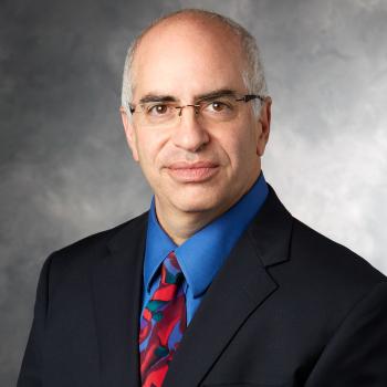 David N. Rosenthal合作论文数Pediatrics Department, Stanford University18
 Min Yao合作论文数18Eric S. Paulson合作论文数18Stuart Wong合作论文数17
 X. Allen Li合作论文数Department of Radiation Oncology, Medical College of Wisconsin16Monica Shukla合作论文数15
 Adam S. Garden合作论文数Department of Radiation Oncology, Division of Radiation Oncology, The University of Texas MD Anderson Cancer Center15
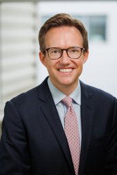 William A Hall合作论文数Departments of Therapeutic Radiology and Neurosurgery, University of Minnesota Hospitals and Clinic14
 Shreenivas Aditya Varnam合作论文数Long island Jewish Hospital, Hofstra Northshore-LIJ School of Medicine13Becky Massey合作论文数12Machtay M合作论文数12Heather a Himburg合作论文数11
 Mohamed Abdallah合作论文数10Straza Michael W合作论文数10
 Kristin Higgins合作论文数City of Hope10
 Shlomo Koyfman合作论文数Department of Radiation Oncology, Cleveland Clinic;Department of Medicine, School of Medicine, Case Western Reserve University;Case Comprehensive Cancer Center, Case Western Reserve University10
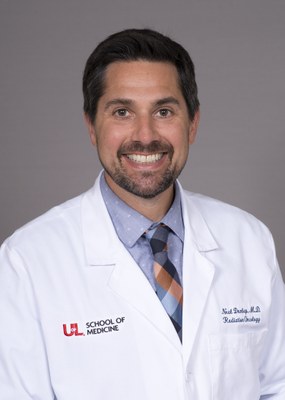 Neal E. Dunlap合作论文数Department of Radiation Oncology, School of Medicine, University of Louisville9
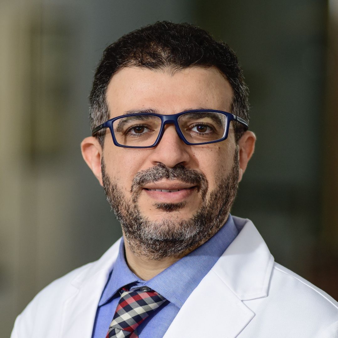 Abdallah Sherif Radwan Mohamed合作论文数Department of Radiation Oncology, Baylor College of Medicine9
 Nancy Y. Lee合作论文数Department of Radiation Oncology, Memorial Sloan-Kettering Cancer Center9
 Farzan Siddiqui合作论文数9
 Matthew Christopher Ward合作论文数8
 Irene Gottlob合作论文数Department of Neuroscience, Psychology & Behaviour, University of Leicester, University of Leicester8
 Jonathan J Beitler合作论文数Medical College of Wisconsin8Michael E. Stadler合作论文数8
 Jayashree Kalpathy-Cramer合作论文数Harvard Medical School;Massachusetts General Hospital8
 Andy Trotti合作论文数Center for Infection Research in Cancer, Moffitt Cancer Center8
 Stuart J. Wong合作论文数Division of Hematology and Oncology, Department of Medicine, Medical College of Wisconsin;Cancer Center, Froedtert Hospital8
 Nadeem Riaz合作论文数Memorial Sloan Kettering Cancer Center8Mitchell Machtay合作论文数8
 Derek Isrow合作论文数Department of Chemistry, University of Miami8J. Zenga合作论文数7D.I. Rosenthal合作论文数7
 Steven Frank合作论文数Department of Anesthesiology and Critical Care Medicine, School of Medicine, Johns Hopkins University7
 James Caudell合作论文数Moffitt Cancer Center7
 Jack Phan合作论文数7Frei Anne C合作论文数7
 Bruce H. Campbell合作论文数Department of Otolaryngology, Medical College of Wisconsin7
 Laurence E. Court合作论文数Department of Radiation Physics, Division of Radiation Oncology, The University of Texas MD Anderson Cancer Center6
 Beth Beadle合作论文数Department of Radiation Oncology, Stanford University6
 Kocak-Uzel Esengul合作论文数6Baron Charles A合作论文数6Ergun Emin Ahunbay合作论文数6
 Stephen Y. Lai合作论文数Department of Head and Neck Surgery, Division of Surgery, The University of Texas MD Anderson Cancer Center;Department of Radiation Oncology, Division of Radiation Oncology, The University of Texas MD Anderson Cancer Center;Department of Molecular and Cellular Oncology, Division of Basic Science Research, The University of Texas MD Anderson Cancer Center6S.J. Zakem合作论文数5Jennifer Bruening合作论文数5




