Spleen cells obtained from mice immunized with partially purified human coagulation Factor V were fused with NS-I mouse myeloma cells, and hybrids were selected. Culture media were screened for anti-Factor V activity, and an antibody- positive clone was obtained and passaged as an ascites tumor in mice. The ascitic fluid from the hybridoma-bearing mouse could be diluted 1:106 before losing reactivity in an anti-Factor V ra- dioimmunoassay. When immobilized on agarose, the monoclonal antibody quantitatively removed Factor V activity from human plasma.' Factor V activity could be eluted with 1.2 M NaCI at pH 6.5. Homogeneous Factor V was isolated by chromatography of barium citrate-adsorbed, polyethylene glycol 6000 precipitated plasma on the antibody column followed by chromatography on phenyl-Sepharose. The isolated Factor V exhibited a single band upon gel electrophoresis in sodium dodecyl sulfate with an ap- parent Mr comparable to that of bovine Factor V (330,000). Upon exposure to thrombin, the activity of Factor V increased 53-fold when measured in Factor V-deficient plasma. This increased ac- tivity was associated with discrete proteolytic cleavages of the par- ent molecule.
Laccases produced by white rot fungi have been extensively evaluated for their potential to decolorize textile wastewaters which contain salts like sodium chloride and sodium sulfate. The effect of sodium chloride and sodium sulfate on Trametes versicolor laccase during the decolorization of an anthraquinone dye (Reactive Blue 19) and the oxidation of 2,2′-azino-bis(3-ethylbenzothiazoline-6-sulfonic acid) (ABTS) were evaluated by steady-state kinetic analysis. The results showed that, while sodium sulfate did not affect laccase activity, sodium chloride inhibited both ABTS oxidation and dye decolorization. However, the type of inhibition was substrate-dependent: it was hyperbolic, noncompetitive with ABTS and parabolic, noncompetitive with Reactive Blue 19. Furthermore, the results suggested that two chlorides may bind to laccase in the presence of the dye unlike recent inhibition models which suggest that there is only one inhibition site. This investigation is the first to provide evidence for and to propose a two-site model of laccase inhibition, providing new insight into NaCl inhibition of laccase. The proposed model is also useful to predict decolorization rates in the presence of sodium chloride and to determine operating conditions that will minimize inhibition.
Individuals with haemophilia A exhibit bleeding tendencies that are not always predicted by their factor (F)VIII level. It has been suggested that bleeding in haemophilia is due not only to defective prothrombin activation but also aberrant fibrinolysis. Thrombin activatable fibrinolysis inhibitor (TAFI) activation was measured in tissue factor (TF)-initiated blood coagulation in blood samples of 28 haemophiliacs and five controls. Reactions were quenched over time with FPRck and citrate and assayed for TAFIa and thrombin-antithrombin (TAT). The TAFIa potential (TP), TAFI activation rate and the TAFIa level at 20 min (TAFIa(20 min)) was extracted from the TAFI activation progress curve. In general, the time course of TAFI activation follows thrombin generation regardless of FVIII activity and as expected the rate of TAFI activation and TP decreases as FVIII decreases. The magnitude of TP was similar among the control subjects and subjects with <11% FVIII. In severe subjects with <1% FVIII at the time of blood collection, the TAFIa(20 min) was inversely and significantly correlated with haemarthrosis (-0.77, P = 0.03) and total bleeds (-0.75, P = 0.03). In all cases, TAFIa(20 min) was more strongly correlated with bleeding than TAT levels at 20 min. Overall, this study shows that TAFI activation in whole blood can be quantified and related to the clinical bleeding phenotype. Measuring TAFIa along with thrombin generation can potentially be useful to evaluate the differential bleeding phenotype in haemophilia A.
Solulin is a soluble form of thrombomodulin that is resistant to proteolysis and oxidation. It has been shown to increase the clot lysis time in factor VIII (fVIII)-deficient plasma by an activated thrombin-activatable fibrinolysis inhibitor (TAFIa)-dependent mechanism. In the present study, blood was drawn from humans and dogs with hemophilia, and thromboelastography was used to measure tissue factor-initiated fibrin formation and tissue-plasminogen activator-induced fibrinolysis. The kinetics of TAFI and protein C activation by the thrombin-Solulin complex were determined to describe the relative extent of anticoagulation and antifibrinolysis. In severe hemophilia A, clot stability increased by > 4-fold in the presence of Solulin while minimally affecting clot lysis time. Patients receiving fVIII/fIX prophylaxis showed a similar trend of increased clot stability in the presence of Solulin. The catalytic efficiencies of TAFI and protein C activation by the thrombin-Solulin complex were determined to be 1.53 and 0.02/μM/s, respectively, explaining its preference for antifibrinolysis over anticoagulation at low concentrations. Finally, hemophilic dogs given Solulin had improved clot strength in thromboelastography assays. In conclusion, the antifibrinolytic properties of Solulin are exhibited in hemophilic human (in vitro) and dog (in vivo/ex vivo) blood at low concentrations. Our findings suggest the therapeutic utility of Solulin at a range of very low doses.
Activated thrombin-activatable fibrinolysis inhibitor (TAFIa or CPU) is a carboxypeptidase that is able to attenuate fibrinolysis. Although its role in fibrinolysis and inflammation has been studied extensively in vitro, its levels and subsequent effect in vivo has not been studied to the same extent. Using our recently developed assay that is specific for TAFIa, we were able to quantify its levels in plasma samples obtained from an Escherichia coli (E. coli) challenged baboon sepsis model. TAFIa levels accumulated appeared to be E. coli dose dependent, where the lethal dose of 1010 CFU/kg generated a peak TAFIa level of 24 nM by 2 h, which represents almost 32% of total plasma level of its precursor, thrombin-activatable fibrinolysis inhibitor (TAFI or proCPU). Furthermore, our data suggest that there is continual TAFI activation under lethal level of E. coli as the apparent half-life of TAFIa is increased from 8 min to 2.2 h. Two sublethal doses of 108 and 106 CFU/kg generated peak TAFIa levels of 1.1 and 0.4 nM, respectively, both by 6 h. Taken together, our data show that TAFIa is generated at systemic levels, in a dose-dependent manner, that can substantially affect both fibrinolysis and inflammatory response in the E. coli challenged baboon sepsis model.
Prothrombin is activated to thrombin by the prothrombinase complex through sequential cleavage at two distinct sites. This occurs at sites of vascular injury in a highly regulated cascade of serine protease and cofactor activation, where activated platelets provide a suitable surface for protease/cofactor/substrate assembly. The precise structural and conformational changes undergone during the transition from prothrombin to thrombin have been studied for decades, and several structures of prothrombin fragments along the activation pathway have been solved. Here we present a new structure analyzed in context of other recent structures and biochemical studies. What emerges is an unexpected mechanism that involves a change in the mode of binding of the F2 domain (fragment 2) on the catalytic domain after cleavage at Arg320, and a subsequent reorientation of the linker between the F2 and catalytic domain to present the Arg271 site for cleavage.
The cysteinyl leukotrienes (cysLTs) LTC4, LTD4, and LTE4 are lipid mediators with physiological and pathophysiological functions. They exert their effects through G protein-coupled receptors (GPCRs), most notably via CysLT1 and CysLT2 receptor. The roles of the CysLT2 receptor are beginning to emerge. Both LTC4 and LTD4 are potent agonists for the CysLT2 receptor; however, LTC4 is rapidly converted to LTD4, which is also the main endogenous ligand for the CysLT1 receptor. A selective and potent agonist at the CysLT2 receptor would facilitate studies to discern between receptor subtypes. We show here that N-methyl LTC4 (NMLTC4), a metabolically stable LTC4 mimetic, is a potent and selective CysLT2 receptor agonist. Two expression systems were used to evaluate the functional activity of NMLTC4 at human and/or mouse CysLT1 and CysLT2 receptors. Through the aequorin cell-based assay for calcium-coupled GPCRs, NMLTC4 was almost equipotent to LTC4 at CysLT2 receptors but was the least efficacious at CysLT1 receptors. In a β-galactosidase–β-arrestin complementation assay, the human (h) CysLT2 receptor can couple with β-arrestin-2, and NMLTC4 is slightly more potent for eliciting β-arrestin-2 binding compared with cysLTs. Furthermore, LTE4 is nearly inactive in this assay compared with its weak partial agonist activity in the aequorin system. In a vascular leakage assay, NMLTC4 is potent and active in mice overexpressing hCysLT2 receptor in endothelium, whereas the response is abrogated in CysLT2 receptor knockout mice. Therefore, NMLTC4 is a potent subtype selective agonist for the CysLT2 receptor in vitro and in vivo, and it will be useful to elucidate its biological roles.
Partial digestion of fibrin by plasmin exposes C-terminal lysine residues which comprise new binding sites for both plasminogen and tissuetype plasminogen activator (tPA). This binding increases the catalytic efficiency of plasminogen activation 3000-fold compared to tPA alone. TAFIa attenuates fibrinolysis by removing these residues, which causes a 97% reduction in tPA catalytic efficiency. The aim of these studies was to determine the kinetics of TAFIacatalyzed lysine cleavage from fibrin degradation products and the kinetics of loss of plasminogen binding sites. We show that the kcat and Km of Glu-Pg binding site removal are 2.34 s and 142.6 nM, respectively, implying a catalytic efficiency of 16.21 μMs. The corresponding values of Lys-Pg binding site removal are 0.89 s and 96 nM, respectively, implying a catalytic efficiency of 9.23 μMs. These catalytic efficiencies of plasminogen binding site removal by TAFIa are the highest of any TAFIa-catalyzed reaction with a biological substrate reported to date and suggest that plasmin-modified fibrin is a primary physiological substrate for TAFIa. We also show that the catalytic efficiency of cleavage of all C-terminal lysine residues, whether they are involved in plasminogen binding or not, is 1.32 μMs. Interestingly, this value increases to 3.85 and 3.35 μMs in the presence of Glu-Pg. These changes are due to a decrease in Km. This suggests that an interaction between TAFIa and plasminogen comprises a component of the reaction mechanism, the plausibility of which was established by showing that TAFIa binds both Gluand Lys plasminogen. The fibrinolytic system is initiated upon release of tissue plasminogen activator (tPA) from endothelial cells at the site of vascular injury which is usually where an insoluble fibrin clot is formed (1). Initially, native or Glu-Plasminogen (Glu-Pg) weakly binds intact fibrin and is subsequently activated to plasmin (Pn) by tPA or by urokinase-type plasminogen activator (uPA). Plasmin has two major functions in fibrinolysis. It directly degrades fibrin by cleaving after specific lysine and arginine residues thus creating soluble fibrin degradation products (FDP) and solubilizing the clot. It also enhances fibrinolysis by converting Glu-Pg to Lys-Pg (2). Both Glu-Pg and Lys-Pg bind intact fibrin, but with very different affinities. Lys-Pg binds intact fibrin with a Kd of 0.15μM and Glu-Pg binds with a Kd of 30μM (3). TAFIa (activated thrombin-activatable fibrinolysis inhibitor also known as carboxypeptidase U [CPU]) is central in regulating plasminogen activation and therefore fibrinolysis. TAFIa is the active form of the plasma zymogen TAFI which is activated by thrombin, plasmin or the thrombin-thrombomodulin complex (4-6). TAFIa is a plasma carboxypeptidase B-like enzyme that removes C-terminal lysine and arginine residues from plasmin-modified fibrin in order to suppress plasminogen activation and clot dissolution (7). Since thrombin clots fibrinogen and then attenuates fibrin degradation by activating TAFI, TAFI represents a link between coagulation and fibrinolysis (6). TAFIa regulates tPA dependent fibrinolysis half maximally at 1nM,
BACKGROUND:Thrombin activatable fibrinolysis inhibitor (TAFI) has been reported to be involved in the pathogenesis and progression of inflammatory bowel disease (IBD). Activated TAFI (TAFIa) attenuates fibrinolysis by cleaving C-terminal lysine residues thus down-regulating plasminogen activation. To date, no reports on TAFIa in IBD have been published.METHODS:Plasma levels of TAFIa were measured using a functional assay in 55 consecutive patients with ulcerative colitis (UC) and 50 with Crohn's disease (CD). Associations of TAFIa with disease activity, hemostatic variables and inflammatory markers were assessed.RESULTS:Plasma TAFIa was higher in CD patients than in those with UC. The disease activity correlated positively with TAFIa levels in the UC group, but not in the CD group. In UC patients, there were positive correlations of TAFIa with white blood cells, C-reactive protein and fibrinogen and an inverse correlation with albumin. In the CD group, a positive correlation was shown for C-reactive protein, fibrinogen and platelet count, while a negative correlation was noted for albumin.CONCLUSIONS:This study is the first to show that TAFIa is increased in CD patients compared with UC and its levels are associated with inflammatory markers in both forms of IBD. These findings fit in the hypothesis that TAFIa may be a marker of active IBD, and in particular of active UC.
Partial digestion of fibrin by plasmin exposes C-terminal lysine residues, which comprise new binding sites for both plasminogen and tissue-type plasminogen activator (tPA). This binding increases the catalytic efficiency of plasminogen activation by 3000-fold compared with tPA alone. The activated thrombin-activatable fibrinolysis inhibitor (TAFIa) attenuates fibrinolysis by removing these residues, which causes a 97% reduction in tPA catalytic efficiency. The aim of this study was to determine the kinetics of TAFIa-catalyzed lysine cleavage from fibrin degradation products and the kinetics of loss of plasminogen-binding sites. We show that the k(cat) and K-m of Glu(1)-plasminogen (Glu-Pg)-binding site removal are 2.34 s(-1) and 142.6 nM, respectively, implying a catalytic efficiency of 16.21 mu M-1 s(-1). The corresponding values of Lys(77)/Lys(78)-plasminogen (Lys-Pg)-binding site removal are 0.89 s(-1) and 96 nM implying a catalytic efficiency of 9.23 mu M-1 s(-1). These catalytic efficiencies of plasminogen-binding site removal by TAFIa are the highest of any TAFIa-catalyzed reaction with a biological substrate reported to date and suggest that plasmin-modified fibrin is a primary physiological substrate for TAFIa. We also show that the catalytic efficiency of cleavage of all C-terminal lysine residues, whether they are involved in plasminogen binding or not, is 1.10 mu M-1 s(-1). Interestingly, this value increases to 3.85 mu M-1 s(-1) in the presence of Glu-Pg. These changes are due to a decrease in K-m. This suggests that an interaction between TAFIa and plasminogen comprises a component of the reaction mechanism, the plausibility of which was established by showing that TAFIa binds both Glu-Pg and Lys-Pg.
The immune and coagulation systems are both implicated in the pathogenesis of rheumatoid arthritis (RA). Plasma carboxypeptidase B (CPB), which is activated by the thrombin/thrombomodulin complex, plays a procoagulant role during fibrin clot formation. However, an antiinflammatory role for CPB is suggested by the recent observation that CPB can cleave proinflammatory mediators, such as C5a, bradykinin, and osteopontin. Here, we show that CPB plays a central role in downregulating C5a-mediated inflammatory responses in autoimmune arthritis. CPB deficiency exacerbated inflammatory arthritis in a mouse model of RA, and cleavage of C5a by CPB suppressed the ability of C5a to recruit immune cells in vivo. In human patients with RA, genotyping of nonsynonymous SNPs in the CPB-encoding gene revealed that the allele encoding a CPB variant with longer half-life was associated with a lower risk of developing radiographically severe RA. Functionally, this CPB variant was more effective at abrogating the proinflammatory properties of C5a. Additionally, expression of both CPB and C5a in synovial fluid was higher in patients with RA than in those with osteoarthritis. These findings suggest that CPB plays a critical role in dampening local, C5a-mediated inflammation and represents a molecular link between inflammation and coagulation in autoimmune arthritis.
Laccase is a multi-copper oxidase that can decolorize textile dyes. There is increasing interest in its use to treat textile wastewaters which also contain auxiliary chemicals such as surfactants and salts. This investigation examines the effect of Merpol, a non-ionic surfactant, on the decolorization of Reactive blue 19, an anthraquinone dye, and on the oxidation of 2,2′-azino-bis(3-ethylbenzothiazoline-6-sulfonic acid) (ABTS) by Trametes versicolor laccase. The results show that the surfactant had little effect on the enzyme or on ABTS oxidation which followed Michaelis–Menten kinetics. However, Reactive blue 19 decolorization was inhibited with increasing Merpol concentration. Spectroscopic analysis of the dye with Merpol and analysis of the kinetic data show that decolorization rates depended on an interaction between the dye and the surfactant. The proposed inhibition by a substrate depletion model in which the dye concentration decreases as a dye molecule binds to a surfactant molecule and/or is sequestered into micelles fits the data suitably and the model was validated by estimating the inhibition constant from independent saturation equilibrium binding assays. This study is the first to investigate the kinetic effect of a surfactant on the enzymatic dye decolorization and to show the depleting effect of Merpol on Reactive blue 19.
Abstract Abstract 2203 Blood coagulation is initiated after vascular injury, promoting formation of the fibrin plug. The prothrombinase complex plays a crucial role during activation of prothrombin (Pro) to thrombin. The complex is composed of the enzyme, factor Xa (fXa), along with its non-enzymatic cofactor, factor Va (fVa), in the presence of calcium on a phospholipid surface. The incorporation of fVa into the prothrombinase complex results in a 300,000-fold increase in the catalytic efficiency of fXa for thrombin generation. Prothrombinase activates prothrombin through initial cleavage at Arg320 followed by cleavage at Arg271 to yield human alpha-thrombin. This pathway is responsible for the generation of a transient catalytically active intermediate, meizothrombin. Recent data has suggested a differential effect of bovine and human factor Va on prothrombin-1 (Pre-1) activation by prothrombinase. This difference was localized within the last ten amino acids from the carboxyl-terminal region of fVa heavy chain. The only amino acid difference between the two cofactor molecules is localized at position 700–701 where the Asn-Arg dipeptide in the fVa of human origin is replaced by the Asp-Glu sequence in the carboxyl-terminal region of the cofactor of bovine origin. We have therefore constructed a recombinant human mutant fVa molecule with these amino acids mutated to their bovine counterpart. We have created a recombinant fVa molecule with the mutation700NR701 →DE. This recombinant cofactor molecule (fVDE) along with wild type factor V (fVWT) were transiently expressed in COS7 cells, purified to homogeneity, and assessed for their capability to by assembled in prothrombinase and promote Pro activation. Thrombin generation was evaluated by SDS-PAGE in a system using all proteins of human origin and the kinetic parameters of the reactions were determined using a chromogenic substrate to assess for thrombin activity. Kinetic analyses revealed that the Kd of fVaDE for human fXa, as well as the kcat and Km values of prothrombinase assembled with fVaDE for human Pro activation were similar to the values obtained following Pro activation by prothrombinase assembled with fVaWT. Surprisingly, SDS-PAGE analyses of prothrombin activation time courses revealed that the overall rate of cleavage of Pro by prothrombinase assembled with fVaDE was significantly delayed with significant accumulation of the intermediate meizothrombin, and delayed thrombin generation when compared to the rate of activation of Pro by prothrombinase assembled with fVaWT. Two-stage clotting assays (PT times) also revealed that fVaDE had reduced clotting activity when compared to fVaWT. Comparison of the rate of cleavage of two recombinant Pro mutant molecules, rMZ-II a recombinant Pro molecule that cannot be cleaved at Arg271 and rP2-II a recombinant Pro molecule that cannot be cleaved at Arg320, by prothrombinase assembled with fVaDE demonstrated impaired rate of cleavage of both substrates when compared to the rate of cleavage of the mutant recombinant Pro molecules by prothrombinase assembled with fVaWT. These findings were verified by experiments using active-site blocked purified human meizothrombin (FPR-meizo). Prothrombinase assembled with fVaDE was considerably impaired in its ability to cleave FPR-meizo at Arg271 as compared to the ability of prothrombinase assembled with fVaWT for the same cleavage. In fact, gel electrophoresis analyses demonstrated that prothrombinase assembled with fVaDE cleaves FPR-meizo with a rate similar to the cleavage of FPR-meizo by fXa alone. All these data together strongly suggest that the 700NR701 portion of the COOH-terminus of the fVa heavy chain plays a significant role in enzyme-substrate recognition/interaction during Pro activation by prothrombinase and thus regulates the rates of thrombin formation locally at the place of vascular injury. Disclosures: No relevant conflicts of interest to declare.
 Paul Kim合作论文数20
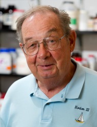 Kenneth Mann合作论文数Department of Biochemistry, Larner College of Medicine, University of Vermont;Haematologic Technologies, Inc.15
 Foley Jonathan H合作论文数12
 Marlys L. Koschinsky合作论文数Department of Physiology & Pharmacology, Schulich School of Medicine & Dentistry, Western University;Robarts Research Institute, Western University9
 John A Samis合作论文数7
 Michael B. Boffa合作论文数Robarts Research Institute, Western University;University of Western Ontario7Kg Mann合作论文数6
 Michael Kalafatis合作论文数Department of Chemistry;Cleveland State University;Department of Chemistry, Cleveland State University6
 Paula Babiarz Tracy合作论文数5Michael A. Bukys合作论文数5Chengliang Wu合作论文数4Tracy Paula B合作论文数4Paul Declerck合作论文数Therapeutic and Diagnostic Antibodies, Department of Pharmaceutical and Pharmacological Sciences, University of Leuven, Leuven, Belgium4Colin Downey合作论文数4
 Cheng Hock Toh合作论文数4Ann Gils合作论文数Department of Pharmaceutical and Pharmacological Sciences, KU Leuven, Leuven, Belgium4Liliana Tejidor合作论文数4Ka Stewart合作论文数3John Walker合作论文数3Alan Giles合作论文数3Becker Lev合作论文数3Fletcher B. Taylor合作论文数3Jerry A. Katzmann合作论文数3timothy j fischer合作论文数2Wim Houdijk合作论文数2RA HENRIKSEN合作论文数2Wg Owen合作论文数2N Brufatto合作论文数2Mark Schneider合作论文数2Greg Jones合作论文数Inivata Inc.2R Wenstone合作论文数2Rea Catherine J合作论文数2
 Jamila Hirbawi合作论文数2Manuel R P合作论文数2
 Lillicrap David合作论文数Department of Pathology and Molecular Medicine.Queen’s University2
 Paul F. Cook合作论文数University of Oklahoma Department of Chemistry and Biochemistry2R. Manuel合作论文数2A. Campbell合作论文数2John A. Samis合作论文数2Bruce A. Ramsay合作论文数Polyferm Canada Inc.2John Morser合作论文数2Karl-Uwe Petersen合作论文数2Tivadar Orban合作论文数2Pascale Champagne合作论文数2VM DOCTOR合作论文数2Nazareth Bastajian合作论文数2K. U. Petersen合作论文数2O.P. Malhotra合作论文数1LS HIBBARD合作论文数1




