AIM:This study aims to comprehensively investigate the dynamic expression pattern of CXCR4 in human dental pulp stromal cells (hDPSCs) and stromal cells from the apical papilla (SCAPs) and assess the impact of different cell dissociation methods on its precise assessment. On this basis, a standardised CXCR4 detection and cell sorting strategy can be established to ensure the high purity and viability of the sorted population. METHODOLOGY:hDPSCs and SCAPs were collected by the outgrowth method from human third molars from three independent healthy (ASA I) patients (< 18 years old). Cells were characterised as mesenchymal stromal cells (MSCs) and expanded to passage 3-6 for experiments. Three different dissociation methods (Enzymatic digestion, Non-enzymatic digestion, Mechanical collection) were used to collect single-cell suspensions from culture flasks. Afterwards, the cells were resuspended in culture medium and allowed to recover at 37°C/5% CO2 for 0-4 h. Surface expression of CXCR4 on viable cells was assessed using flow cytometry. Cell metabolic activity and senescence after dissociation treatment were tested by XTT Assay and senescence staining. Cell migration capacity was evaluated by wound healing assay. The data were statistically analysed using p < 0.05 as a statistical significance reference. RESULTS:hDPSCs and SCAPs exhibited typical MSC profiles and comparable cell viability under the same dissociation methods and time points. hDPSCs showed higher CXCR4 expression than SCAPs. CXCR4 expression displayed time-dependent fluctuations in both cell types and cell viability was significantly affected by dissociation method and recovery period. Enzymatic digestion resulted in higher CXCR4 expression after 2-h recovery and preserved over 90% cell viability. In contrast, non-enzymatic dissociation and mechanical scraping impaired cell metabolic activity, migration capacity, and induced premature senescence. CONCLUSION:In in vitro expanded hDPSCs and SCAPs, the cell-detachment method and recovery time can significantly affect the CXCR4 membrane detection and subsequent cell-survival performance. To balance optimal cell viability and detection reliability, enzymatic cell dissociation followed by a 2-h recovery time seems to be the best protocol for CXCR4 detection and subsequent downstream experiments within the 0-4 h time frame.
OBJECTIVE:To systematically evaluate current Artificial Intelligence (AI) based approaches for the diagnosis of impacted teeth other than third molars, and to assess their diagnostic performance, clinical relevance, and existing limitations. METHODS:PubMed, Web of Science, Cochrane Library, Embase, and Scopus were used to identify relevant studies. Study methodology, dataset preparation, and key metrics were collected from each included article. The Quality Assessment of Diagnostic Accuracy Studies (QUADAS-2) was applied. RESULTS:Of the 30 included studies, 15 focused on mesiodens and 15 on impacted canines. No AI-based diagnostic studies on impacted premolars or incisors were identified. Panoramic radiographs were the most commonly used imaging modality, followed by CBCT and periapical radiographs. Across detection and classification tasks, most AI models demonstrated good to excellent diagnostic performance, with more than 70% of reported metrics exceeding 0.80. YOLO- and ResNet-based architectures were most frequently applied, with YOLO models generally achieving comparatively higher accuracy. Segmentation models showed high spatial agreement, with Dice similarity coefficients frequently exceeding 0.90. CBCT-based studies demonstrated strong performance for three-dimensional assessment, particularly for segmentation tasks, while alternative diagnostic materials and ANN-based approaches yielded moderate to high predictive accuracy for unerupted tooth size. CONCLUSIONS:AI-based diagnostic systems show promising potential in the detection, classification, segmentation and prediction of impacted teeth other than third molars, offering valuable support for early intervention and personalized treatment planning. Future research should aim to include larger datasets, potentially through multi-center collaboration, as well as following standardized evaluation protocols, using more easily interpretable AI techniques (such as Grad-CAM, SHAP) to ensure robust, transparent, and clinically reliable implementation. CLINICAL SIGNIFICANCE:AI provides accurate and efficient tools for detecting and classifying impacted teeth, enabling early diagnosis and personalized treatment planning. Its integration may improve diagnostic Accuracy and streamline orthodontic workflows, but broader adoption requires multi-center validation and explainable models to ensure reliability.
The history of stromal-derived factor-1 (SDF-1), alias CXCL12, started serendipitously and relatively late in the cytokine cDNA cloning era (1975-2000) and evolved at the biological level from progenitor cell-specific chemokine in the bone marrow to multifunctional cytokine with growth factor-like and tissue-regenerative activities. This evolution was parallelled by the integration of SDF-1/CXCL12 within the protein families of chemokines, cytokines and cell growth-promoting recombinant products having the potential for clinical applications. Here, we use this central position of CXCL12 as small signaling protein as an example for future developments in regenerative medicine. We provide context about SDF-1 biology within the field of skin wound healing research and how this compares with studies of other cytokines and growth factors. We also discuss whether SDF-1 formulations may be exemplary for other cytokines used for tissue regeneration. Normal skin wound healing is fraught with delays and complications in patients with specific underlying diseases, such as diabetes, hypertension and other elderly-related comorbidities, skin infections and accidental physical insults. Except for platelet-derived growth factor (PDGF), many cytokines, including vascular endothelial growth factor (VEGF) and epidermal growth factor (EGF), have failed so far in clinical studies of skin wound healing. This is in part due to the fact that (i) the biology of tissue regeneration is complex and insufficiently studied, (ii) in vitro approaches hardly mimic in vivo situations and (iii) commonly used animal models of acute and chronic wounding do not perfectly match human skin wound regeneration. A review of critical cells and molecules in normal skin and their actions in wounded tissue and a balanced comparison of the recent literature are preambles for progress in wound repair. We define advantages and limitations of recent approaches and appeal for more research. In particular, the possibilities of cellular immunomodulation mediated by endogenous and exogenous SDF-1/CXCL12 as a key molecule for skin regeneration are reviewed. Furthermore, biomaterials and scaffolds for the delivery and use of cytokines in precision medicine and aspects of their biofabrication are outlined with SDF-1 as an example. Finally, we indicate how applications of dermatological SDF-1 formulations for skin wound healing may be tailored for applications in other acute and chronic inflammatory conditions and regenerative medicine. Thereby, SDF-1/CXCL12 is placed at the crossroads between recombinant products, cytokines, chemokines and growth factors and occupies a central position between regenerative biology and medicine.
OBJECTIVES:To longitudinally follow up a cohort of patients with autotransplanted teeth, assessing how the timing of starting orthodontic treatment impacts root length. MATERIALS AND METHODS:Patients under 18 with at least one open-apex autotransplanted premolar (AP) replaced to a central incisor position were included. Root/crown ratio (RCR) was calculated on periapical radiographs taken at intervals of 3-6 months after transplantation. The RCR of AP in patients without orthodontic treatment and patients where orthodontic treatment was initiated at different time points (3,6,9,12,18 months) were compared. RESULTS:52 autotransplanted premolars were included and 315 peri-apical radiographs were collected. The median timespan between autotransplantation and the start of orthodontic force (OF) application was 8.5 (IQR 2-23) months, with a mean follow-up period of 32 (IQR 9-46) months. Early OF post-autotransplantation correlated with early stagnation of root development. When OF was delayed, AP tended to show more root lengthening. Delayed OF was linked to a higher incidence of External Apical Root Resorption (EARR) once forces were introduced, ultimately resulting in root lengths comparable to those with early OF. The presence or absence of OF is the factor most notably influencing the final root length of AP. CONCLUSIONS:The timing of OF application did not compromise the final root length of autotransplanted teeth. Early OF to prevent ankylosis is feasible without affecting root length. Future research should utilize 3D imaging to provide a deeper understanding of orthodontic treatment's effects on autotransplanted teeth. CLINICAL RELEVANCE:The study underscores the importance of tailored orthodontic intervention timing in autotransplanted teeth, balancing early force application to avoid ankylosis against the risk of EARR.
Orofacial bone tissue engineering addresses bone loss caused by trauma, malformations, or tumors, enabling restoration and implant rehabilitation. Angiogenesis plays a crucial role in osteogenesis by ensuring nutrient and oxygen transport essential for bone regeneration. Preclinical large animal models are vital for translational research and require noninvasive, nondestructive methods aligned with 3Rs principles (Replacement, Reduction, and Refinement) to assess angiogenesis. This study proposes high-resolution cone-beam computed tomography subtraction angiography (HR-CBCT-SA) adapted for the orofacial region as an innovative method for monitoring angiogenesis during jawbone regeneration. Three Yucatan minipigs with a surgically created buccal wall jawbone defect per hemimandible were followed for 90 days by CBCT-SA to assess vascular remodeling. Morphometric parameters, including vessel number, node count, radius, and length, were analyzed and validated against histological morphometry. CBCT-SA revealed vascular dynamics during healing. By day 10, increased vessel and node counts along with reduced vessel radius and length indicated neoangiogenesis. At day 30, vessel maturation was aligned with transition of fibrous tissue to osteoid matrix deposition. By day 90, vascular metrics stabilized, reflecting bone remodeling phases characterized by replacement of lamellar and medullary bone replacement. Extrabony vascular networks underwent more pronounced changes than intrabony vessels, underscoring the leading role of periosteum in regeneration. Histology validated CBCT-SA findings, although resolution limitations prevented detection of vessels smaller than 500 µm. Nevertheless, CBCT-SA captured angiogenic changes over time and supported nondestructive monitoring without compromising tissue integrity. This study establishes HR-CBCT-SA as a reliable, nondestructive imaging technique for assessing vascular changes during jawbone regeneration in preclinical models. It demonstrates significant translational potential because of the clinically validated use of CBCT-angiography. Advances in artificial intelligence (AI)-driven image analysis are expected to enhance sensitivity and accuracy, improving vascular assessment. Moreover, this approach can be extended for investigating vascular-related oral pathologies (e.g., radiochemical osteonecrosis of the jaws), offering valuable tool to advance research in jawbone regeneration.
Orthodontically induced inflammatory root resorption (OIIRR) is one of the most common complications in dentistry. It is considered irreversible and harmful to tooth integrity. The narrow, irregular morphology and complex, layered structure of periodontal and periapical tissues, combined with a potentially bacteria-laden inflammatory environment, make treatment and prevention an immense challenge. This study presents a biomimetic-engineered thermoresponsive hydrogel with distinct physicochemical properties that serves as a biological scaffold to support the growth and differentiation of periodontal ligament stem cells (PDLSCs), while maintaining optimal flow for injectability and diffusion. Additionally, it provides strong antibacterial protection and excellent biocompatibility, allowing in vivo tracking of stem cells. Furthermore, it mimics the native stem cell niche, with slow-release rapamycin inducing autophagy and orchestrating cascades that modulate adipogenic and osteogenic differentiation, mechanical stress response, migration, redox homeostasis, inflammation, and stress adaptation. This enables encapsulated PDLSCs to thrive even in inflammatory, stressful environments, preventing excessive resorption in the periodontal-apical complex in an animal model. This innovative approach pioneers a new frontier in the prevention of root resorption, offering a potential game-changer for dentistry. The hydrogel design further opens exciting possibilities for regenerative treatments targeting other systemic inflammatory diseases.
Nanoparticles are emerging as transformative agents in endodontics, addressing challenges in treating the dentin-pulp complex. This scoping review aims to explore multifunctional applications of nanoparticles in endodontics, with a focus on their roles in promoting tissue regeneration through therapeutic effects, enhancing material properties, and serving a carrier function. Following PRISMA-Scoping Review guidelines, a comprehensive literature search was conducted across Web of Science, PubMed, and Scopus. A total of 490 articles were initially identified, of which 92 met the preliminary eligibility criteria. Following full-text screening, 70 studies were included in the qualitative synthesis. Key findings from both in vitro and in vivo studies are summarized in tabular form. Results reveal a notable imbalance in the types of nanomaterials studied: inorganic nanomaterials were reported in 77
Bone disorders represent a significant global burden. Currently, animal models are used to develop and screen novel treatments. However, interspecies variations and ethical concerns highlight the need for a more complex 3D bone model. In this study, we developed a simplified in vitro bone-like model using a U-CUP perfusion-based bioreactor system, designed to provide continuous nutrient flow and mechanostimulation through 3D cultures. An immortalized human fetal osteoblastic cell line was seeded on collagen scaffolds and cultured for 21 days in both a perfusion bioreactor system and in static cultures. PrestoBlue™ assay, scanning electron microscopy, and proteomics allowed monitoring of metabolic activity and compared morphological and proteome differences between both conditions. Results indicated an altered cellular morphology in the bioreactor compared to the static cultures and identified a total of 3494 proteins. Of these, 105 proteins exhibited significant upregulation in the static culture, while 86 proteins displayed significant downregulation. Enrichment analyses of these proteins revealed ten significant pathways including epithelial-mesenchymal transition, TNF-alpha signaling via NF-kB, and KRAS pathway. The current data indicated of osteogenic differentiation enhancement within the bioreactor on day 21 compared to static cultures. In conclusion, the U-CUP perfusion bioreactor is beneficial for facilitating osteogenic differentiation in 3D cultures.
Introduction: As personalized medicine advances, there is an escalating need for sophisticated tools to understand complex biomechanical phenomena in clinical research. Recognizing a signi ficant gap, this study pioneers the development of patient-speci fic in silico models for tooth autotransplantation (TAT), setting a new standard for predictive accuracy and reliability in evaluating TAT outcomes. Methods: Development of the models relied on 6 consecutive cases of young patients (mean age 11.66 years +/- 0.79), all undergoing TAT procedures. The development process involved creating detailed in silico replicas of patient oral structures, focusing on transplanting upper premolars to central incisors. These models underpinned finite element analysis simulations, testing various masticatory and traumatic scenarios. Results: The models highlighted critical biomechanical insights. The finite element models indicated homogeneous stress distribution in control teeth, contrasted by shapedependent stress patterns in transplanted teeth. The surface deviation in the postoperative year for the transplanted elements showed a mean deviation of 0.33 mm (+/- 0.28), signi ficantly higher than their contralateral counterparts at 0.05 mm (+/- 0.04). Conclusions: By developing advanced patient-speci fic in silico models, we are ushering in a transformative era in TAT research and practice. These models are not just analytical tools; they are predictive instruments capturing patient uniqueness, including anatomical, masticatory, and tissue variables, essential for understanding biomechanical responses in TAT. This foundational work paves the way for future studies, where applying these models to larger cohorts will further validate their predictive capabilities and in fluence on TAT success parameters. (J Endod 2024;50:820-826.)
Traumatic dental injuries (TDIs) are a frequent challenge in pediatric dentistry, requiring a coordinated interdisciplinary approach for successful management. These injuries can affect various dental tissues and pose significant diagnostic and therapeutic challenges, particularly in young patients with developing dentition. This review aims to explore the complexities of managing TDIs in young permanent dentition, focusing on the role of orthodontics, endodontics, and other specialties in providing comprehensive care. We highlight the importance of interdisciplinary collaboration in achieving optimal functional and aesthetic outcomes, emphasizing the need for updated diagnostic tools and treatment protocols tailored to the growing child. Specific recommendations are provided for clinicians on integrating orthodontic and endodontic management in trauma cases, focusing on minimizing complications and ensuring long-term prognosis.
Background: To investigate the 2-year radiographic outcome of cone-beam computed tomography (CBCT)-guided tooth autotransplantation (TAT) of a premolar to the maxillary incisor region in children. Methods: Eighteen paediatric patients with a missing maxillary incisor were recruited for CBCT-guided premolar transplantation to the maxillary central incisor region (TAT group), neighbouring a healthy contralateral central incisor (control group). Radiological parameters, assessed at 2-year follow-up, included periodontal ligament space (PLS), the outline of lamina dura, the vertical distance between the cementoenamel junction and crestal bone (CEJ-CB), pulp obliteration, radiographic root area (RRA) and length (RRL) and crown-root ratio (CRR). Results: Positive root growth in 89% of the TAT cases, with an average increase in RRA by 27% compared to the control. Pulp obliteration was observed in 83% of the TAT cases (partial: 55.6%; complete: 27.8%). The average crown-to-root ratio was 0.72 and 1.01 for the control and TAT group, respectively. No significant differences existed between TAT and control concerning PLS (p=0.09), lamina dura (p=0.12), and CEJ-CB distance mesially (p=0.17) or distally (p=0.28). Conclusions: CBCT-guided TAT might act as a viable treatment option for rehabilitating missing maxillary (centrally) incisor teeth in paediatric patients when osseointegration may not be the preferred option. (Clinical trial center and ethical board University Hospitals, KU Leuven: S55287; ClinicalTrials.gov Identifier: NCT02464202)
Incorporation of growth factors, signaling molecules and drugs can be vital for the success of tissue engineering in complex structures such as the dentoalveolar region. This has led to the development of a variety of drug release systems. This study aimed to develop pNIPAM-methylcellulose microgels with different synthesis parameters based on a 23 full factorial design of experiments for this application. Microgel properties, including volume phase transition temperature (VPTT), hydrodynamic size, drug loading and release, and cytocompatibility were systematically evaluated. The results demonstrated successful copolymerization and development of the microgels, a hydrodynamic size ranging from -200 to -500 nm, and VPTT in the range of 34-39 degrees C. Furthermore, loading of genipin, capable of inducing odontoblastic differentiation, and its sustained release over a week was shown in all formulations. Together, this can serve as a solid basis for the development of tunable drug-delivering pNIPAM-methylcellulose microgels for specific tissue engineering applications.
Objective This article reviews the applications of Finite Element Models (FEMs) in personalized dentistry, focusing on treatment planning, material selection, and CAD-CAM processes. It also discusses the challenges and future directions of using Finite Element Analysis (FEA) in dental care. Data This study synthesizes current literature and case studies on FEMs in personalized dentistry, analyzing research articles, clinical reports, and technical papers on the application of FEA in dental biomechanics. Sources Sources for this review include peer-reviewed journals, academic publications, clinical case studies, and technical papers on dental biomechanics and Finite Element Analysis. Key databases such as PubMed, Scopus, Embase, and ArXiv were used to identify relevant studies. Study Selection Studies were selected based on their relevance to the application of FEMs in personalized dentistry. Inclusion criteria were studies that discussed the use of FEA in treatment planning, material selection, and CAD-CAM processes in dentistry. Exclusion criteria included studies that did not focus on personalized dental treatments or did not utilize FEMs as a primary tool. Conclusions FEMs are essential for personalized dentistry, offering a versatile platform for in-silico dental biomechanics modeling. They can help predict biomechanical behavior, optimize treatment outcomes, and minimize clinical complications. Despite needing further advancements, FEMs could help significantly enhance treatment precision and efficacy in personalized dental care. Clinical Significance FEMs in personalized dentistry hold the potential to significantly improve treatment precision and efficacy, optimizing outcomes and reducing complications. Their integration underscores the need for interdisciplinary collaboration and advancements in computational techniques to enhance personalized dental care.
SDF-1/CXCL12 is a unique chemotactic factor with multiple functions on various types of precursor cells, all carrying the cognate receptor CXCR4. Whereas individual biological functions of SDF-1/CXCL12 have been well documented, practical applications in medicine are insufficiently studied. This is explained by the complex multifunctional biology of SDF-1 with systemic and local effects, critical dependence of SDF-1 activity on aminoterminal proteolytic processing and limited knowledge of applicable modulators of its activity. We here present new insights into modulation of SDF-1 activity in vitro and in vivo by a macromolecular compound, chlorite-oxidized oxyamylose (COAM). COAM prevented the proteolytic inactivation of SDF-1 by two inflammation-associated proteases: matrix metalloproteinase-9/MMP-9 and dipeptidylpeptidase IV/DPPIV/CD26. The inhibition of proteolytic inactivation was functionally measured by receptor-mediated effects, including intracellular calcium mobilization, ERK1/2 phosphorylation, receptor internalization and chemotaxis of CXCR4-positive cells. Protection of SDF-1/CXCL12 against proteolysis was dependent on electrostatic COAM-SDF-1 interactions. By in vivo experiments in mice, we showed that the combination of COAM with SDF-1 delivered through physiological fibrin hydrogel had beneficial effect for the healing of skin wounds. Collectively, we show that COAM protects SDF-1 from proteolytic inactivation, maintaining SDF-1 biological activities. Thus, protection from proteolysis by COAM represents a therapeutic strategy to prolong SDF-1 bioavailability for wound healing applications.
While available treatments have addressed a variety of complications in the dentoalveolar region, associated challenges have resulted in exploration of tissue engineering techniques. Often, scaffold biomaterials with specific properties are required for such strategies to be successful, development of which is an active area of research. This study focuses on the development of a copolymer of poly (N-isopropylacrylamide) (pNIPAM) and chitosan, used for 3D printing of scaffolds for dentoalveolar regeneration. The synthesized material was characterized by Fourier transform infrared spectroscopy, and the possibility of printing was evaluated through various printability tests. The rate of degradation and swelling was analyzed through gravimetry, and surface morphology was characterized by scanning electron microscopy. Viability of dental pulp stem cells seeded on the scaffolds was evaluated by live/dead analysis and DNA quantification. The results demonstrated successful copolymerization, and three formulations among various synthesized formulations were successfully 3D printed. Up to 35% degradability was confirmed within 7 days, and a maximum swelling of approximately 1200% was achieved. Furthermore, initial assessment of cell viability demonstrated biocompatibility of the developed scaffolds. While further studies are required to achieve the tissue engineering goals, the present results tend to indicate that the proposed hydrogel might be a valid candidate for scaffold fabrication serving dentoalveolar tissue engineering through 3D printing.
Introduction:Wound healing is a complex process to restore homeostasis after injury and insufficient skin wound healing is a considerable problem in medicine. Whereas many attempts of regenerative medicine have been made for wound healing with growth factors and cell therapies, simple pharmacological and immunological studies are lagging behind. We investigated how fibrin hydrogels modulate immune cells and molecules in skin wound healing in mice.Methods:Physiological fibrin hydrogels (3.5 mg/mL fibrinogen) were generated, biophysically analyzed for stiffness and protein contents and were structurally studied by scanning electron microscopy. Physiological fibrin hydrogels were applied to full thickness skin wounds and, after 3 days, cells and molecules in wound tissues were analyzed. Leukocytes, endothelial cells, fibroblasts and keratinocytes were explored with the use of Flow Cytometry, whereas cytokines and matrix metalloproteinases were analyzed with the use of qPCR, ELISAs and zymography. Skin wound healing was analyzed microscopically at day 3, macroscopically followed daily during repair in mice and compared with commercially available fibrin sealant Tisseel.Results:Exogenous fibrin at physiological concentrations decreased neutrophil and increased non-classical Ly6Clow monocyte and resolutive macrophage (CD206+ and CX3CR1+) populations, at day 3 after injury. Fibrin hydrogel reduced the expression of pro-inflammatory cytokines and increased IL-10 levels. In line with these findings, gelatinase B/MMP-9 was decreased, whereas gelatinase A/MMP-2 levels remained unaltered. Frequencies of dermal endothelial cells, fibroblasts and keratinocytes were increased and keratinocyte migration was enhanced by fibrin hydrogel. Importantly, physiological fibrin accelerated the healing of skin wounds in contrast to the highly concentrated fibrin sealant Tisseel, which delayed wound repair and possessed a higher fiber density.Conclusion:Collectively, we show that adding a tailored fibrin hydrogel scaffold to a wound bed positively influences the healing process, modulating leukocyte populations and inflammatory responses towards a faster wound repair.
Oral health is essential for a good overall health. Dento-alveolar conditions have a high prevalence, ranging from tooth decay periodontitis to alveolar bone resorption. However, oral tissues exhibit a limited regenerative capacity, and full recovery is challenging. Therefore, regenerative therapies for dento-alveolar tissue (e.g., alveolar bone, periodontal membrane, dentin-pulp complex) have gained much attention, and novel approaches have been proposed in recent decades. This review focuses on the cells, biomaterials and the biofabrication methods used to develop therapies for tooth root bioengineering. Examples of the techniques covered are the multitude of additive manufacturing techniques and bioprinting approaches used to create scaffolds or tissue constructs. Furthermore, biomaterials and stem cells utilized during biofabrication will also be described for different target tissues. As these new therapies gradually become a reality in the lab, the translation to the clinic is still minute, with a further need to overcome multiple challenges and broaden the clinical application of these alternatives.
Introduction: Understanding the healing process of dental pulp after tooth autotransplantation (TAT) and regenerative endodontic treatment (RET) of immature teeth is important both clinically and scientifically. This study aimed to characterize the pattern of dental pulp healing in human teeth that underwent TAT and RET using state-of-the-art im-aging techniques. Materials and Methods: This study examined 4 human teeth, 2 premolars that underwent TAT, and 2 central incisors that received RET. The premolars were extracted after 1 year (case 1) and 2 years (case 2) due to ankylosis, while the central incisors were extracted after 3 years (cases 3 and 4) for orthodontic reasons. Nanofocus x-ray computed tomography was used to image the samples before being processed for histological and immunohistochemical analysis. Laser scanning confocal second harmonic generation imaging (SHG) was used to examine the patterns of collagen deposition. A maturity-matched premolar was included as a negative control for the histological and SHG analysis. Results: Analysis of the 4 cases revealed different patterns of dental pulp healing. Similarities were observed in the progressive obliteration of the root canal space. However, a striking loss of typical pulpal architecture was observed in the TAT cases, while a pulp-like tissue was observed in one of the RET cases. Odontoblast-like cells were observed in cases 1 and 3. Conclusions: This study provided insights into the patterns of dental pulp healing after TAT and RET. The SHG imaging sheds light on the patterns of collagen deposition during reparative dentin formation.
The biomechanics of transplanted teeth remain poorly understood due to a lack of models. In this context, finite element (FE) analysis has been used to evaluate the influence of occlusal morphology and root form on the biomechanical behavior of the transplanted tooth, but the construction of a FE model is extremely time-consuming. Model order reduction (MOR) techniques have been used in the medical field to reduce computing time, and the present study aimed to develop a reduced model of a transplanted tooth using the higher-order proper generalized decomposition method. The FE model of a previous study was used to learn von Mises root stress, and axial and lateral forces were used to simulate different occlusions between 75 and 175N. The error of the reduced model varied between 0.1% and 5.9% according to the subdomain, and was the highest for the highest lateral forces. The time for the FE simulation varied between 2.3 and 7.2 h. In comparison, the reduced model was built in 17s and interpolation of new results took approximately 2.10−2s. The use of MOR reduced the time for delivering the root stresses by a mean 5.9 h. The biomechanical behavior of a transplanted tooth simulated by FE models was accurately captured with a significant decrease of computing time. Future studies could include using jaw tracking devices for clinical use and the development of more realistic real-time simulations of tooth autotransplantation surgery.
The aim of this study was to compare the quantification of hard-tissue debris by using micro-computed tomography (micro-CT) and nano-focus computed tomography (nano-CT) after root canal instrumentation. Ten mandibular molars containing an isthmus in the mesial root were scanned in a SkyScan 1172 micro-CT device with a voxel size of 12.8 µm and in a NanoTom nano-CT device with 5.5 µm. The mesial root canals were irrigated with 5 mL of saline solution at the orifice level, instrumented with Reciproc R25 files and a second scanning was performed by micro-CT and nano-CT devices for post-instrumentation images. DataViewer software was used for registering the pre- and post-operative micro-CT and nano-CT images. The root canal and the debris were segmented for quantitative analysis of the volume of the canal and volume of debris using CTAn software. Statistical analysis was performed using the T test for comparison between volume of the canal after instrumentation and volume of debris in both image modalities. The level of significance was set at p < 0.05. Nano-CT images showed higher values of debris when compared with micro-CT (p < 0.05) after root canal instrumentation. No difference was observed between the volume of the root canal after instrumentation in the two imaging methods used (p > 0.05). Nano-CT technology can be recommended as a more precise method for quantitative analysis of hard-tissue debris. Moreover, in Endodontic research it is a promising method, as it is capable of providing higher spatial and contrast resolution, faster scanning and higher image quality.
 Reinhilde Jacobs合作论文数DentoMaxillofacial Radiology Center, University Hospital Leuven;Department of Imaging and Pathology, Faculty of Movement and Rehabilitation Sciences, Katholieke Universiteit Leuven;Faculty of Medicine, Katholieke Universiteit Leuven;Department of Dental Medicine, Karolinska Institutet45
 Constantinus Politis合作论文数Faculty of Medicine, KU Leuven;Department of Oral and Maxillofacial Surgery, Universitaire Ziekenhuizen Leuven11
 Ivo Lambrichts合作论文数Biomedical Research Institute, Hasselt University;Laboratory for Histology and Regeneration, Hasselt University8
 Pierre Lahoud合作论文数UZ Leuven, Katholieke Univ Leuven6
 Ghislain Opdenakker合作论文数Department of Microbiology, Immunology and Transplantation, Faculty of Medicine, KU Leuven6
 Van Gorp Gertrude合作论文数5
 Annabel Braem合作论文数CERN5
 Paul Lambrechts合作论文数Department of Oral Health Science, Faculty of Medicine, Katholieke Universiteit Leuven5Guy Willems合作论文数5E Shaheen合作论文数4
 Mehdi Salar Amoli合作论文数Oral & Maxillofacial Surg, Univ Hosp Leuven4
 Mario Tanomaru Filho合作论文数Departamento de Odontologia Restauradora, Faculdade de Odontologia de Araraquara, Universidade Estadual Paulista Júlio de Mesquita Filho4
 Veerle Bloemen合作论文数4
 Nastaran Meschi合作论文数2 Department of Oral Health Sciences, University Hospitals Leuven4
 Karla de Faria Vasconcelos合作论文数OMFS IMPATH Research Group, Department of Imaging & Pathology, Faculty of Medicine, University of Leuven and Oral & Maxillofacial Surgery, University Hospitals Leuven, Leuven, Belgium
Department of Oral Diagnosis, Division of Oral Radiology, Piracicaba Dental School, State University of Campinas, Piracicaba, Brazil4
Karla de Faria Vasconcelos合作论文数OMFS IMPATH Research Group, Department of Imaging & Pathology, Faculty of Medicine, University of Leuven and Oral & Maxillofacial Surgery, University Hospitals Leuven, Leuven, Belgium
Department of Oral Diagnosis, Division of Oral Radiology, Piracicaba Dental School, State University of Campinas, Piracicaba, Brazil4 Maria Cadenas de Llano Perula合作论文数4Vital Sibylle Opsahl合作论文数4Benjamin Salmon合作论文数4
 Catherine Chaussain合作论文数Faculté de Santé, Université Paris Cité;Assistance Publique Hôpitaux de Paris;Lab Oral Health, Dental School, Université Paris Cité4
 Eman Shaheen合作论文数Department of Radiology, University Hospitals Leuven4
 Elke Van De Casteele合作论文数4
 Juliane Maria Guerreiro Tanomaru合作论文数3
 Resmi Anand合作论文数3
 Fernanda Ferrari Esteves Torres合作论文数3Ruchika Joon合作论文数3
 Bernardo Camargo dos Santos合作论文数3
 Raphael Richert合作论文数Université Claude Bernard Lyon 1, Université de Lyon3
 Zohre Mousavi Nejad合作论文数Biotechnol Res Ctr, Shiraz Univ Med Sci3
 Maxime Ducret合作论文数Faculty of Dentistry, McGill University3
 Yi Sun合作论文数3
 Annelies Bronckaers合作论文数3
 Claire Bardet合作论文数3
 Ruparel Nikita B合作论文数Department of Endodontics, University of Texas Health Science Center at San Antonio3
 Austah Obadah N合作论文数3
 Vanessa Chrepa合作论文数Department of Endodontics, University of Texas Health Science Center at San Antonio3
 Van Dessel Jeroen合作论文数Faculty of Medicine, University Hospitals Leuven3
 Sohaib Shujaat合作论文数College of Dentistry, University of Dammam3
 Pereira Rafaela Vaz合作论文数Laboratório de Imunobiofotônica, UFMG3Wim Coucke合作论文数3
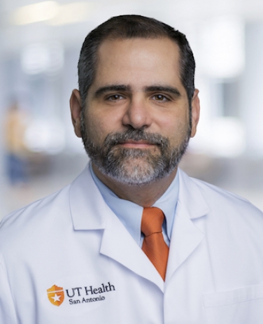 Anibal Diogenes合作论文数Department of Cell Systems & Anatomy, The University of Texas Health Science Center at San Antonio;Department of Endodontics, The University of Texas Health Science Center at San Antonio3Maryam Shahbazian合作论文数3L. Daems合作论文数2F Mangione合作论文数2J Lesieur合作论文数2Andres Eduardo Torres Garcia合作论文数2wael khalil合作论文数2Weston Fath合作论文数2
 Francesca Mangione合作论文数2Ali Al-Rimawi合作论文数2




