Amaç: Bu çalışmada beyin ölümü gerçekleşen hastalarda organ bağışının seyrini araştırmak, organ bağışından organ nakline kadar olan aşamaları incelemek, kullanılmayan veya reddedilen vericilerin nedenlerini ortaya çıkarmak ve çözüm önerileri ortaya konması amaçlandı. Gereç ve Yöntemler: Ocak 2015-Aralık 2016 tarihleri arasında, aile izinleri olan tüm kadavra bağışçılarının Sağlık Bakanlığı Ulusal Koordinasyon Merkezi Başkanlığı arşivlerindeki donör kayıt verilerini inceledik. Donör adaylarının demografik verileri, yoğun bakım kalış süresindeki verileri ve donör olarak kullanılmama nedenleri incelendi. Sonuçlar: 985 donör incelendi; %65'i erkek, ortalama yaş 47.5 (0-96) idi ve donörlerin %65'inde nörolojik nedenlerle beyin ölümü gerçekleşmişti. Ortalama beyin ölümü rapor süresi 5.4 ± 9.3 (1-169) gündü. Donörlerin 169’unda (%17.1) enfeksiyon tespit edildi. Ortalama yoğun bakımda kalış süresi 8,55 ± 4,42 (2-38) gündü. Donörlerin sadece 159’undan (%16,2) kalp grefti alındı. 495 (%50,2) donör tıbbi nedenlerle reddedildi, 64 (%6,5) donör, alıcı merkezler tarafından reddedildiği için kalp nakli için uygun değildi. Potansiyel bağışçıların %20'sinde kalp bağışı için aile onayı alınmadı. Aile onayı olmayan bağışçıların %70'i 65 yaşın altındaydı ve %67'sinin greft olarak kalp kullanımından kaçınmak için herhangi bir tıbbi sorunu yoktu. Kalp onayı olmayan donörlerde greft olarak karaciğer kullanım oranı %71, böbrek için kullanım oranı %70 idi. Sonuç: Beyin ölümü tanısından donörlerin bakımına kadar olan süreci yürütecek ve yönlendirecek organ nakli ekiplerine ihtiyacımız vardır.
Objectives: Although the mortality rate of primary ventricular septal defect (VSD) closure surgery has been reduced to <0.5% in many centers, low cardiac output syndrome and multi-organ failure syndrome development in the post-operative period are still the most common causes of mortality and morbidity. The use of biochemical markers in the early period can be used as effective predictors for reducing mortality and morbidity. In this study, the effect of post-operative serum lactate and cardiac troponin I (cTNI) levels on hospital mortality in patients who underwent primary closure of VSD was investigated retrospectively. Methods: The effect of lactate and cTNI values on hospital mortality in 52 patients who underwent surgical repair for VSD was investigated. Serum lactate levels of all patients in the first 6 h after the operation; on the first post-operative day, cTNI values were examined retrospectively. Serum lactate levels exceeding 3.5 mmol/ lt and cTNI values exceeding 35 ng/mL were determined as the cut-off points. Results: Serum lactate level and cTNI increase were found to be statistically different in the mortality group (p<0.05). It was shown that increased cTNI was associated with weight, left ventricular end-diastolic diameter, and cardiopulmonary bypass time (p<0.05). Serum lactate levels were 4.1±0.5 mmol/lt in the non-mortality group and 15.3±8.3 mmol/lt in the mortality group, and the difference was statistically significant (p<0.05). Hospital mortality was observed in 6 patients. Conclusion: In patients undergoing VSD closure surgery, serum lactate levels measured in the first 6 h postoperatively and cTNI levels measured on the first post-operative day are associated with hospital mortality. Concomitant elevations of lactate and cTNI values in patients after VSD closure should suggest to take caution and early supportive treatments to reduce mortality.
Purpose: The aim of this study was to analyze neurocognitive function in patients who underwent continuous flow left ventricular assist device (LVAD) implantation. Material and Method: This cross-sectional study included three groups: LVAD (n = 31), heart failure patients (n = 26), and healthy volunteers (n = 27). The Rey Auditory-Verbal Learning Test (RAVLT), Judgement of Line Orientation Test (JLOT), Trail Making Test (TMT), Stroop Color-Word Interference Test (SCWIT), Verbal Fluency Test (VFT), Symbol-Digit Modality Test (SDMT) were used to assess the neurocognitive functions. Data were analyzed at a median 12 (3-47) months after LVAD implantation. The LVAD patients were also divided by aortic valve opening (AVO) into three subgroups as "closed" (n = 9), "1-6" (n = 8) and "7-10" (n = 14) opening per ten beats and data were re-analyzed accordingly. Results: There was no significant difference among the groups according to SCWIT, JLOT, SDMT, TMT, and VFT scores. Post-hoc analyzes of RAVLT scores showed significant differences between the LVAD and the other two groups in favor of the LVAD group. Also, the patients with AVO "7-10" the response times were longer and learning scores were found to be lower than those without AVO. Conclusion: With continuous-flow LVAD, neurocognitive functions were not impaired. The learning performance was better in cases where there was no AVO and flow was completely device dependent. We may speculate that neurocognitive functions are not worsening with continuous cerebral blood flow and even it may improve learning performance.
BACKGROUND: Heart transplantation is limited by the scarcity of suitable donors. Patients with advanced biventricular failure may require biventricular support to provide optimal cardiac output and end-organ perfusion. We highlight the outcomes of using the HeartWare HVAD System (HVAD) in a biventricular configuration. METHODS: This retrospective study included patients implanted with HVAD as a biventricular assist device (BiVAD) between 2009 and 2017 at 12 participating centers. When used as a right ventricular assist device (VAD) (RVAD), the HVAD can be attached to the right ventricle (RV) or the right atrium (RA). Kaplan-Meier survival estimates were calculated comparing the 2 RVAD implant locations. Comparisons were also made between the timing of RVAD implantation (primary vs staged) on adverse event (AE) profiles and survival. RESULTS: Among the 93 patients who were implanted with a HVAD BiVAD, Kaplan-Meier survivals at 1-year and 2-year were 56% and 47%, respectively. Survival was independent of the location of the HVAD RVAD implant or whether there was an interval between left VAD and RVAD implantation. The most common AEs were bleeding (35.5%), infection (25.8%), and respiratory failure (20.4%). CONCLUSIONS: This study illustrated similar survival in patients receiving a primary or staged HVAD BiVAD implant at 1 year and 2 years. This study also established that the locations of the RVAD implant (RV or RA) result in similar AE profiles. (C) 2020 International Society for Heart and Lung Transplantation. All rights reserved.
Background: This study aims to evaluate quality of life among patients with an implanted left ventricular assist device in two different populations. Methods: This cross-sectional study included a total of 20 patients (15 males, 5 females; mean age 41.8 +/- 13.4 years; range, 20 to 65 years) from Turkey and 40 patients (33 males, 7 females; mean age 55.1 +/- 11.6 years; range, 21 to 76 years) from Germany who underwent left ventricular assist device implantation and reached their third month of treatment between January 2016 and August 2016. The Euro Quality of Life-5 Dimension Questionnaire and Kansas City Cardiomyopathy Questionnaire-12 were used to assess quality of life of both groups. Results: Sixty-five percent of the German patients returned to work, while none of the Turkish patients returned after implantation (p=0.000). The Turkish (70%) and German (40%) patients reported that they had difficulty in living with a left ventricular assist device (p=0.028). The mean Euro Quality of Life-5 Dimension Questionnaire visual analog scale scores of the Turkish and German patients were 70.50% and 62.38%, respectively. The mean Kansas City Cardiomyopathy Questionnaire-12 scores of the Turkish patients were significantly higher than those of the German patients (p=0.01). Conclusion: Left ventricular assist device implantation improved the quality of life of both Turkish and German patients. Turkish patients with left ventricular assist device should be encouraged to adopt a more independent life, not to limit their lives to home and to return to work according to their functional capacity.
AIM:Pump thrombosis (PT) is a detrimental complication of left ventricular assist device (LVAD) therapy. There is no consensus on optimal PT treatment. The aim of this study was to present a treatment strategy for patients with PT.METHOD:The hospital records of patients who underwent isolated LVAD implantation between May 2013 and October 2018 were retrospectively evaluated. Pump thrombosis was suspected in the setting of impaired flow/power parameters and haemolysis. Protocols for the management of suspected PT varied by patient presentation. Parameters that increased the PT risk were investigated by dividing the patients into two groups according to the presence of PT. Preoperative and operative data were analysed.RESULTS:Pump thrombosis was observed in 20 of 81 patients. All patients with PT presented elevated lactate dehydrogenase levels and higher power and/or low-/high-flow alarm at admission. All patients were treated initially with intravenous unfractionated heparin infusion; three patients did not require further treatment, one patient died due to sudden cardiac arrest, and three patients underwent urgent surgery for LVAD exchange. Thirteen (13) patients received tissue plasminogen activator infusion; eight were discharged without any signs of thrombosis, and three were bridged to transplant. One (1) major bleeding event leading to death was observed. Freedom from second PT was found in 91% cases at 6 months and in 68.2% at 1 year. We found that a larger left ventricle and the type of pump determined the risk of PT.CONCLUSIONS:Low-dose thrombolytic therapy should be considered as a feasible treatment option for patients with PT.
Continuous-flow left ventricular assist devices (LVADs) reduce peak systolic flow, increase diastolic flow, and eliminate pulsatility of circulation. Altered blood flow may lead to a change in end-organ perfusion. Analysis of the flow dynamics of the arteries of end organs, such as the brain, may indicate whether an organ is perfused sufficiently. The aim of this study is to evaluate and identify the flow pattern changes of carotid (CA) and middle cerebral arteries (MCA) in LVAD patients and to compare with heart failure patients and healthy volunteers. Eighty-nine individuals were included in this cross-sectional study. Participants were divided into three groups: LVAD patients (n = 31), heart failure patients (n = 26), and healthy volunteers (n = 27). Carotid and transcranial Doppler ultrasonography were performed for all study groups for peak systolic velocity (PSV), end-diastolic velocity (EDV), pulsatility (PI), and resistive (RI) indices of CA and MCA. Flow dynamics were compared between the groups. Doppler ultrasonographic data were analyzed at a median 12 (3-47) months after LVAD implantation. CA-PSV was lower in LVAD group compared with the other two groups (P < .001), MCA-PSV of LVAD and heart failure groups were similar and lower than healthy volunteers (P < .05). The highest values for CA-EDV were found in the LVAD group (P < .05). MCA-EDV values were found to be lowest in heart failure group (P < .05). For PI and RI, in all CA and MCA, the LVAD group had lower indices compared with the other two groups (P < .001). In addition, MCA flow analysis in patients with LVADs was identified for the first time with this study.
The gold standard treatment for end-stage heart failure is heart transplantation; however, the rate of transplantation remains inadequate because of the paucity of organ donation. The left ventricular assist device (LVAD) has been used as a bridge therapy before transplantation. The LVAD is being used increasingly because it reduces mortality despite the accompanying morbidities. Therefore, the anesthetic management of LVAD-related morbidities is important and requires experience and knowledge. Herein, we describe a 60-year-old male patient with an LVAD with complaints of right hemiparesis, dysphasia, and facial paralysis. We aim to present the anesthetic management of a patient with intracranial hemorrhage who underwent LVAD exchange due to thrombosis.
Purpose There is a growing trend toward the use of less invasive, non-sternotomy approaches in all fields of cardiac surgery, including implantation of left ventricular assist devices (LVADs). The less invasive, thoracotomy procedure has been successfully used worldwide, with an estimated 25% of implants performed internationally using this approach. This approach is also used more frequently in varying patient populations, including those coming into the procedure on ECMO. With the new UNOS transplant listing criteria, ECMO use may change, and understanding the impact on subsequent procedures in this population is of increasingly important. We present here a comparison of outcomes of HVAD implants by a thoracotomy approach in patients either on or off ECMO at baseline. Methods This Retrospectively analysis of clinical data and outcomes of 281 patients implanted with the HVAD Pump via a thoracotomy approach at high volume thoracotomy implanting centers in Europe in a real-world setting. Implants were performed both on and off pump, and with standard and alternative outflow graft locations. Patients were implanted directly from ECMO in 17%. Results Compared to patients implanted with an HVAD without baseline ECMO, those implanted directly from ECMO experienced a higher rate of bleeding events (0.30 vs. 0.11 events per patient year (EPPY), p value = 0.02, respectively), more renal dysfunction (0.19 vs. 0.05 EPPY, p value = 0.03) and a trend toward few cardiac arrythmias (0.19 vs. 0.07 EPPY, p value = 0.08). Overall survival at one year post implant was 81% in the non-ECMO thoracotomy group, compared to 56% in the group implanted directly from ECMO (p value = 0.0003). Conclusion The analysis of real world use of the HVAD implanted using a thoracotomy approach demonstrates that there is significantly worse survival and adverse event profile in patients undergoing implants off ECMO. However, these results do not appear to be worse than previous reports of LVAD implants off ECMO via a sternotomy approach.
ÖZ ALCAPA (Anomalous origin of the left coronary artery from pulmonary artery)Sendromu (Bland-White-Garland Sendromu) anormal yerleşimli sol koroner arterin aorta yerine pulmoner arterden çıkışı ile karakterize, ender görülen bir konjenital koroner arter anomalisidir.Anestezi indüksiyonu ve idamesi pek çok nedenle özellik gösterir.Bu olgu sunumu ile sık rastlanmayan bu sendromda anestezi yönetiminin tartışılması ve intraoperatif transözofageal ekokardiyografi bulgularının paylaşılması, kullanımının önemine dikkat çekilmesi amaçlanmıştır.
Purpose Minimally invasive implantation strategies have been reported for implantation of the new generation ventricular assist device (LVAD). However, mid-term follow up of minimally invasive LVAD implantation is largely unknown. Here we report mild-term outcomes of minimally invasive surgery Methods From March 2015 to October 2018, 49 patients underwent LVAD (17 paitents Heartmate3, 32 patient HeartWare) implantation at our department. Apical cannulation was performed via a left lateral minithoracotomy and the outflow graft anastomosis was performed to the ascending aorta via a mini sternotomy approach. All patients were evaluated overall inhospital and mid-term mortality, inotropes, intra-operative blood product transfusion, intensive care unit length of stay, total time on mechanical ventilation, postoperative and late right ventricular failure and adverse events. Results Mean age was 45,2 ± 15,1 years (range:19-77); male 69,3%; dilated cardiomyopathy 61,2%. Eleven patients (22,4%) had intraortic baloon pump preoperatif period. TAPSE was 13.4±3.2 (7-20) mm, right ventricle FAC was 25 % ± 7 (10-40). Mean INTERMACS was 2,8±0,7 (1-4). The average cardiopulmonary bypass durations was 86,5,2+/-29,7 min (range: 12-141). Peroperative red blood cell transfusion 1.6 ± 1.4 (0-5) Unite, early right ventricular failure was 10.1%, mean mechanical ventilation time 2.1 ± 2,5 8 1-15 days, intensive care unit length of stay was 7,3±4,2 (1-16) days. At 443,7 days mean follow up time 82,5% survival was observed. The most common adverse event on device was driveline infection (18,3%). Gastrointestinal bleeding was 2%, hemorrhagic cerebrovascular event was 8%, pump trombosis 10,2%, late right ventricle failure 5%, hemorrhagic cerebrovascular event was 8%. Eleven patients (22,4%) were transplanted. Conclusion Minimally invasive implantation of left ventricular assist devices both of two pump HeartWar (HVAD) and Heartmate 3 is safely feasible. After a very good initial experience with this technique it has become the method of choice in our department.
 Umit Kervan合作论文数Department of Cardiovascular Surgery, Yuksek Ihtisas Hospital of Turkey60
 Feyza Aysenur Pac合作论文数Department of Pediatric Cardiology Kizilay S, Turkiye Yuksek Ihtisas Education and Research Hospital47
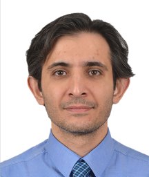 Dogan Emre Sert合作论文数39Sinan Sabit Kocabeyoglu合作论文数37
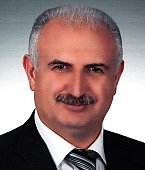 Hikmet Koçak合作论文数Faculty of Medicine, Atatürk University32
 Irfan Tasoglu合作论文数Department of Cardiovascular Surgery, Ankara City Hospital21
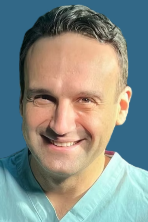 Ertekin Utku Unal合作论文数Ufuk Üniversitesi21erdal ege合作论文数18
 Seref Alp Kucuker合作论文数Dept Cirurgia Cardiovasc, Hosp Cidade Ancara16
 Karahan Mehmet合作论文数Department of Cardiovascular Surgery, Turkiye Yuksek Ihtisas Heart Education and Research Hospital15
 Zeki Catav合作论文数Department of Thoracic and Cardiovascular Surgery;Bayindir Hospital;Department of Thoracic and Cardiovascular Surgery, Bayindir Hospital15
 Ahmet Basoglu合作论文数14Burcu Demirkan合作论文数14Ibrahim Yekeler合作论文数13
 Sevket Balli合作论文数13
 Oner Gulcan合作论文数13Omer Soysal合作论文数12Oflaz Mehmet Burhan合作论文数12
 Ibrahim Ece合作论文数11Ahmet Saritas合作论文数11Metin Gülcüler合作论文数10Koca Serhat合作论文数Department of Pediatric Cardiology, Yuksek Ihtisas Heart-Education and Research Hospital10
 Ahmet Tulga Ulus合作论文数9Emre Aygun合作论文数9Hasan Berat Cihan合作论文数9Okan Yurdakok合作论文数8Cemal Levent Birincioglu合作论文数8
 Aysegul Ozgok合作论文数Department of Anesthesiology, Yuksek Ihtisas Educational and Research Hospital8
 Tugcin Bora Polat合作论文数Department of Pediatric Cardiology Kizilay S, Turkiye Yuksek Ihtisas Education and Research Hospital8Mustafa Emmiler合作论文数8
 Serhat Bahadir Genc合作论文数Dept Pediat Cardiac Surg, Univ Hlth Sci Istanbul7
 Demir Aslı合作论文数Anesthesiology Department, Türkiye Yüksek İhtisas Education and Research Hospital7
 Ali Umit Yener合作论文数7Kerim Cagli合作论文数7Ahmet Vedat Kavurt合作论文数Dept Pediat Cardiol, Ankara City Hosp6E. Aygun合作论文数6
 Caliskan Aytac合作论文数Department of Cardiovascular Surgery, Turkey Yuksek Ihtisas Training and Research Hospital6
 Hasan Ozdemir合作论文数Department of Chemical Engineering, Istanbul University6
 Riza Turkoz合作论文数Adana Teaching and Medical Research Center, Baskent University6Sabit Kocabeyoglu合作论文数6Mehmet Ali Ozatik合作论文数Cardiovascular Surgery Clinic;Psychology Department;Hacettepe University;Psychology Department, Hacettepe University6Azman Ateş合作论文数6Denizhan Bagrul合作论文数Minist Hlth, Ankara City Hosp5
 Anıl Ozen合作论文数Dept Cardiovasc Surg, Ankara City Hosp5
 Tütün Ufuk合作论文数Clinic of Cardiovascular Surgery, Türkiye Yüksek Ihtisas Hospital5
 Ayse Esin Kibar合作论文数Department of Pediatric Cardiology, Mersin Women's and Children's Hospital5
 Jan Schmitto合作论文数Department of Cardiothoracic, Transplantation and Vascular Surgery, Hannover Medical School5
 Adnan Yalcinkaya合作论文数5




