Deep reinforcement learning (DRL) has emerged as a powerful framework in medical image analysis, enabling sequential decision-making for tasks such as annotation, diagnosis, localization, and segmentation. Despite numerous studies in this domain, there is still a lack of systematic understanding of methodological choices, data types, and task-specific approaches. In this survey, we provide a structured analysis and multi-dimensional taxonomy of DRL applications in medical imaging, highlighting the advantages and limitations of existing approaches. In particular, we organize the literature based on multiple dimensions, including medical data type, task category, and DRL techniques, offering an integrated perspective that supports better understanding and comparison of current methods. This work aims to provide practical insights and guidance for future research and development in DRL-based healthcare systems.
تعاني خدمة الطاقة الكهربائية في الجمهورية العربية السورية من العديد من الصعوبات الناتجة عن نقص الموارد (الفيول) بالإضافة إلى التخريب الذي تعرضت له العديد من مراكز التوليد من قبل المجموعات الإرهابية، ترافق ذلك مع حصار جائر تعرضت له بلدنا أدى إلى تخفيض كميات وقود التشغيل الذي تزود به محطات التوليد, وقد تسبب كل ماسبق إلى تطبيق برامج التقنين في المحافظات وفقاً لاستهلاك تلك المحافظات ومراكز الإنتاج الموجودة فيها (مصانع، مراكز ضخ، مستشفيات وعدد السكان). كما يتطلب التنبؤ باستهلاك الطاقة الكهربائية معرفة كميات الاستهلاك اليومية وأوقات الاستهلاك وغيرها من العوامل المؤثرة والتي تشكل كميات كبيرة من البيانات [1]. ولا يزال التنبؤ الدقيق بالحمل الكهربائي يمثل مهمة صعبة بسبب العديد من المشاكل مثل الطابع غير الخطي للسلسلة الزمنية أو الأنماط الموسمية التي يعرضها، والتي تستغرق وقتاً كبيراً كما تؤثر على دقة الأداء في التنبؤ. يمكن تحسين العملية باستخدام شبكاتRNN . [2] بدايةً، تم تحديد الاستهلاك المثالي والمناسب للمنطقة ومقارنته مع الانتاج وإمكانية تمرير الفائض لعمليات احتياطية أخرى أو تزويد مراكز الانتاج بالفائض الذي يمكن الحصول عليه من خلال عملية التنبؤ السابقة. كما تم استخدام الشبكات العصبية التكرارية RNN (Recurrent Neural Network) وهي عبارة عن سلاسل زمنية تعتمد على تسلسل البيانات وفقاً لدلائل زمنية وقدرتها على التنبؤ بالقيم المستقبلية اعتماداً على البيانات السابقة. ثم تم مقارنة أداء تلك الشبكات مع شبكات DNN (Dense Neural Network) للحصول على تنبؤ مستقبلي أمثل قابل لخدمة وزارة الكهرباء في الجمهورية العربية السورية وحل مشكلة التنبؤ بالحمل الكهربائي بالمقارنة مع الدراسات السابقة. تم أيضاً اعتماد طريقة التقسيم المتتالي القائم على الوقت، والتي لها القدرة على العمل بصورة أعلى دقة بالنسبة للبيانات ذات العينات العشوائية. وبالنسبة لحالات انخفاض تنظيم البيانات الساعية لاستهلاك القدرة الكهربائية، يمكن لنا أخذ عينات لمجموعة من البيانات بالنسبة للزمن وأخذ 20 بالمئة من البيانات على سبيل المثال كعينات تدريب واختبار. بناءً على قيم التنبؤ الناتجة عن هذه الدراسة يتم العمل على توزيع الطاقة الكهربائية بالشكل الأنسب وبما يتوافق مع أهمية الاستخدام الأعلى.
يتميز عصرنا الراهن بالانتشار الواسع النطاق للبيانات على اختلاف أنواعها حتى أضحى من المستحيل على المحللين استخلاص معلومات ذات معنى باللجوء فقط إلى المداخل التقليدية للتحليل التمهيدي للبيانات. مع وجود كميات كبيرة من البيانات المخزنة ازدادت الحاجة إلى تطوير أدوات تمتاز بالقوة والسرعة لتحليلها، واستخراج المعلومات والمعارف منها، ومن هنا ظهرت تقنيات التنقيب في البيانات (Data Mining) كتقنيات تهدف إلى استخراج المعرفة من كميات هائلة من البيانات (تعرف بدورها بالبيانات الضخمة Big Data) [1]. يتطلب التنبؤ باستهلاك الطاقة الكهربائية معرفة كميات الاستهلاك اليومية وأوقات الاستهلاك وغيرها من العوامل المؤثرة والتي تشكل كميات كبيرة من البيانات يمكن تحليلها باستخدام خوارميات تنقيب البيانات. ولا يزال التنبؤ الدقيق بالحمل الكهربائي يمثل مهمة صعبة بسبب العديد من المشاكل مثل الطابع غير الخطي للسلسلة الزمنية أو الأنماط الموسمية التي يعرضها، والتي تستغرق وقتاً كبيراً كما تؤثر على دقة الأداء في التنبؤ. يمكن تحسين العملية باستخدام خوارزمية شعاع الدعم (SVM) .[2] بدايةً، تم دراسة العوامل المناخية ونوع وموقع وفترة الاستهلاك ومجموعة أخرى من العوامل المؤثرة وذلك لتحسين أداء عملية التنبؤ باستهلاك الطاقة الكهربائية. كما تم تحليل خوارزميات العنقدة والتصنيف التالية للتنبؤ باستهلاك الطاقة الكهربائية، حيث تم اقتراح استخدام خوارزمية SVM لحل مشكلة التنبؤ بالحمل الكهربائي، والتي قدمت حلاً لمشاكل التصنيف والانحدار كما أنها ساعدت في تصنيف البيانات الفئوية وأعطت حلاً أمثلياً مستقل من النموذج بالمقارنة مع الدراسات السابقة. تم أيضاً استخدام مجموعة من المصنفات مثل الغابات العشوائية، آلة شعاع الدعم والشبكات العصبية وغيرها للوصول إلى نتائج دقيقة تساعد على اتخاذ القرارات المناسبة في مجال التنبؤ باستهلاك الكهرباء. وفي المرحلة الأخيرة، بناءً على قيم التنبؤ الناتجة عن هذه الدراسة تم العمل على توزيع الطاقة الكهربائية بالشكل الأنسب وبما يتوافق مع أهمية الاستخدام الأعلى بحيث يصبح لدينا القدرة على تشغيل مصادر الطاقة في أوقات محددة وبكميات مناسبة لمحاولة تقليل الهدر الناجم عن تشغيل المصادر الغير ضرورية.
إن الحاجة المتزايدة لاسترجاع الصور من قواعد البيانات الضخمة جعلت مجال استرجاع الصور بالاعتماد على المحتوى Content-Based Image Retrieval (CBIR) مجالاً ملحاً وضرورياً للبحث. اقترح الباحثون الكثير من خوارزميات استرجاع الصور من خلال استخراج السمات الهامة والمميزة من المحتوى المرئي للصورة لأهمية السمات المستخرجة في تحسين دقة أنظمة الاسترجاع. وفي هذا البحث، تم إجراء مقارنة لست خوارزميات محلية مشهورة على مدى عقد من الزمن (LBP, LTP, LTrP, MMCM, COALTP and LMP) واختبار هذه الخوارزميات باستخدام نوعين مختلفين من قواعد البيانات: قواعد بيانات الصور الملونة(Color image database) و قواعد بيانات النسجة(Texture database) وباستخدام أربعة مقاييس للمسافات(L1, Euclidean, Cityblock and Cosine) لاسترجاع الصور الأكثر مطابقة لصورة الاستعلام من خلال اختيار الصور ذات المسافة الأقصر. تم تقييم أداء الخوارزميات المدروسة باستخدام ثلاثة مقاييس: متوسط دقة الاسترجاع (Average Retrieval Precision) ARP، متوسط الاسترداد Average Recall ومتوسط معدل الاسترجاع (Average Retrieval Rate) ARR، كشفت هذه الدراسة تفوق خوارزمية COALTP على الخوارزميات الأخرى المختبرة، وبالإضافة إلى ذلك، أظهرت النتائج أن خوارزميات الأنماط المحلية أكثر كفاءة في استرجاع الصور من قواعد بيانات النسجة مقارنة مع قواعد البيانات الملونة.
In this paper, a new method for object detection and pose estimation in a monocular image is proposed based on FDCM method. it can detect object with high speed running time, even if the object was under the partial occlusion or in bad illumination. In addition, It requires only single template without any training process. The Modied FDCM based on FDCM with improvments, the LSD method was used in MFDCM instead of the line tting method, besides the integral distance transform was replaced with a distance transform image, and using an angular Voronoi diagram. In addition, the search process depends on Line segments based search instead of the sliding window search in FDCM. The MFDCM was evaluated by comparing it with FDCM in dierent scenarios and with other four methods: COF, HALCON, LINE2D, and BOLD using D-textureless dataset. The comparison results show that MFDCM was at least 14 times faster than FDCM in tested scenarios. Furthermore, it has the highest correct detection rate among all tested method with small advantage from COF and BLOD methods, while it was a little slower than LINE2D which was the fasted method among compared methods. The results proves that MFDCM able to detect and pose estimation of the objects in the clear or clustered background from a monocular image with high speed running time, even if the object was under the partial occlusion which makes it robust and reliable for real-time applications.
The current research introduces a novel method for fracture detection and classification. The basic stages of fracture detection includes preprocessing of bone image and morphological operations to obtain the ROI region which is manipulated by a post processing stage to remove non-fracture pixels. The suggested approach extract three features from bone image which are transverse, cracks and divergence features in order to define the fracture type or integrity of bone image. The designed systems detect the different fracture types correctly beside the hybrid fracture type. We applied experiments on a dataset consisting of 155 bone images including 100 fracture images, 30 hand-fingers images and 25 normal images. The systems achieved 92% true detection rate for general bone fractures, 93.33% true detection rate for finger bone fractures and 93.33% true rejection rate. The experiments showed that the inner false detection rates between each type and the others are less than 4%.
ISSN: 2347-8578 www.ijcstjournal.org Page 78 Hybrid Recognition System under Feature Selection and Fusion Eng. Ali M Mayya , Dr. Mariam M Saii [2] PhD Student , Professor Assistance [2] Department of Computer Engineering University of Tishreen, Lattakia Syria ABSTRACT The study suggests a hybrid human recognition system based on face ear and palm print images. The aim of our study is to show the importance of biometric fusion for enhancement of the recognition rate. The system takes face images and segment them into face and ear. Then face, ear and palm images are extracted and fused. The FFBPNN followed by mahalanobis classifier is used in classification stage. The Experiments is applied on 5 databases with different illumination and pose variations and the best result obtained from the face-ear-palm fusion features with 97.5% recognition rate compared with 94.33% for face-ear fusion.
Multimodal biometric systems are more promised and accurate than unimodal ones. Beside increase the performance, the multimodal system minimize the universality problem. The current research introduces a new iris and palm fusion system. The feature of palm print is extracted using connectivity points and lifelines orientations, while features of iris is extracted using wavelet transform. The classification method was the distance classifier. The score level fusion is applied using modified version of majority voter. The system accuracy was 97.29% for palm, 71.97% and 98.54% for fusion.
The visual speech modality plays an important role in the perception and production of speech. Although not purely confined to the mouth, it is generally agreed that the large proportion of speech information conveyed in the visual modality stems from the mouth region of interest (ROI). To this end, it is imperative that an audio-visual speech processing (AVSP) system be able to accurately detect, track and normalize the mouth of a subject within a video sequence. This task is referred to as facial feature detection (FFD). The goal of FFD is to detect the presence and location of features, such as eyes, nose, nostrils, eyebrow, mouth, lips, ears, etc., with the assumption that there is only one face in an image. This differs slightly to the task of facial feature location which assumes the feature is present and only requires its location. Facial feature tracking is an extension to the task of location in that it incorporates temporal information in a video sequence to follow the location of a facial feature as time progresses. Throughout this article the tasks of facial feature detection, location and tracking are all thought to be encapsulated under the broad banner of FFD.
Visual recognition systems based on the movement of speaker lips are one of the most modern and important recognition systems currently and have received a great deal of attention in the last decade for their potential use in the latest applications that rely on speech recognition systems. It is one of the modern researches used to build systems for understanding or interpreting Speech without hearing it for speakers. The research suggests using a hybrid system for visual recognition of isolated words spoken in Arabic. This system is a supervised and an offline system. It passes through several stages, beginning with identifying facial features followed by detection and identification of the lips, and then reading the lips to extract a set of visual features that indicate the spoken word. Hidden Markov models were used in the classification phase. The proposed system was tested on 4155 samples. The results showed that the proposed hybrid system led to rate of up to 73% achieving an increase of up to 12% from the research based on the same database Which can be considered as a starting point in building recognition systems that adopt the integration of audio and visual features together.
The research offers a fully automatic method for tumor segmentation on Magnetic Resonance Images MRI. In this method, at first in the preprocessing level, anisotropic diffusion filter is applied to the image by 8-connected neighborhood for removing noise from it. In the second step, using Support Vector Machine SVM Classifier for tumor detection accurately. After creating the appropriate mask image, based on the symmetry property in axial and coronary magnetic resonance images. The tumor detected and segmented (Dice coefficient > 0.90) in a few seconds. The method applied on several MR images with different types regardless of the degree of complexity in those images.
The proposed method has implemented series of steps. First the ratio of existing noise is reduced depending on Wiener Filter, followed by the stage of image borders removing in order to enrich high-density primary regions and suppress remaining ones. Thresholding stage has converted the image into a binary form, then the small foreground elements are removed in a binary image, after that the Skeletonization stage is applied in order to reduce the data points, then a matrix containing all data points which are lying on the target ellipse to be drawn. Detection of the ellipse corresponding to the limits of the fetus head in the input ultrasound image is done via the Least Square Fitting (LSF) algorithm, which returns the ellipse parameters including the minor axis which corresponds to the bi-parietal distance measure. The results of auto-measurements are compared with manual measurements of a specialist to improve the effective of the proposed method. (C) 2017 Ain Shams University.
Kidney Detection and Segmentation in MR images allows extracting meaningful information for nephrologists, also for practical use in clinical routine, thus we should apply an fast, automatic and robust algorithm. We demonstrate the possibility of construct an algorithm that achieve these requirements. Therefore, a novel kidney segmentation algorithm was created depending on multiple stages. The Region of Interest (ROI) is extracted after we convert the input image to binary one via specific thresholding level yields from K Mean Clustering algorithm. The resulted binary image contain both of kidneys as the biggest regions, so we can isolate them after we calculate the objects areas in labeled image. Finally we can use some morphological operation to remove small objects surrounding the kidney region. The effectiveness of this method is demonstrated through experimental results on complex MR slices. Kidneys were accurately detected and segmented in a few seconds.
A new approach of human recognition using ear images is introduced. It consists of two basic steps which are the ear segmentation and ear recognition. In the first one, Likelihood skin detector is used to determine the skin areas in the side face images. Then, some of the morphological operations are applied to determine the ear region. This region is extracted using image processing techniques. The ear recognition step depends on the segmented ear images as inputs. A hybrid PCA_Wavelet algorithm is used to extract the ear features from ear. Finally, the feed forwarding back propagation neural network is trained using the feature vectors. Tests which applied on 460 images, which have been taken during 4 months and under different illumination and pose variations, show that the system achieved a rate of 96.73% for ear extraction and 98.9% for recognition. More experiments are done to specify the best wavelet level, the best number of features, the best classification method, and the best threshold value. The study is also compared with other ones at the area of ear recognition. Correspondence to: Ali Mahmoud Mayya, Computer and Automatic Control Engineering department, Tishreen University, Syria, E-mail: alimia1988@yahoo.com
The research offers an innovative way to represent the fetus in three-dimensions. This is done by transferring scan fetus video to a series of grayscale images. Each image processed individually in order to obtain binary image represents a fetus object. Although the ultrasound image segmentation is difficult, but we were finding two ways to isolate the fetal region of the image. First we improve the contrast and enhance the edges. After that we extract the fetal area by applying Region Growing algorithm, or using Thresholding followed by Morphological operations. Before the final step, we apply the Edge Refining to the resulted binary images. Then we get the three-dimensional formation of surface through overlaying the resulting images on top of each other . This paper is important because it helps doctor to detect potential abnormalities or in estimation the gender of fetus .
This research offers an innovative way to reduce the speckle noise which associated with the ultrasound imaging. This in turn leads to improving the input image and raise the human diagnostic performance. It uses an Undecimated Wavelet Transform UWT to decompose the image signal to the coefficients, to take the diagonal coefficient and applied Independent Directional Mask after replacing the value of each pixel in this coefficient with the corresponding pixel in the original image, this result yields after applying median filter in case the value of the pixel is smaller than the threshold value which is pre-specified. Then we apply the Smoothing Directional filter and re-directed the formation of the new diagonal coefficient, and then make the inverse undecimated wavelet transform IUWT to form the resulting image after reducing the noise of it (De-speckled Image). The used method showed a clear improvement on several ultrasound images via traditional statistical measurements (PSNR and MSE) and outperformed the other methods which used in common to reduce the speckle noise.
This paper proposes a new speed approach for the segmentation of the lung images in order to detect and extract the tumor region. The approach consists of two main stages, which are the preprocessing stage, marker watershed stage and the tumor detection stage. The preprocessing consists of laplacian filtering to enhance edges and make the next stages more efficient. The marker watershed step applies the Sobel gradient function on the foreground and background markers to get the possible tumor region. The post processing stage consists of tumor detection and segmentation in which the area of the tumor is calculated. The results are done on a medical lung database obtained from Tishreen hospital (in Lattakia, Syria) which consists of 59 images from 10 persons. The result shows robustness of the system in detecting and segmenting tumor region in different depths. The designed GUI supplies user with tumor region and area, and time of each stage.
The virtual texture is due to regular or random variation in the gray level or color in an image. Features based on texture are often useful in automatically distinguishing between objects and in finding boundaries between regions. New features that are based on texture analysis of the face skin are proposed as efficient tools for face recognition. In the preprocessing step, the analyzed face region is detected. Then, the texture features of this region, namely, energy, entropy and homogeneity, are extracted. In order to test the performance of the skin texture based features in face recognition we combine them with our previously introduced statistical features that are extracted from a coded image which is obtained from the edge detection of a binary version of the original gray scale image. The statistical features and skin texture parameters are fed to a FBP neural network for face recognition. Computer simulation results with 100 test images of 10 persons (the images of each person in various poses, facial expression, and facial details) show that the proposed skin texture features highly enhance the recognition rate.
Recognition method of human face using statistical analysis feature extraction and a neural network algorithm is proposed. In the preprocessing step we detect the edges of the face image by using the Sobel algorithm. Then we propose a new method to transform the two-dimension black and white image to a one-dimension vector. Finally, based on the statistical analysis, we extract seven features. In the recognition step we use the fast backpropagation (FBP) algorithm. Computer simulation results with 100 test images of 10 persons (the images of each person in a various pauses, facial expression, and facial details) show that the proposed method yields a high recognition rate
 Zaid Kraitem合作论文数Dept Comp Engn, Tishreen Univ5
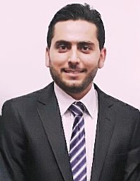 Ali Mahmoud Mayya合作论文数Department of Computer Engineering, Tishreen University5
 Oulfat Jolaha合作论文数2
 Jafar Alkheir合作论文数2
 Said El-Khamy合作论文数Dept. of Electrical Engineering, Faculty of Engineering, Alexandria University2Jaber Hanna合作论文数1Nour Sami Ghadban合作论文数1Abdel-Alim, O.合作论文数1
 Issa Ibraheem合作论文数1Rabie Habib合作论文数1




