The mechanisms of intracranial solute transport are fundamental to human brain health. However, our understanding of their interplay remains incomplete, in part due to the complexity of integrating insights across scales, between species, and from different modalities. In this work, we combine mixed-dimensional mathematical modeling, multi-modal magnetic resonance images, and high performance computing to present a computational framework for calculating the spatiotemporal spreading of a solute across the subarachnoid space, ventricular system, and brain parenchyma, including surface perivascular spaces. To illustrate applicability, we simulate a series of scenarios relating to intrathecal tracer injections, including variations in cerebrospinal fluid production, intracranial pulsatility, perivascular fluid flow and -structure. This openly available framework allows for evaluating the integrated effect of different transport mechanisms, bridging from the vascular mesoscale to the organ-level macroscale and across time scales of seconds to hours, in subject-specific geometries. This work introduces a multiscale, integrative modeling framework for brain fluid flow and solute transport, bridging mesoscale perivascular networks with the organ-level macroscale and timescales of seconds to hours in subject-specific geometries.
Over the last three decades, advances in bioimaging have revolutionized our ability to study the structure and function of living systems. At the nano- to microscale, the new capabilities of cellular microscopy have resulted in the generation and wide availability of large-scale high-resolution imaging data, as strikingly exemplified by increasingly complex dense reconstructions of cortical tissue. In parallel, research in scientific computing and computational mathematics have led to the development of highly accurate, efficient and scalable numerical algorithms and their realization in open scientific software. In this talk, I will show how we, in the last few years, have developed high-fidelity computational models of cellular and subcellular physiology, incorporating detailed geometric structure, biophysical dynamics and advanced cell imaging data. I will highlight key numerical challenges - some that we have tackled and some open problems - and point at future directions.
Skeletal muscle displays remarkable plasticity, adapting its size and strength in response to mechanical loading, especially, from exercise. This process, known as hypertrophy, is fundamental to athletic training and rehabilitation, but is challenging to quantitatively predict due to its multifactorial, multiscale nature. Specifically, skeletal muscle hypertrophy results from an integration of macroscopic mechanical stimuli with the intracellular signaling pathways that govern muscle growth. In this work, we present a multiscale computational model that mechanistically integrates these mechanical and biochemical stimuli and offers a framework for predicting the outcomes of different types of exercise on skeletal muscle growth. The framework couples a transversely isotropic hyperelastic model for tissue-level mechanics with a system of ordinary differential equations representing the IGF1-AKT-mTOR-FOXO signaling pathway, a key regulator of protein synthesis and degradation. We link these scales using a volumetric growth model, where the signaling dynamics inform a growth tensor that drives changes in muscle cross-sectional area. This approach enables the simulation of long-term muscle adaptation, providing a mechanistic tool to investigate how different exercise protocols lead to macroscopic hypertrophy. Simulations from our model capture the temporal dynamics of hypertrophy under varying load protocols and highlight how feedback between protein synthesis and muscle growth regulates the dose-response relationship to prevent unbounded growth. Using muscle geometries derived from the Visible Human dataset, we study how human variations in muscle geometry affect hypertrophy. Finally, we demonstrate that the mechanochemical coupling between muscle geometry and signaling not only predicts macroscopic shape changes but also provides buffering from local signaling heterogeneity. Ultimately, this framework offers a predictive computational tool for optimizing training regimens and understanding the multiscale determinants of muscle adaptations.
Astrocyte endfeet form a near-continuous sheath around the brain's vasculature, defining the perivascular spaces (PVS) that are crucial for brain fluid flow and solute transport. Yet, their precise physiological role remains poorly understood. Using 3D electron microscopy data, we created a high-fidelity poroelastic computational model of an arteriole segment with surrounding endfeet and parenchyma to investigate tissue displacement and fluid flow within the PVS, endfeet, and extracellular space in response to blood vessel pulsations. Our model predicts that arteriole dilations compress the PVS while expanding the overall endfoot sheath volume due to tangential stretch. Moreover, fluid exchange primarily occurs through inter-endfoot gaps, driven by pressure differences, rather than across the aquaporin-4 (AQP4) rich endfoot membrane. PVS stiffness critically modulates these dynamics: Increased stiffness of the PVS, for instance, due to vessel pathology or aging, would minimize or even reverse fluid exchange at the gliovascular interface. While AQP4 mediated water movement has a negligible impact on pulsation-driven mechanics, it significantly enhances osmotically driven fluid flow. Overall, our findings elucidate the complex balance of forces governing gliovascular mechanics and suggest that PVS composition strongly influences endfoot-parenchymal fluid exchange.
The mechanisms of intracranial solute transport are fundamental to human brain health, with alterations often linked to disease and functional impairment, and with distinct opportunities for personalized diagnostics and treatment. However, our understanding of these mechanisms and their interplay remains incomplete, in part due to the complexity of integrating insights across scales, between species and from different modalities. Here, we combine mixed-dimensional modelling, multi-modal magnetic resonance images, and high performance computing to construct and explore a high-fidelity in-silico model of human intracranial molecular enrichment. This model predicts the temporo-spatial spreading of a solute within an image-derived geometric representation of the subarachnoid space, ventricular system and brain parenchyma, including networks of surface perivascular spaces (PVSs). Our findings highlight the significant impact of cerebrospinal fluid (CSF) production and intracranial pulsatility on molecular enrichment following intrathecal tracer injection. We demonstrate that low-frequency vasomotion induces moderate CSF flow in surface PVS networks which substantially enhances tracer enrichment, and that impaired enrichment is a direct natural consequence of enlarged PVSs. This openly available technology platform thus provides an opportunity for integrating separate observations on diffusion in neuropil, vascular dynamics, intracranial pulsatility, CSF production, and efflux, and for exploring drug delivery and clearance in the human brain. ### Competing Interest Statement The authors have declared no competing interest.
Recent advances in microscopy and 3D reconstruction methods have allowed for characterization of cellular morphology in unprecedented detail, including the irregular geometries of intracellular subcompartments such as membrane-bound organelles. These geometries are now compatible with predictive modeling of cellular function. Biological cells respond to stimuli through sequences of chemical reactions generally referred to as cell signaling pathways. The propagation and reaction of chemical substances in cell signaling pathways can be represented by coupled nonlinear systems of reaction-transport equations. These reaction pathways include numerous chemical species that react across boundaries or interfaces (e.g., the cell membrane and membranes of organelles within the cell) and domains (e.g., the bulk cell volume and the interior of organelles). Such systems of multi-dimensional partial differential equations (PDEs) are notoriously difficult to solve because of their high dimensionality, non-linearities, strong coupling, stiffness, and potential instabilities. In this work, we describe Spatial Modeling Algorithms for Reactions and Transport (SMART), a high-performance finite-element-based simulation package for model specification and numerical simulation of spatially-varying reaction-transport processes. SMART is based on the FEniCS finite element library, provides a symbolic representation framework for specifying reaction pathways, and supports geometries in 2D and 3D including large and irregular cell geometries obtained from modern ultrastructural characterization methods.
Neurotoxic protein fragments such as amyloid-beta and tau accumulate in characteristic staging patterns in Alzheimer’s disease (AD). The brain clears such metabolic substances via multiple different systems, including via the glymphatic (extracellular/extravascular) pathway. Here, we ask how the distinct features that characterize human glymphatic function would affect the prion-like cascade of protein invasion associated with AD. To address this question, we extract and analyze individual clearance rates from human glymphatic MRI (gMRI) data sets. These clearance rates define subject-specific maps of glymphatic clearance that vary both across cortical lobes and Braak staging regions. We apply these clearance maps as initial states in a computational network model linking misfolded proteins, tissue damage, and local clearance to simulate a series of individual proteinopathy trajectories. Our results show that the spatial heterogeneity in initial clearance induces characteristic propagation patterns, delaying and redirecting the disease progression. Moreover, reducing this spatial heterogeneity accelerates disease progression and induces staging patterns typically associated with AD. A comparison between well-rested subjects and subjects who underwent a single night of sleep deprivation did not reveal differences in initial clearance maps nor in simulated disease progression. These findings suggest that spatial heterogeneity in brain clearance may be a key factor for neurodegenerative resilience. ### Competing Interest Statement The authors have declared no competing interest. EPSRC, EP/L015803/1, EP/R020205/1 NSF, 2325276 ERC, 714892 Research Council of Norway, 324239
Excitable tissue is fundamental to brain function, yet its study is complicated by extreme morphological complexity and the physiological processes governing its dynamics. Consequently, detailed computational modeling of this tissue represents a formidable task, requiring both efficient numerical methods and robust implementations. Meanwhile, efficient and robust methods for image segmentation and meshing are needed to provide realistic geometries for which numerical solutions are tractable. Here, we present a computational framework that models electrodiffusion in excitable cerebral tissue, together with realistic geometries generated from electron microscopy data. To demonstrate a possible application of the framework, we simulate electrodiffusive dynamics in cerebral tissue during neuronal activity. Our results and findings highlight the numerical and computational challenges associated with modeling and simulation of electrodiffusion and other multiphysics in dense reconstructions of cerebral tissue.
Cerebrospinal fluid (CSF) is integral to brain function. CSF provides mechanical support for the brain and helps distribute nutrients, neurotransmitters and metabolites throughout the central nervous system. CSF flow is driven by several processes, including the beating of motile cilia located on the walls of the brain ventricles. Despite the physiological importance of CSF, the underlying mechanisms of CSF flow and solute transport in the brain ventricles remain to be comprehensively resolved. This study analyzes and evaluates specifically the role of motile cilia in CSF flow and transport. We developed finite element methods for modeling flow and transport using the geometry of embryonic zebrafish brain ventricles, for which we have detailed knowledge of cilia properties and CSF motion. The computational model is validated by in vivo experiments that monitor transport of a photoconvertible protein secreted in the brain ventricles. Our results show that while cilia contribute to advection of large particles, diffusion plays a significant role in the transport of small solutes. We also demonstrate how cilia location and the geometry of the ventricular system impact solute distribution. Altogether, this work presents a computational framework that can be applied to other ventricular systems, together with new concepts of how molecules are transported within the brain and its ventricles.
Alzheimer's disease, the most common form of dementia, is a systemic neurological disorder associated with the formation of toxic, pathological aggregates of proteins within the brain that lead to severe cognitive decline, and eventually, death. In normal physiological conditions, the brain rids itself of toxic proteins using various clearance mechanisms. The efficacy of brain clearance can be adversely affected by the presence of toxic proteins and is also known to decline with age. Motivated by recent findings, such as the connection between brain cerebrospinal fluid clearance and sleep, we propose a mathematical model coupling the progression of toxic proteins over the brain's structural network and protein clearance. The model is used to study the interplay between clearance in the brain, toxic seeding, brain network connectivity, aging, and progression in neurodegenerative diseases such as Alzheimer's disease. Our findings provide a theoretical framework for the growing body of medical research showing that clearance plays an important role in the etiology, progression, and treatment of Alzheimer's disease.
The EMI (Extracellular-Membrane-Intracellular) model describes electrical activity in excitable tissue, where the extracellular and intracellular spaces and cellular membrane are explicitly represented. The model couples a system of partial differential equations in the intracellular and extracellular spaces with a system of ordinary differential equations on the membrane. A key challenge for the EMI model is the generation of high-quality meshes conforming to the complex geometries of brain cells. To overcome this challenge, we propose a novel cut finite element method (CutFEM) where the membrane geometry can be represented independently of a structured and easy-to-generated background mesh for the remaining computational domain. Starting from a Godunov splitting scheme, the EMI model is split into separate PDE and ODE parts. The resulting PDE part is a non-standard elliptic interface problem, for which we devise two different CutFEM formulations: one single-dimensional formulation with the intra/extracellular electrical potentials as unknowns, and a multi-dimensional formulation that also introduces the electrical current over the membrane as an additional unknown leading to a penalized saddle point problem. Both formulations are augmented by suitably designed ghost penalties to ensure stability and convergence properties that are insensitive to how the membrane surface mesh cuts the background mesh. For the ODE part, we introduce a new unfitted discretization to solve the membrane bound ODEs on a membrane interface that is not aligned with the background mesh. Finally, we perform extensive numerical experiments to demonstrate that CutFEM is a promising approach to efficiently simulate electrical activity in geometrically resolved brain cells.
In this work, we are interested in solving large linear systems stemming from the extra–membrane–intra model, which is employed for simulating excitable tissues at a cellular scale. After setting the related systems of partial differential equations equipped with proper boundary conditions, we provide its finite element discretization and focus on the resulting large linear systems. We first give a relatively complete spectral analysis using tools from the theory of Generalized Locally Toeplitz matrix sequences. The obtained spectral information is used for designing appropriate preconditioned Krylov solvers. Through numerical experiments, we show that the presented solution strategy is robust w.r.t. problem and discretization parameters, efficient and scalable.
Flow of cerebrospinal fluid through perivascular pathways in and around the brain may play a crucial role in brain metabolite clearance. While the driving forces of such flows remain enigmatic, experiments have shown that pulsatility is central. In this work, we present a novel network model for simulating pulsatile fluid flow in perivascular networks, taking the form of a system of Stokes-Brinkman equations posed over a perivascular graph. We apply this model to study physiological questions concerning the mechanisms governing perivascular fluid flow in branching vascular networks. Notably, our findings reveal that even long wavelength arterial pulsations can induce directional flow in asymmetric, branching perivascular networks. In addition, we establish fundamental mathematical and numerical properties of these Stokes-Brinkman network models, with particular attention to increasing graph order and complexity. By introducing weighted norms, we show the well-posedness and stability of primal and dual variational formulations of these equations, and that of mixed finite element discretizations.
The activity and dynamics of excitable cells are fundamentally regulated and moderated by extracellular and intracellular ion concentrations and their electric potentials. The increasing availability of dense reconstructions of excitable tissue at extreme geometric detail pose a new and clear scientific computing challenge for computational modelling of ion dynamics and transport. In this paper, we design, develop and evaluate a scalable numerical algorithm for solving the time-dependent and nonlinear KNP-EMI equations describing ionic electrodiffusion for excitable cells with an explicit geometric representation of intracellular and extracellular compartments and interior interfaces. We also introduce and specify a set of model scenarios of increasing complexity suitable for benchmarking. Our solution strategy is based on an implicit-explicit discretization and linearization in time, a mixed finite element discretization of ion concentrations and electric potentials in intracellular and extracellular domains, and an algebraic multigrid-based, inexact block-diagonal preconditioner for GMRES. Numerical experiments with up to 10^8 unknowns per time step and up to 256 cores demonstrate that this solution strategy is robust and scalable with respect to the problem size, time discretization and number of cores.
 Kent-Andre Mardal合作论文数Simula Research Laboratory Lysaker 134 1325 Norway26
 Anders Logg合作论文数Chalmers University of Technology18
 Vegard Vinje合作论文数Simula Research Laboratory16
 Lars Magnus Valnes合作论文数11Ada Johanne Ellingsrud合作论文数Simula Res Lab, Dept Numer Anal & Sci Comp, N-0164 Oslo, Norway11
 Travis Thompson合作论文数Univ Oxford, Math Inst, Oxford, England11
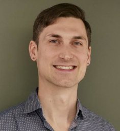 Gabriel Balaban合作论文数Univ Oslo, Dept Informat, Oslo, Norway8
 Miroslav Kuchta合作论文数Simula Research Laboratory8
 Martin Sandve Alnæs合作论文数8
 Garth N. Wells合作论文数Department of Engineering, University of Cambridge7
 Joakim Sundnes合作论文数Simula Research Laboratory, Norway7
 Cecile Daversin-Catty合作论文数6
 Henrik Finsberg合作论文数Department of Computational Physiology, Simula Research Laboratory, Fornebu, Norway.6
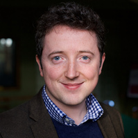 Patrick E. Farrell合作论文数Mathematical Institute, University of Oxford;Oriel College, University of Oxford6
 Andre Massing合作论文数6
 Simon W. Funke合作论文数6
 Jan Blechta合作论文数Fac Math, Tech Univ Chemnitz5
 Mats G. Larson合作论文数CHALMERS UNIVERSITY OF TECHNOLOGY5
 Geir Halnes合作论文数Royal Institute of Technology, AlbaNova University5
 Samuel Wall合作论文数5
 Ingeborg G Gjerde合作论文数Dept Math, Univ Bergen4
 Andrew T. T. McRae合作论文数4Per Kristian Eide合作论文数4
 Geir Andre Ringstad合作论文数Oslo University Hospital4
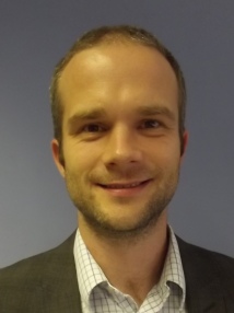 Colin Cotter合作论文数Applied Mathematics and Mathematical Physics Section, Department of Mathematics, Faculty of Natural Sciences, Imperial College London4
 Chris N. Richardson合作论文数4
 David A. Ham合作论文数Department of Earth Science and Engineering, Imperial College London4Miklós Homolya合作论文数3
 Eleonora Piersanti合作论文数Simula Research Laboratory3Xiaoran Lai合作论文数Faculty of Medicine, University of Oslo3
 Padmini Rangamani合作论文数Department of Bioengineering, Jacobs School of Engineering, University of California, San Diego;Department of Mechanical and Aerospace Engineering, University of California San Diego3
 Alvaro Köhn-Luque合作论文数Oslo Ctr Biostat & Epidemiol, Univ Oslo3
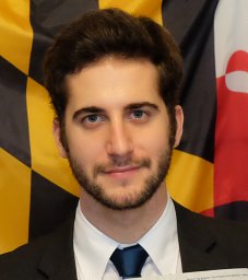 Matteo Croci合作论文数o=Oden Institute for Computational Engineering and Sciences, The University of Texas at Austin3
 Maleckar Mary M合作论文数Center for Biomedical Computing, Simula Research Laboratory, Oslo, Norway;and Center for Cardiological Innovation, Oslo University Hospital;Center for Cardiological Innovation, Oslo University Hospital3
 Stian Ross合作论文数3Lizao Li合作论文数3
 Lawrence Mitchell合作论文数3
 Robert C. Kirby合作论文数Department Mathematics, Baylor University3
 Florian Rathgeber合作论文数INSTITUT F 3Kristian B. Ølgaard合作论文数Delft University of Technology3
 Arnoldo Frigessi合作论文数Oslo Centre of Biostatistics and Epidemiology, Institute of Basic Medical Sciences, University of Oslo;INTEGREAT3
 Hans Henrik Odland合作论文数3
 Gaute Einevoll合作论文数University of Oslo;Department of Physics, Norwegian University of Life Sciences;International Neuroinformatics Coordinating Facility3Graham Markall合作论文数2
 Anders Eklund合作论文数2
 Karen-Helene Støverud合作论文数SINTEF Digital2Marius Causemann合作论文数2Benjamin Kehlet合作论文数2
 Hadrien Oliveri合作论文数University of Oxford2




