Objectives:Lower limb ischemia after extracorporeal life support for refractory cardiac arrest may be prevented by effective distal perfusion catheter (DPC) placement. We describe the prevalence of cannulated lower limb ischemia, the DPC technique and its improvements over time, and the blood flow velocities by doppler ultrasound associated with ischemia. Methods:Retrospective single-centre study including patients ≥18 years-old receiving extracorporeal life support under cardiopulmonary resuscitation. Data is expressed as frequencies (percentages), compared using Fisher's exact test. Logistic regression evaluated technical improvements associated with absence of ischemia. Blood flow velocity cutoffs associated with ischemia were determined according to receiver operator characteristics curves. Results:We included 338 patients, 60[50-66] years-old. Survival to discharge was 24%.DPC was placed under ultrasound guidance using stiff wires: initial technique. This was improved resulting in the optimized technique by distal perfusion catheter heparin perfusion, using >12 cm-long sheaths, and using braided sheaths to avoid kinking. Cannulated lower limb ischemia occurred in 23/338 [6.8 %] in the overall population, in 10/77[13 %] with the initial technique and 13/261[5 %] with the optimized technique, p = 0.01. Braided sheaths were associated with absence of ischemia in multivariable analysis, odds ratio 0.18, 95 % confidence interval [0.045-0.65], p = 0.001. Blood flow velocity associated with ischemia was ≤17 cm/s in the mid-superficial femoral artery, and ≤12 cm/s in the more distal arteries. Conclusions:Using the optimized distal perfusion catheter technique, ischemia prevalence was 5 %. Braided catheters were associated with absence of lower limb ischemia. Blood flow velocity thresholds associated with cannulated lower limb ischemia were ≤17 cm/s.
AIMS:This study explores the association between time from cardiac arrest to extracorporeal membrane oxygenation (ECMO) initiation and survival in patients with in-hospital cardiac arrest (IHCA) treated with extracorporeal cardiopulmonary resuscitation (ECPR). METHODS:This retrospective study included IHCA patients who received ECPR at the University of Minnesota Medical Center from 2016 to 2024. The primary outcome was survival to discharge; the secondary outcome was favorable neurological status, defined as Cerebral Performance Category score of 1-2. The association between time from arrest to ECPR, referred to as resuscitation time, and survival was evaluated with logistic regression analyses. RESULTS:A total of 100 patients were included. The median age was 61 years and the median resuscitation time was 33 min. Overall, 30 % of patients survived to discharge and 25 % had favorable neurological outcomes. Survival was significantly higher in patients with resuscitation duration less than 30 min compared to those with duration over 30 min (53.1 % vs 19.1 %, p = 0.001). Survivors had a significantly shorter median time from arrest to ECPR than non-survivors (26 vs. 35 min, p = 0.005). In a multivariable analysis, resuscitation duration remained independently associated with survival to discharge (p = 0.04). CONCLUSION:Shorter resuscitation time is a key determinant of survival in IHCA patients, with optimal outcomes achieved when ECMO is initiated within 30 min of arrest.
Sudden cardiac arrest, and in particular sudden out-of-hospital cardiac arrest (OHCA) remains a major public health concern in which survival statistics, and in particular neurologically intact survival statistics, have remained largely unimproved over many decades. Overall survival remains approximately 10%, being somewhat better in victims receiving bystander cardiopulmonary resuscitation (CPR), and those who are found to have a shockable rhythm (i.e., VT or VF). CPR and defibrillation (especially public-access defibrillation) remain the essential immediate management tools. However, recent research has introduced several novel adjunctive interventions (e.g., mechanical compression-decompression devices, 'head-up' CPR methodology, portable extra-corporeal circulatory assistance [ECPR]) that will hopefully impact survival positively. In any case, it is apparent that no single resuscitative tool will be sufficient to markedly improve OHCA survival; the combined application of a multi-faceted strategy is needed. This might comprise bystander CPR, combined use of 'head-up' CPR along with impedance threshold valve [ITD] and active compression-decompression mechanical chest compression devices. Application of mobile ECPR devices as early as possible during resuscitation appears to improve outcomes albeit expensive and complex to deploy broadly. Employed together, these novel steps, offer the possibility of moving the survival needle in a positive direction.
Aims:The study explores the association between race, survival and neurological outcomes among out-of-hospital cardiac arrest (OHCA) patients listed in Minnesota metro and the University of Minnesota Extracorporeal Cardiopulmonary Resuscitation (UMN-ECPR) program. Methods:This retrospective study included OHCA patients with initial shockable rhythm from two distinct cohorts: the Minnesota metro CARES cohort, treated with conventional CPR and the UMN-ECPR database (2016-2023). Race was categorized as white or non-white. Good neurological outcome was defined as a Cerebral-Performance-Category score of 1-2. Logistic regression analyses examined survival by race, with primary models adjusted for age and gender and exploratory models further adjusted for witnessed status, location, bystander CPR, return-of-spontaneous-circulation, CPR duration. Results:Of 2,700 OHCA patients in the CARES cohort, primarily treated with conventional CPR, 16.5 % were non-white. Compared to white patients, non-whites were younger (mean age 54.0 vs. 64.4 years), more often female (32.8 % vs. 23.6 %), and less likely to receive bystander CPR (52.2 % vs. 60 %). Non-white patients had lower age- and gender-adjusted odds of survival to discharge (OR: 0.64; 95 % CI, 0.5-0.82; p < 0.001) and favorable neurological outcome (OR: 0.48; 95 % CI, 0.35-0.64; p < 0.001). Among 414 ECPR patients (22.7 % non-white), non-white patients were younger (mean age 51 vs. 58.8 years) with lower bystander CPR rates (65.2 % vs. 74.8 %). There were no significant differences in age- and gender-adjusted survival (OR: 1.17; 95 % CI, 0.69-2; p = 0.554) or neurological outcome (OR: 1.07; 95 % CI, 0.61-1.88; p = 0.818). Conclusion:Non-white race was linked to worse outcomes in the conventional CPR cohort but not in the ECPR cohort.
Heart failure (HF) is a complex clinical syndrome that represents one of the leading causes of morbidity and mortality in developed nations. It is well established that every HF-related hospital admission leads to worsened quality of life for the patient and their caregiver and also imposes a significant financial burden on society. Therefore, reducing hospital admissions for this population has emerged as a critical tactic over the past decades. Initial attempts at remote monitoring focused on self-reported vital signs and symptoms, yet these proved ineffective. Meanwhile, subsequent technological advancements have enabled the development of miniature sensors capable of detecting and monitoring a wide range of physiologically relevant parameters; some of these advancements have been integrated into implantable devices, such as pacemakers and defibrillators. However, noninvasive monitoring has recently emerged as an alternative option for patients with HF, offering early congestion detection without requiring an invasive procedure. This review aims to summarize implanted and noninvasive devices, their characteristics, monitored parameters, and potential limitations and challenges around their integration into routine clinical practices.
Voluntary stretching of upper back and shoulder muscles is often associated with a sense of well-being of unknown cause. The goal of this study was to examine the impact of shoulder/upper back stretching on heart rate (HR) and blood pressure (BP) responses in healthy individuals. Twenty-four healthy individuals underwent continuous beat-to-beat HR and BP monitoring during active standing (AS) and during shoulder/upper back extension stretching. Measurements were compared using appropriate statistical tests. With AS, HR increased (median 24 bpm) and systolic BP decreased (median - 28 mmHg). Shoulder/upper back stretching elicited a similar BP drop but a lesser HR increment (p < 0.001). The HR increase per mmHg BP fall (∆HR/∆BP) was significantly lower during stretch than during AS (0.34 vs. 1.1 beats/min/mmHg; p < 0.001). Thus, the HR response with stretch-induced BP fall averaged only 30.9% of that seen with AS. Shoulder/upper back muscle stretching induces transient BP reduction with only limited compensatory tachycardia compared to hypotension during AS. These findings suggest a neural reflex mechanism possibly initiated by muscle mechanoreceptors, with predominant vasodepression that may contribute to relaxation and a sense of well-being.
BACKGROUND:Stretch-induced syncope (SIS) is a poorly understood condition that we hypothesized may be due to a neural reflex hypotensive response triggered by stretching of shoulder/upper back muscles. OBJECTIVE:This study compared the impact of shoulder/upper back stretching on heart rate (HR) and blood pressure (BP) responses in patients with SIS, with the findings in controls evaluated for symptoms unrelated to stretching. METHODS:The study population comprised 33 individuals: 9 otherwise healthy patients with SIS and 24 healthy controls. Beat-to-beat HR and systolic BP (SBP) and mean arterial pressure (MAP) responses were recorded during active standing (AS), Valsalva maneuver, and respiratory sinus arrhythmia. Patients with SIS also underwent carotid sinus massage while seated. In addition, all subjects undertook an active shoulder/upper back extension maneuver for approximately 10-15 seconds while keeping forearms still and breathing normally. RESULTS:Stretch elicited a drop in BP to nadir values of SBP and MAP (95.9 ± 24.2 and 76.2 ± 17.3 mm Hg in patients with SIS and controls, respectively). However, stretch-induced SBP and MAP decrease was greater in patients with SIS (P=.003 and P=.013). Further, the ratio of the ΔHR increase to ΔBP drop was lower (P=.001) during stretch-induced hypotension than during comparable hypotension induced immediately after AS. CONCLUSION:Shoulder/upper back stretching induces a transient hypotensive response in humans, with BP fall greater in patients with SIS than in controls. Further, compensatory HR increment associated with stretch-induced hypotension was less in both patients with SIS and controls than comparable transient BP fall with AS, suggesting chronotropic restraint. Thus, SIS results from exaggerated stretch-induced vasodepression with limited compensatory tachycardia favoring a neural reflex mechanism.
INTRODUCTION:The haemodynamic effects veno-arterial extracorporeal membrane oxygenation (VA-ECMO) remain inadequately understood. We investigated invasive left ventricular (LV) haemodynamics in patients who underwent treatment with an intensive care strategy involving extracorporeal cardiopulmonary resuscitation (ECPR). METHODS:We conducted invasive haemodynamic assessments on 15 patients who underwent ECPR and achieved return of spontaneous circulation. Left ventricular end-diastolic pressure (LVEDP), ejection fraction (LVEF), end-diastolic volume (LVEDV), and stroke work (LVSW) were evaluated using simultaneous invasive left heart catheterization and 3D echocardiography. Paired comparisons between high and low VA-ECMO flow were performed. RESULTS:Invasive haemodynamic studies were performed in 15 patients aged 58 (43,65) years at 3.0 (2.0, 4.0) days after cannulation. Six patients survived the index hospitalization, and 9 expired during the index hospitalization. Among the total cohort, transitioning from the highest VA-ECMO flow (median 4.0 L/min) to the lowest VA-ECMO flow (median 2.0 L/min) led to increases in LVEDV from 85 (68,125) mL to 106 (70,153) mL (p = 0.005) and LVEDP from 14 (8,23) mmHg to 17 (12,30) mmHg (p = 0.001), respectively. Similarly, the LVSW increased from 2051 ± 1525 mL*mmHg at the highest level of VA-ECMO flow to 2627 ± 1559 at the lowest VA-ECMO flow (p = 0.01). CONCLUSION:High VA-ECMO flow significantly reduced LVEDP, LVEDV, and LVSW compared to low VA-ECMO flow.
Introduction: Epinephrine has been the main drug recommended for decades during cardiopulmonary resuscitation (CPR). But epinephrine's I3-adrenergic effects might increase myocardial oxygen consumption and may cause arrythmias after ROSC. Norepinephrine has a weaker I3-adrenergic effect and could be useful during CPR. Studies on norepinephrine's effect on hemodynamic parameters and cerebral perfusion are scarce. This study aimed to assess norepinephrine's hemodynamic impact in an experimental model of cardiac arrest. Methods: After an initial dose study to determine the optimal dose, we conducted a prospective randomized study with 19 pigs. After 3 minutes of untreated ventricular fibrillation, animals received boluses of 0.5 mg Epinephrine (EPI) or 1 mg Norepinephrine (NE) every 5 minutes during CPR. Coronary perfusion pressure (CPP), carotid blood flow (CBF) and cerebral perfusion pressure (CePP) were evaluated. Results: At baseline, hemodynamic parameters did not differ between the two groups. During CPR, CPP and CBF were similar: 17.3 (12.8; 31.8) in the EPI group vs 16.0 (11.1; 37.7) in the NE group, p = 0.9 and 28.4 (22.0; 54.8) vs 30.8 (12.2; 56.3) respectively, p = 0.9. CePP was not significantly lower during resuscitation in the NE group compared to the EPI group: 12.2 (-8.2; 42.2) vs 7.8 (-2.0; 32.0) p = 0.4. Survival rate was low with only one animal in the EPI group and 2 in the NE group. Conclusion: Cerebral perfusion pressure, coronary perfusion pressure and carotid blood flow during CPR did not significantly differ between the norepinephrine group and the epinephrine group. Further investigations should evaluate different options such as a continuous NE infusion.
Background The role of hypothermia in post-arrest neuroprotection is controversial. Animal studies suggest potential benefits with lower temperatures, but high-fidelity ECPR models evaluating temperatures below 30 °C are lacking. Objectives To determine whether rapid cooling to 24 °C initiated upon reperfusion reduces brain injury compared to 34 °C in a swine model of ECPR. Methods Twenty-four female pigs had electrically induced VF and mechanical CPR for 30 min. Animals were cannulated for VA-ECMO and cooled to either 34 °C for 4 h (n = 8), 24 °C for 1 h with rewarming to 34 °C over 3 h (n = 7), or 24 °C for 4 h without rewarming (n = 9). Cooling was initiated upon VA-ECMO reperfusion by circulating ice water through the oxygenator. Brain temperature and cerebral and systemic hemodynamics were continuously monitored. After four hours on VA-ECMO, brain tissue was obtained for examination. Results Target brain temperature was achieved within 30 min of reperfusion (p = 0.74). Carotid blood flow was higher in the 24 °C without rewarming group throughout the VA-ECMO period compared to 34 °C and 24 °C with rewarming (p < 0.001). Vasopressin requirement was higher in animals treated with 24 °C without rewarming (p = 0.07). Compared to 34 °C, animals treated with 24 °C with rewarming were less coagulopathic and had less immunohistochemistry-detected neurologic injury. There were no differences in global brain injury score. Conclusions Despite improvement in carotid blood flow and immunohistochemistry detected neurologic injury, reperfusion at 24 °C with or without rewarming did not reduce early global brain injury compared to 34 °C in a swine model of ECPR.
Introduction: Veno-arterial extracorporeal membrane oxygenation (VA-ECMO) is a frequently used hemodynamic support strategy, but considerable debate exists about its hemodynamic effects. We evaluated changes in left ventricular (LV) function, volumes, and work in patients treated with VA-ECMO using invasive LV catheterization and three-dimensional echocardiographic volumes. Methods: In this case series, patients underwent evaluation due to persistent vasoplegia or poor LV function despite treatment with VA-ECMO. Hemodynamic parameters were reported as medians with interquartile ranges. Paired comparisons were done to evaluate hemodynamics at the baseline (highest) and lowest tolerated levels of VA-ECMO support. Results: Six patients aged 53.5 (41.8, 57.8) years were included. Three patients received VA-ECMO for refractory cardiogenic shock and three patients for extracorporeal cardiopulmonary resuscitation. The baseline LV ejection fraction was 22.1% (19.0%, 24.7%). The baseline and lowest VA-ECMO flows were 4.0 (4.0, 4.0) L/min and 1.0 (1.0, 1.5) L/min, respectively. Compared to the lowest flow, full VA-ECMO support reduced LV end-diastolic volume [116 (90, 153) versus 94 (58, 119) mL, p=0.03], LV end-diastolic pressure [16 (12, 24) versus 14 (9,15) mmHg, p=0.03], LV stroke work [2640 (1800, 4275) versus 1953 (759, 2179) mL*mmHg, p=0.03], and pressure-volume area [6864 (4038, 7715) versus 4575 (3142, 5888) mL*mmHg; p=0.046], respectively. The pressure-volume curves at the highest and lowest VA-ECMO flows are represented in blue and red, respectively ( Figure ). Mean arterial pressure and heart rate were similar at the lowest and highest flows (p=0.17 and p=0.60, respectively). All patients were decannulated from VA-ECMO. Four survived the index hospitalization. Conclusion: High flow VA-ECMO support significantly reduced LV end-diastolic volume, end-diastolic pressure, stroke work, and pressure-volume area compared to low flow.
 Demetris Yannopoulos合作论文数Departments of Neurology and Medicine—Cardiovascular Division, University of Minnesota Medical Cente53
 Jason A Bartos合作论文数Dept Med, Div Cardiol, Univ Minnesota41
 Rajat Kalra合作论文数Medical School, University of Birmingham36
 Tamas Alexy合作论文数Department of Physiology;University of Southern California;School of Medicine;School of Medicine, University of Southern California23Deborah Jaeger合作论文数18Ganesh Raveendran合作论文数Cardiovascular Division, University of Minnesota16
 Christopher Gaisendrees合作论文数15
 Andrea Elliott合作论文数14Gurevich Sergey合作论文数10Gutiérrez Alejandra合作论文数Research Department of Molecular Cardiology, Cleveland Clinic9
 Georgios Georgiopoulos合作论文数8Alejandra Gutierrez Bernal合作论文数8Pierre S. Sebastian合作论文数8Alexandra M Marquez合作论文数8
 Christos Kontogiannis合作论文数Alexandra General Hospital, National and Kapodistrian University of Athens;St George's University Hospital8
 David Benditt合作论文数Cardiovascular Division, Medical School, University of Minnesota8
 Adamantios Tsangaris合作论文数7
 Ranjit John合作论文数Division of Cardiothoracic Surgery, Department of Surgery, Medical School, University of Minnesota7Tomaž Goslar合作论文数7Sebastian Voicu合作论文数7Chatzidou Sofia合作论文数6
 Tom P. Aufderheide合作论文数Department of Emergency Medicine, Medical College of Wisconsin6Andrew Shaffer合作论文数5
 Tahar Chouihed合作论文数5Despoina Koukousaki合作论文数4Adrian Ripeckyj合作论文数4Emily Walser合作论文数4
 Gerasimos I. Siasos合作论文数4
 Diamantis I Tsilimigras合作论文数Department of Surgery, The Ohio State University Wexner Medical Center4
 Michael D. Spartalis合作论文数4
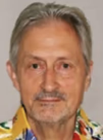 Richard Sutton合作论文数National Heart & Lung Institute, Imperial College London4
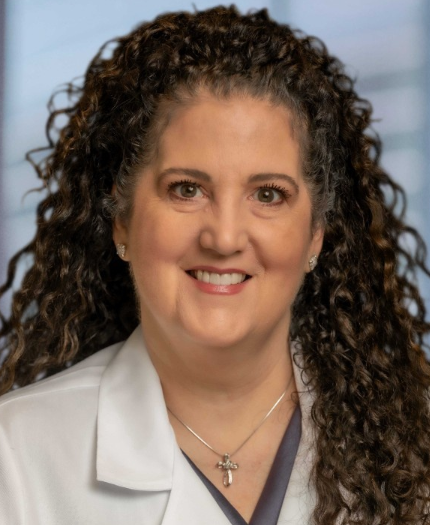 Cindy M. Martin合作论文数Division of Advanced Heart Failure, Houston Methodist;Academic Institute, Houston Methodist;DeBakey Heart & Vascular Center, Houston Methodist4Keith Wesley合作论文数3
 Manan Gandhi合作论文数Autonomous Control & Decis Syst Lab, Georgia Inst Technol3Claire Carlson合作论文数3
 Georg Schlachtenberger合作论文数3
 Johnny Chahine合作论文数Department of Internal Medicine, Cleveland Clinic3
 Thomas Murray合作论文数Department of Pediatrics, Yale School of Medicine, Yale University3
 Marc Richard Conterato合作论文数3
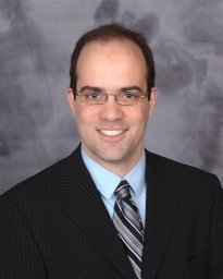 Prisco Anthony R合作论文数Biotechnology and Bioengineering Center, Medical College of Wisconsin;Department of Physiology, Medical College of Wisconsin;Medical College of Wisconsin, Medical College of Wisconsin3
 Schizas Dimitrios合作论文数3
 Thorsten Wahlers合作论文数Department of Cardiac and Thoracic Surgery, University Hospital Cologne3
 Evangelos Theodorou合作论文数Daniel Guggenheim School of Aerospace Engineering, College of Engineering, Georgia Institute of Technology3
 Ralph Frascone合作论文数Department of Emergency Medicine, Regions Hospital3Ioannis Paraskevaidis合作论文数Second Department of Cardiology and Heart Failure Clinic, Attikon University Hospital3
 Guillaume Debaty合作论文数pôle urgences Samu-Smur, CHU de Grenoble3
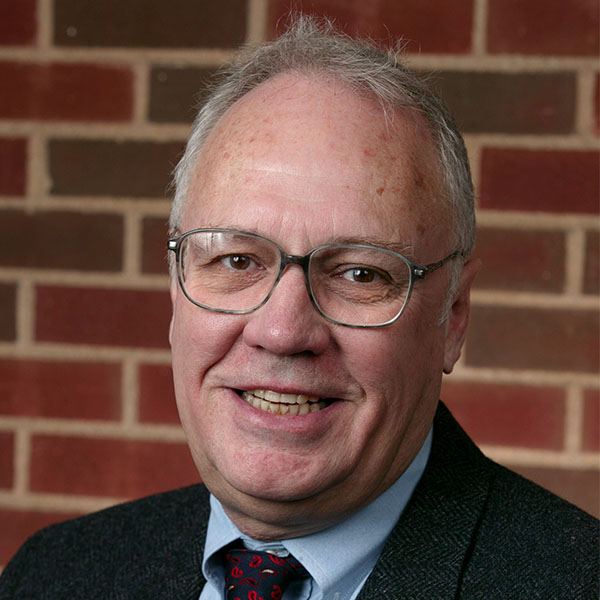 John E. Connett合作论文数Division of Biostatistics, School of Public Health, University of Minnesota3
 Kimon Stamatelopoulos合作论文数Medical School, National & Kapodistrian University of Athens;Department of Clinical Therapeutics, Alexandra hospital3
 Christina Piperi合作论文数Laboratory of Biological Chemistry, University of Athens Medical School3




