Cigarette smoking induces epigenetic changes that can cause degenerative heterogeneity with aging and disease. In disease such as age--related macular degeneration (AMD), the leading worldwide cause of blindness among the elderly, retinal pigment epithelial (RPE) cell heterogeneity is a key change. Since smoking is a powerful risk factor for AMD, we hypothesized that smoke induces epigenetic--mediated degenerative RPE heterogeneity. We administered cigarette smoke condensate (CSC) to young and aged mice. Using snRNA-seq and single nuclear ATAC sequencing, we identified distinct healthy and dedifferentiated RPE clusters in both aged vehicle-and young CSC--treated mice. Dedifferentiated RPE had globally decreased chromatin accessibility and expression of genes linked to "hallmarks of aging." Notably, young, dedifferentiated RPE also exhibited a compensatory upregulation of hallmarks of aging--related genes including mitochondrial function and proteostasis while aged dedifferentiated RPE did not, which decreased their survival following CSC treatment, as experimentally verified with TUNEL labeling. Similar populations of dedifferentiated and healthy RPE were identified both in mice exposed to cigarette smoke for 4 mo and in macular RPE from a donor who smoked and another with early AMD, but not from a nonsmoker donor. Degenerative cellular heterogeneity that includes an abnormal cluster can jeopardize cell survival and represents a hallmark of ocular aging.
Environmental exposure such as cigarette smoke induces epigenetic changes that can induce degenerative heterogeneity and accelerate aging. In early age-related macular degeneration (AMD), the leading worldwide cause of blindness among the elderly, retinal pigment epithelial (RPE) cell heterogeneity is a key change. Since smoking is the strongest environmental risk factor for AMD, we hypothesized that cigarette smoke induces degenerative RPE heterogeneity through epigenetic changes that are distinct from aging, and that with aging, the RPE becomes vulnerable to cigarette smoke insult. We administered cigarette smoke condensate (CSC) intravitreally to young and aged mice and performed snRNA-seq and snATAC-seq on the RPE/choroid. This analysis identified separate cell clusters corresponding to healthy and abnormal, dedifferentiated RPE in both aged vehicle-treated and young CSC-treated mice. The dedifferentiated RPE were characterized by a global decrease in chromatin accessibility and decreased expression of genes in functional categories that were linked to hallmarks of aging. Notably, young, dedifferentiated RPE also exhibited a compensatory upregulation of hallmarks of aging-related genes, specifically those related to mitochondrial function and proteostasis. In contrast, aged dedifferentiated RPE did not express these compensatory changes, and did not survive CSC treatment, as experimentally verified with TUNEL labeling. These changes are relevant to early AMD because we identified through scRNA-seq, similar dedifferentiated and healthy macular RPE clusters in a donor who smoked and another with early AMD, but not from a nonsmoker. Degenerative cellular heterogeneity can include an abnormal cluster that jeopardizes cell survival and may represent an additional hallmark of ocular aging.
PURPOSE:Ultrahigh-dose-rate radiation therapy (FLASH RT) has not previously been studied for the treatment of ocular targets. This work describes the feasibility of ocular FLASH irradiation of a single mouse eye using kilovoltage x-ray sources with rotating anode technology. METHODS AND MATERIALS:A 75-kW-powered rotating anode kilovoltage x-ray source was implemented for FLASH and conventional (CONV) dose-rate irradiation of superficial targets in small animals. A docking immobilization device was designed to facilitate irradiation of an individual mouse eye. Mouse positioning reproducibility was assessed via Hausdorff distance and Dice similarity coefficients of features from cone beam computed tomographies (CT) of immobilized animals. Thermoluminescent dosimeters and Gafchromic film dosimeters were used to characterize output (dose rate), depth dose rate, and to assess geometric parameters of the beam. In a pilot study of ocular irradiation, healthy C57BL/6J mouse eyes were irradiated to doses of up to 26 Gy at either FLASH or CONV dose rates. Retinal function was assessed 2 months following irradiation using electroretinography. Morphologic changes were evaluated via histopathology. RESULTS:Animal setup was highly reproducible, with typical Hausdorff distances between bony features of 0.34 mm and Dice similarity coefficient of 0.92 for surface features. At FLASH irradiation settings, dose rates of 52.6 ± 2.6 Gy/s were measured using in vivo thermoluminescent dosimeters. CONV dose rates of 1.1 ± 0.1 Gy/s were achievable with the same setup by minimizing the input current. Following irradiation to 26 Gy, retinal function was preserved for FLASH-irradiated eyes but significantly impaired in CONV-irradiated eyes. Histopathology assessment confirmed significant inner retinal atrophy in CONV-irradiated eyes that was absent in FLASH-irradiated eyes. CONCLUSIONS:Irradiation of the mouse eye was feasible at FLASH and CONV dose rates by the orthovoltage x-ray using the rotating anode x-ray source. A pilot study of ocular irradiation in healthy mice demonstrated sparing effects of FLASH on the mouse retina.
Background: Age-related macular degeneration (AMD) is a major cause of irreversible vision loss in the developed world, and the approved products for geographic atrophy (GA), a late-stage form of dry AMD, have shown limited efficacy and require frequent administration. Therefore, longer-lasting therapies with improved efficacy would be a welcome addition to AMD treatment. One potential therapeutic is ONL1204, a small peptide inhibitor of the Fas receptor that has prevented cell death and inflammation in retinal disease models. This study characterizes the pharmacokinetics (PK) and durability of protection conferred by ONL1204. Methods: Ocular pharmacokinetic profiles were generated over 3 months in rabbit and minipig following a single intravitreal (IVT) injection of ONL1204 at multiple doses. Ocular pharmacodynamics were evaluated in two models: a rabbit model using a single IVT injection of ONL1204 with a delayed sodium iodate challenge coupled with fluorescein angiography to quantify RPE loss, and a chronic mouse model that reflects key features of dry AMD disease pathology to assess the efficacy of repeat IVT administrations of ONL1204. Results: ONL1204 had prolonged residence in the ocular tissues of rabbit and minipig, with a vitreous humor half-life of over 100 days. ONL1204 demonstrated significant protection of the retinal pigment epithelium (RPE) in the rabbit sodium iodate model. In the chronic mouse model, two administrations of ONL1204 preserved RPE morphology, reduced caspase-8 activity, and decreased inflammation. Conclusions: These data represent key characteristics of ONL1204, highlighting its clinical potential as a therapeutic for chronic retinal diseases, including GA.
PURPOSE To determine if aqueous humor lactic acid levels, a surrogate marker for tumor cell metabolic activity, are elevated in uveal melanoma (UM) and whether L-lactate levels correlate with clinical, pathologic, and molecular tumor characteristics. DESIGN Case-control study. SUBJECTS Eyes with UM and control eyes undergoing cataract surgery. METHODS L-Lactate levels were measured from aqueous humor taken at the time of enucleation for UM (n = 34) or controls (n = 13). Twenty-three (68%) UM patients were followed for at least 5 years. Clinical information was gathered by retrospective chart review. Tumor samples were profiled by RT-qPCR and factor analysis to emulate prognostic classification. MAIN OUTCOME MEASURE Aqueous humor lactate levels. RESULTS Aqueous L-lactate concentration in UM eyes was higher than controls (P < .01). The L-lactate concentration correlated with increasing mitotic activity on pathologic examination (P = .02), but not with tumor size, prior brachytherapy, predominant histologic cell type, or metastasis. L-Lactate levels did not correlate with gene expression profile. CONCLUSION L-Lactate levels in aqueous samples from patients with uveal melanoma were higher than controls, which lays the foundation for future studies into whether this may be helpful for diagnosing suspicious uveal lesions.
An 11-step synthesis of the octacyclic-fused dimeric phenylphenalenone anigorootin (phytoalexin in banana plants) is reported. The synthetic strategy uses an electrochemical dimerization as the key step, which stereospecifically installs the four asymmetric centers. Mechanistic aspects of the dimerization process are discussed.
Maternal smoking during pregnancy (MSDP) is associated with significant cognitive and behavioral effects on offspring. While neurodevelopmental outcomes have been studied for prenatal exposure to nicotine, the main psychoactive component of cigarette smoke, its contribution to MSDP effects has never been explored. Comparing the effects of these substances on molecular signaling in the prenatal and adult brain may provide insights into nicotinic and broader tobacco consequences that are developmental-stage specific or age-independent. Pregnant mice were administered nicotine or exposed to chronic cigarette smoke, and RNA-sequencing was performed on frontal cortices of postnatal day 0 pups born to these mice, as well as on frontal cortices and blood of the adult dams. We identified 1,010 and 4,165 differentially expressed genes (DEGs) in nicotine and smoking-exposed pup brains, respectively (FDR<0.05, Ns = 19 nicotine-exposed vs 23 vehicle-exposed; 46 smoking-exposed vs 49 controls). Prenatal nicotine exposure (PNE) alone was related to dopaminergic synapses and long-term synaptic depression, whereas MSDP was associated with the SNARE complex and vesicle transport. Both substances affected SMN-Sm protein complexes and postsynaptic endosomes. Analyses at the transcript, exon, and exon-exon junction levels supported gene level results and revealed additional smoking-affected processes. No DEGs at FDR<0.05 were found in adult mouse brain for any substance (12 nicotine-administered vs 11 vehicle-administered; 12 smoking-exposed vs 12 controls), nor in adult blood (12 smoking-exposed vs 12 controls), and only 3% and 6.41% of the DEGs in smoking-exposed pup brain replicated in smoking-exposed blood and human prenatal brain, respectively. Together, these results demonstrate variable but overlapping molecular effects of PNE and MSDP on the developing brain, and attenuated effects of both smoking and nicotine on adult versus fetal brain.
Age-related macular degeneration (AMD) is a leading cause of irreversible blindness in the developed world. While great advances have been made in the treatment of the neovascular ("wet") form of the disease, there is still a significant need for therapies that prevent the vision loss associated with the advanced forms of dry, atrophic AMD. In this atrophic form, retinal pigment epithelial (RPE) and photoreceptor cell death is the ultimate cause of vision loss. In this review, we summarize the cell death pathways and their relation to RPE and retinal cell death in AMD. We review the data that support targeting programmed cell death through inhibition of the Fas receptor as a novel approach to preserve these structures and that this effect results from inhibiting both canonical death pathway activation and reducing the associated inflammatory response. These data lay the groundwork for current clinical strategies targeting the Fas pathway in this devastating disease.
Purpose/Objective(s) Normal tissue sparing effects from FLASH radiotherapy (RT) are potentially transformative for the treatment of ocular cancers. Reducing damage to the abundant at-risk structures within the eye can prospectively increase visual retention rates, relax treatment margins, and eliminate the need for motion tracking by surgically implanted fiducials. To date, no prior work has explored FLASH-RT effects in the context of the eye. In this work, we have conducted a pilot study of x-ray FLASH effects for ocular irradiation in a preclinical mouse model by measuring the functional and pathological changes in healthy eyes following FLASH or conventional (CONV) dose rate irradiation. Materials/Methods An immobilization device was designed to enable focal irradiation to a single mouse eye using a FLASH-capable rotating anode x-ray tube. A 5 mm diameter lead collimator was manufactured to encompass the 3 mm target of the entire eye. Dose and dose rate measurements were performed with calibrated radiographic EBT3 films. Cone-beam CTs were acquired of four C57BL6J mice in immobilization to confirm setup reproducibility, quantified by the mean Hausdorff distance between bone segmentations. Healthy 8-week-old C57BL6J mice were irradiated with 150 kVp x-rays to doses of 21 Gy or 34 Gy at FLASH (right eye) and CONV (left eye) dose rates, respectively. Both eyes were irradiated to limit the influence of varying baseline vision between animals in our analysis. Visual acuity was assessed in 3 mice per dose level using scotopic electroretinography (ERG) up to 2 months post irradiation. Histopathological changes were assessed through H&E staining of harvested eyes. Results Mouse setup in our immobilization device was highly reproducible, with a mean Hausdorff distance of 0.34 ± 0.10 mm. Measured dose rates within the field were 67.0 ± 1.9 Gy/s and 1.2 ± 0.1 Gy/s at FLASH and CONV settings, respectively. ERGs revealed that FLASH-irradiated eyes at 21 Gy retained visual function up to 2 months post irradiation, while 21 Gy CONV induced blindness at all sampled time points with more severe surrounding skin effects. At 34 Gy, all eyes were blinded within 1 week, with comparable skin toxicities in the irradiated areas regardless of dose rate. Pathological assessment showed a loss of the photoreceptor layer of the retina from CONV irradiation, which remained intact in FLASH-treated mice. Conclusion We have developed a novel platform to study x-ray FLASH effects from ocular irradiation in mice. Functional ERG assay revealed a preservation of visual function from 21 Gy at FLASH dose rates that was not present from 21 Gy CONV. Differential damages between FLASH and CONV irradiations were confirmed through histopathology. This first demonstration of FLASH normal tissue sparing effects in a mouse eye model presents a unique and promising translation opportunity for clinical FLASH treatment. Further studies are ongoing to explore the long-term effects of FLASH radiation on vision and its efficacy on intraocular tumors.
The purpose of this study was to evaluate the effect of bovine colostrum (BC) in the regeneration of corneal epithelial cells on an ocular alkali burn model. Twenty-four C57BL/6 mice were categorized into two gender/age-matched groups for treatment. Two days after inducing a corneal alkali burn in all left eyes with 4 μl of sodium hydroxide 0.15 mol/l, both eyes of group 1 were treated with BC 4 times per day, and both eyes of group 2 were treated with isotonic saline solution (SS). The epithelial defect was photographed and measured by fluorescein staining on days two, four, seven, and ten. Ocular burn damage was assessed with a pre-established classification in clock hours from the limbus. After 10 days both eyes were processed, half of the group's corneas were assessed histopathologically, and the other half was used for pro/anti-inflammatory cytokine quantification using ELISA. BC treated (Group 1) corneas revealed significantly improved fluorescein staining score for limbal involvement when compared to SS treated (Group 2) corneas at days 4 (p = 0.013), 7 (p < 0.001), and 10 (p < 0.001), respectively. No differences were noted in limbal involvement at day 2 between the two groups (p > 0.99). The overall change (difference in slope) in fluorescein staining for limbal involvement between days 2 and 10 was -0.1669 (p = 0.006). Histologic examinations and cytokine measurements of group 2 demonstrated a strong inflammatory component compared to group 1. Our data indicates that topical application of BC facilitates corneal re-epithelialization and wound healing by suppressing the inflammatory process in an ocular alkali burn model.
The synthesis of 4-phenylphenalenones, a group of phytoalexins exclusive from Musa acuminata (bananas), has been revisited to extend the scope of the derivatives that can be prepared. The discovery that 4-methoxyphenalenone participates in a selective substitution reaction allowed the preparation of 4-bromophenalenone, which proved to be an efficient substrate for Suzuki cross-couplings. The results solve the previously uncovered problem of preparing electron-rich 4-heteroarylphenalenones and provide a general synthesis of this type of phytoalexins.
Purpose: The purpose of this study was to compare the efficacy of high ultraviolet A (UVA) irradiance photoactivation of riboflavin (vitamin B2) versus the standard corneal cross-linking protocol on bacterial viability. Methods: Methicillin-sensitive Staphylococcus aureus (MSSA) Newman strain and methicillin-resistant multidrug-resistant S. aureus (MDR-MRSA) USA300, CA409, CA127, GA656, and NY315 strains were exposed to a UVA energy dose of 5.4 to 6 J/cm2 by 2 high irradiance regimens: A) 30 mW/cm2 for 3 minutes and B) 10 mW/cm2 for 10 minutes with B2 0.1%. Control groups included B2/UVA alone, CA409 exposed to standard B2 0.1% + UVA (3 mW/cm2 for 30 minutes), and an untreated sample. Cell viability was assessed. Triplicate values were obtained. The Mann–Whitney test and Student t test were used for statistical analysis. Results: There was no difference comparing the median bacterial load (log CFU/mL) of the untreated samples versus regimen A: Newman P = 0.7, CA409 P = 0.3, USA300 P = 0.5, CA127 P = 0.6, GA656 P = 0.1, and NY315 P = 0.2 (P ≥ 0.1); and B: Newman P= 0.1, CA409 P = 0.3, USA300 P = 0.4, CA127 P = 0.6, GA656 P = 0.1, and NY315 P = 0.3 (P ≥ 0.1). Standard regimen killed 100% of CA409. Conclusions: Photoactivation of B2 by high UVA irradiance does not seem to be effective for bacterial eradication in this study.
The nuclear factor‐erythroid 2‐related factor‐2 (Nrf2), a major antioxidant transcription factor, is decreased in several age‐related diseases including age‐related macular degeneration (AMD), the most common cause of blindness among the elderly in western society. Since Nrf2’s mito‐protective response is understudied, we investigated its antioxidant response on mitochondria. Control and Nrf2‐deficient retinal pigmented epithelial (RPE) cells were compared after treating with cigarette smoke extract (CSE). Mitochondrial antioxidant abundance and reactive oxygen species (ROS) were quantified. Mitochondrial function was assessed by TMRM assay, NADPH, electron transport chain activity, and Seahorse. Results were corroborated in Nrf2 −/− mice and relevance to AMD was provided by immunohistochemistry of human globes. CSE induced mitochondrial ROS to impair mitochondrial function. H 2 O 2 increase in particular, was magnified by Nrf2 deficiency, and corresponded with exaggerated mitochondrial dysfunction. While Nrf2 did not affect mitochondrial antioxidant abundance, oxidized PRX3 was magnified by Nrf2 deficiency due to decreased NADPH from decreased expression of IDH2 and pentose phosphate pathway (PPP) genes. With severe CSE stress, intrinsic apoptosis was activated to increase cell death. PPP component TALDO1 immunolabeling was decreased in dysmorphic RPE of human AMD globes. Despite limited regulation of mitochondrial antioxidant expression, Nrf2 influences PPP and IDH shuttle activity that indirectly supplies NADPH for the TRX2 system. These results provide insight into how Nrf2 deficiency impacts the mitochondrial antioxidant response, and its role in AMD pathobiology.
The accumulation of lipids within drusen, the epidemiologic link of a high fat diet, and the identification of polymorphisms in genes involved in lipid metabolism that are associated with disease risk, have prompted interest in the role of lipid abnormalities in AMD. Despite intensive investigation, our understanding of how lipid abnormalities contribute to AMD development remains unclear. Lipid metabolism is tightly regulated, and its dysregulation can trigger excess lipid accumulation within the RPE and Bruch's membrane. The high oxidative stress environment of the macula can promote lipid oxidation, impairing their original function as well as producing oxidation-specific epitopes (OSE), which unless neutralized, can induce unwanted inflammation that additionally contributes to AMD progression. Considering the multiple layers of lipid metabolism and inflammation, and the ability to simultaneously target multiple pathways, microRNA (miRNAs) have emerged as important regulators of many age-related diseases including atherosclerosis and Alzheimer's disease. These diseases have similar etiologic characteristics such as lipid-rich deposits, oxidative stress, and inflammation with AMD, which suggests that miRNAs might influence lipid metabolism in AMD. In this review, we discuss the contribution of lipids to AMD pathobiology and introduce how miRNAs might affect lipid metabolism during lesion development. Establishing how miRNAs contribute to lipid accumulation in AMD will help to define the role of lipids in AMD, and open new treatment avenues for this enigmatic disease.
Age-related macular degeneration (AMD) is a leading cause of blindness in the elderly. The extent to which epigenetic changes regulate AMD progression is unclear. Here we globally profiled chromatin accessibility in the retina and retinal pigmented epithelium (RPE) from AMD patients and controls. Global decreases in chromatin accessibility occurr in RPE in early AMD, and in the retina with advanced disease, suggesting that dysfunction in RPE cells drives disease progression. Footprints of photoreceptor and RPE-specific transcription factors are enriched in differentially accessible regions (DARs). Genes associated with DARs show altered expression in AMD. Cigarette smoke treatment of RPE cells recapitulates epigenomic changes seen in AMD, providing an epigenetic link between the known risk factors for AMD and AMD pathology. Finally, overexpression of HDAC11 is partially responsible for the reduction in chromatin accessibility, identifying potential new targets for treatment of AMD.
Age-related macular degeneration (AMD) is a significant cause of vision loss in the elderly. The extent to which epigenetic changes regulate AMD progression is unclear. Here we globally profile chromatin accessibility using ATAC-Seq in the retina and retinal pigmented epithelium (RPE) from AMD and control patients. Global decreases in chromatin accessibility occur in the RPE with early AMD, and in the retina of advanced disease, suggesting that dysfunction in the RPE drives disease onset. Footprints of photoreceptor and RPE-specific transcription factors are enriched in differentially accessible regions (DARs). Genes associated with DARs show altered expression in AMD. Cigarette smoke treatment of RPE cells recapitulates chromatin accessibility changes seen in AMD, providing an epigenetic link between a known risk factor for AMD and AMD pathology. Finally, overexpression of HDAC11 is partially responsible for the observed reduction in chromatin accessibility, suggesting that HDAC11 may be a potential new therapeutic target for AMD.
 James T. Handa合作论文数49
 Peter Louis Gehlbach合作论文数Department of Otolaryngology, School of Medicine, Johns Hopkins University23Lei Wang合作论文数16
 Debasish Sinha合作论文数Department of Ophthalmology,University of Pittsburgh16
 Ashley Behrens合作论文数Department of Otolaryngology-Head and Neck Surgery, School of Medicine, Johns Hopkins University13Ebrahimi Katayoon B合作论文数12
 Sayantan Datta合作论文数Human Genetics Unit, Indian Statistical Institute;Department of Ophthalmology, Johns Hopkins School of Medicine;Indian Statistical Institute, Johns Hopkins School of Medicine12Roy S. Chuck合作论文数10Stacey L. Hose合作论文数7J. Samuel Zigler合作论文数7Andreina Tarff合作论文数6Rebecca Yee合作论文数School of Medicine and Health Sciences, George Washington University6
 Morton F. Goldberg合作论文数Johns Hopkins Hospital6Felipe Otálvaro合作论文数Instituto de Quimica, Universidad de Antioquia6Laura Di Meglio合作论文数5natalia fijalkowski合作论文数5Sonny Dike合作论文数5Praveena K Gupta合作论文数5
 Naoshi Kondo合作论文数5Tongyun Liu合作论文数5
 Keisuke Mori合作论文数Department of Ophthalmology, International University of Health and Welfare5Js Zigler合作论文数4
 William Hidalgo合作论文数Grp Invest Bioquim & Microbiol GIBIM, Univ Ind Santander4
 Thanh V. Hoang合作论文数Solomon H. Snyder Department of Neuroscience, Johns Hopkins University School of Medicine, Baltimore, MD 21205, USA.4
 Seth Blackshaw合作论文数Department of Otolaryngology-Head and Neck Surgery, School of Medicine, Johns Hopkins University4
 Jiang Qian合作论文数Department of Wilmer Eye Institute, School of Medicine, Johns Hopkins University4
 Bernd Schneider合作论文数Max Planck Institute for Chemical Ecology;Leiden University4
 Ying Zhang (张颖)合作论文数The First Affiliated Hospital, School of Medicine, Zhejiang University;School of Public Health, Zhejiang Univeristy4Guillermo Noguera合作论文数4Johann M. G. Reyes合作论文数4
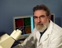 Gerard A. Lutty合作论文数The Wilmer Ophthalmological Institute, Johns Hopkins University School of Medicine4Eric F. Wawrousek合作论文数4
 Choul Yong Park合作论文数4
 Rhonda Grebe合作论文数4Elia J. Duh合作论文数The Wilmer Ophthalmological Institute, The Johns Hopkins University School of Medicine4
 Imran Bhutto合作论文数The Johns Hopkins Hospital, The Wilmer Ophthalmological Institute4Masashi Fujihara合作论文数4William N. May合作论文数4o suwan apichon合作论文数3Margaret Yew合作论文数3Ashkan Pirouzmanesh合作论文数3
 Peng Shang合作论文数Univ Pittsburgh, Sch Med, Dept Ophthalmol, Pittsburgh, PA 15261 USA3
 Shyam S. Biswal合作论文数Environmental Health and Engineering, Bloomberg School of Public Health, Johns Hopkins University3
 Aleksander S. Popel合作论文数Popel Systems Biology Laboratory, Department of Biomedical Engineering, School of Medicine, Johns Hopkins University3
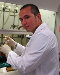 Karagiannis Emmanouil D合作论文数The David H. Koch Institute for Integrative Cancer Research, Massachusetts Institute of Technology3Merbs Shannath L合作论文数3Kocab Andrew J合作论文数3
 Arturo Casadevall合作论文数W. Harry Feinstone Department of Molecular Microbiology and Immunology, Bloomberg School of Public Health, Johns Hopkins University;School of Medicine, Johns Hopkins University3
 Cheng Zhang (张诚)合作论文数Institute of Information and Communication, School of Electronics, School of Electronics Engineering and Computer Science, Peking University3




