Background: Thymic carcinomas are rare aggressive mediastinal tumors with a median survival of 2 years. Observation: We present a pediatric patient who was diagnosed with metastatic thymic carcinoma and showed continuous improvement of his primary mass and lung metastases with a regimen of cisplatin/docetaxel followed by long-term maintenance therapy with sunitinib for over 5 years. Conclusions: This report demonstrates a long-term positive treatment effect using chemotherapy followed by sunitinib in an advanced thymic carcinoma. We are not aware of other reports of pediatric patients with metastatic thymic carcinoma treated with sunitinib maintenance who maintained a durable response for this prolonged period of time.
Background Previous studies suggest that use of artificial intelligence (AI) algorithms as diagnostic aids may improve the quality of skeletal age assessment, though these studies lack evidence from clinical practice. Purpose To compare the accuracy and interpretation time of skeletal age assessment on hand radiograph examinations with and without the use of an AI algorithm as a diagnostic aid. Materials and Methods In this prospective randomized controlled trial, the accuracy of skeletal age assessment on hand radiograph examinations was performed with (n = 792) and without (n = 739) the AI algorithm as a diagnostic aid. For examinations with the AI algorithm, the radiologist was shown the AI interpretation as part of their routine clinical work and was permitted to accept or modify it. Hand radiographs were interpreted by 93 radiologists from six centers. The primary efficacy outcome was the mean absolute difference between the skeletal age dictated into the radiologists’ signed report and the average interpretation of a panel of four radiologists not using a diagnostic aid. The secondary outcome was the interpretation time. A linear mixed-effects regression model with random center- and radiologist-level effects was used to compare the two experimental groups. Results Overall mean absolute difference was lower when radiologists used the AI algorithm compared with when they did not (5.36 months vs 5.95 months; P = .04). The proportions at which the absolute difference exceeded 12 months (9.3% vs 13.0%, P = .02) and 24 months (0.5% vs 1.8%, P = .02) were lower with the AI algorithm than without it. Median radiologist interpretation time was lower with the AI algorithm than without it (102 seconds vs 142 seconds, P = .001). Conclusion Use of an artificial intelligence algorithm improved skeletal age assessment accuracy and reduced interpretation times for radiologists, although differences were observed between centers. Clinical trial registration no. NCT03530098 © RSNA, 2021 Online supplemental material is available for this article. See also the editorial by Rubin in this issue.
Abstract Bacterial osteomyelitis of the scapula is rare in pediatric patients. When it occurs, it usually affects young infants and patients often exhibit fever and functional limitation of the extremity. We present a case of a 12-year-old boy with subacute bacterial scapular osteomyelitis and a normal physical examination. The patient was briefly treated with intravenous antibiotics and then transitioned to oral antibiotics to complete a 4-week course for methicillin-sensitive Staphylococcus aureus osteomyelitis.
The mediastinum is the most common location for intrathoracic masses in children. This chapter reviews an approach to the imaging work-up of mediastinal masses in pediatric patients based on anatomic location, and specific imaging characteristics. Common diseases, imaging pitfalls and select rare entities are described to provide a comrehensive discusion on the subject matter.
Rationale and Objectives: The educational value of the daily resident readout, a vital component of resident training, has been markedly diminished due to a significant decrease in imaging volume and case mix diversity. The goal of this study was to create a "simulated" daily readout (SDR) to restore the educational value of the daily readout. Materials and Methods: To create the SDR the following tasks were performed; selection of cases for a daily worklist for each resident rotation, comprising a combination of normal and abnormal cases; determination of the correct number of cases and the appropriate mix of imaging modalities for each worklist; development of an ''educational'' environment consisting of separate ''instances'' of both our Picture Archive Communication System and reporting systems; and the anonymization of all of the cases on the worklists. Surveys of both residents and faculty involved in the SDR were performed to assess its effectiveness. Results: Thirty-two residents participated in the SDR. The daily worklists for the first 20 days of the SDR included 3682 cases. An average of 480 cases per day was dictated by the residents. Surveys of the residents and the faculty involved in the SDR demonstrated that both agreed that the SDR effectively mimics a resident's daily work on rotations and preserves resident education during the Coronavirus Disease 2019 crisis. Conclusion: The development of the SDR provided an effective method of preserving the educational value of the daily readout experience of radiology residents, despite severe decreases in imaging exam volume and case mix diversity during the Coronavirus Disease 2019 pandemic. (c) 2020 The Association of University Radiologists. Published by Elsevier Inc. All rights reserved.
Magnetic resonance imaging (MRI) assessment for appendicitis is limited by exam time and patient cooperation. The radially sampled 3-dimensional (3-D) T1-weighted, gradient recalled echo sequence (radial GRE) is a free-breathing, motion robust sequence that may be useful in evaluating appendicitis in children. To compare the rate of detection of the normal appendix with contrast-enhanced radial GRE versus contrast-enhanced 3-D GRE and a multi-sequence study including contrast-enhanced radial GRE. This was a retrospective study of patients ages 7–18 years undergoing abdominal-pelvic contrast-enhanced MRI between Jan. 1, 2012, and April 1, 2016. Visualization of the appendix was assessed by consensus between two pediatric radiologists. The rate of detection of the appendix for each sequence and combination of sequences was compared using a McNemar test. The rate of detection of the normal appendix on contrast-enhanced radial GRE was significantly higher than on contrast-enhanced 3-D GRE (76% vs. 57.3%, P=0.003). The rate of detection of the normal appendix with multi-sequence MRI including contrast-enhanced radial GRE was significantly higher than on contrast-enhanced 3-D GRE (81.3% vs. 57%, P<0.001). There was no significant difference between the rate of detection of the normal appendix on contrast-enhanced radial GRE alone and multi-sequence MRI including contrast-enhanced radial GRE (76% vs. 81.3%, P=0.267). Contrast-enhanced radial GRE allows superior detection of the normal appendix compared to contrast-enhanced 3-D GRE. The rate of detection of the normal appendix on contrast-enhanced radial GRE alone is nearly as good as when the contrast-enhanced radial GRE is interpreted with additional sequences.
Non-Cartesian magnetic resonance imaging (MRI) sequences have shown great promise for abdominal examination during free breathing, but break down in the presence of bulk patient motion (i.e. voluntary or involuntary patient movement resulting in translation, rotation or elastic deformations of the body). This work describes a data-consistency-driven image stabilization technique that detects and excludes bulk movements during data acquisition. Bulk motion is identified from changes in the signal intensity distribution across different elements of a multi-channel receive coil array. A short free induction decay signal is acquired after excitation and used as a measure to determine alterations in the load distribution. The technique has been implemented on a clinical MR scanner and evaluated in the abdomen. Six volunteers were scanned and two radiologists scored the reconstructions. To show the applicability to other body areas, additional neck and knee images were acquired. Data corrupted by bulk motion were successfully detected and excluded from image reconstruction. An overall increase in image sharpness and reduction of streaking and shine-through artifacts were seen in the volunteer study, as well as in the neck and knee scans. The proposed technique enables automatic real-time detection and exclusion of bulk motion during MR examinations without user interaction. It may help to improve the reliability of pediatric MRI examinations without the use of sedation.
Low nephron number has been shown to be a risk factor for hypertension (HTN) in adulthood. Kidney volume may serve as a surrogate marker for nephron mass. The relationship between kidney volume and ambulatory blood pressure (BP) in the pediatric population is not known. A retrospective chart review of children younger than 21 years who were evaluated for HTN was performed. Twenty‐four‐hour BP and ultrasonography data were obtained. Multiple regression was used to examine associations between BP and kidney volume. Of 84 children (mean age 13.87 years, 72.6% males), 54 had HTN. Systolic BP index during the awake, sleep, and 24‐hour periods (all P≤.05) was found to be positively correlated with total kidney volume. Greater total kidney volume was found to be a positive predictor of 24‐hour and sleep systolic index (P≤.05). It failed to serve as a predictor of HTN, pre‐HTN, or white‐coat HTN. Contrary to expectation, total kidney volume was positively associated with systolic BP indices.
Previously proposed techniques based on non-Cartesian or modified Cartesian sampling schemes allow for free-breathing acquisitions with solid motion-robustness. However, residual artifacts such as motion blurring often remain. Techniques including self-gating, motion-weighting, and extra-dimensional reconstruction have been proposed to further improve image quality.
Thrombotic disease is rare in neonates. Many of the cases reported in literature are attributed to the placement of central catheters. We report on a case of aortic thrombosis in a newborn infant with significant respiratory distress due to meconium aspiration, necessitating intubation and placement of central catheters. Due to the location and size of the thrombus in our case, various subspecialties were involved, which ultimately guided therapy to anti-coagulate the patient.
terms.php and incorporate the Creative Commons Attribution – Non Commercial (unported, v3.0) License (http://creativecommons.org/licenses/by-nc/3.0/). By accessing the work you hereby accept the Terms. Non-commercial uses of the work are permitted without any further permission from Dove Medical Press Limited, provided the work is properly attributed. For permission for commercial use of this work, please see paragraphs 4.2 and 5 of our Terms (https://www.dovepress.com/terms.php). Journal of Vascular Diagnostics and Interventions 2016:4 23–37 Journal of Vascular Diagnostics and Interventions Dovepress
Boys commonly present with acute scrotal pain to the emergency room. The potentially associated high morbidity necessitates that our attending partners, trainees, and physician extenders in Pediatrics, Radiology, Emergency Medicine, and Urology be knowledgeable and trained in triage management principles. Yet, no electronic resources exist to provide such training. The aims of our study were to determine if an interdisciplinary clinical team (i.e. Pediatric Urology, Radiology, and Emergency Medicine) could co-create a CEVL e-learning platform and to determine its efficacy in educating our healthcare partners about such triage management.
Osteosarcoma (OS) and the Ewing sarcoma family of tumors (ESFT) are the most common malignant bone tumors in children and adolescents. While significant improvements in survival have been seen in other pediatric malignancies the treatment and prognosis for pediatric bone tumors has remained unchanged for the past 3 decades. This review and update of pediatric malignant bone tumors will provide a general overview of osteosarcoma and the Ewing sarcoma family of tumors, discuss bone tumor genomics, current challenges, and emerging drug targets.
Magnetic resonance cholangiopancreatography (MRCP) is commonly performed in the evaluation of known or suspected pancreaticobiliary disease in children. The administration of a negative oral contrast agent can improve the quality of the examination without significant additional cost. We describe our experience with certain brands of acai juice, blueberry juice and pineapple juice as negative oral contrast agents in children. We believe these fruit juices are safe, palatable and may improve MRCP image quality.
A 2 year 9 month-old girl presented with active hemorrhage from a Wilms' tumor. The hemorrhage was controlled with embolization of the left renal artery and a left nephrectomy was subsequently performed. Embolization to control active bleeding in trauma scenarios and as a preoperative intervention to minimize blood loss has been extensively reported in the pediatric population. There are very few case reports of embolization of an actively bleeding tumor.
Hepatic neoplasms constitute approximately 5% to 6% of all pediatric intra-abdominal masses, most of which are malignant. When compared with traditional multi-phase computed tomography protocols that are often used in adults, magnetic resonance (MR) imaging is particularly desirable for evaluating liver lesions in children because of the lack of ionizing radiation and superb soft tissue contrast resolution. The goal of this article is to discuss common benign and malignant pediatric hepatic lesions and their key MR imaging findings. Particular emphasis is placed on the utility of new hepatocyte-specific contrast agents to narrow the differential diagnosis.
Introduction: Crohn’s disease (CD) is a chronic inflammatory bowel disease of unknown etiology which affects between 400,000 and 600,000 people in North America (1). CD can involve any part of the gastrointestinal tract, and has a chronic, relapsing, and remitting clinical course. Long-standing inflammation can result in bowel obstruction, stricture, fistula, and/or abscess. In addition, there is an increased risk for small and large bowel malignancy in areas of chronic inflammation (2). Assessment of inflammatory activity plays a crucial role in the individualized management of patients with CD. Therefore, the need for a reliable, non-invasive imaging evaluation for inflammatory activity is crucial for the effective management of CD. Diffusion weighted MR imaging (DW-MRI) has shown restricted diffusion in inflamed bowel segments (3, 4). These studies, however, were limited to overall diffusivity analysis with the apparent diffusion coefficient (ADC) as the quantitative parameter. Recently, Freiman et al. used the Intra-Voxel Incoherent Motion (IVIM) analysis to show that the restricted diffusion reflected by the ADC analysis is caused by decreased fast-diffusion component rather than to decreased slow-diffusion component associated with tissue cellularity (5). However, the Incoherent Motion (IM) is a property of the tissue, not of the voxels, and does not stop at voxel boundaries. In homogeneous regions, it extends beyond voxel boundaries, and these properties change at changes in tissue microstructure and macrostructure, not at the voxel boundaries. We recently introduced a new model that drops the dependency on the voxel structure, and refer to the Incoherent Motion generating the signal arising from multiple connected voxels (IM-MRI). In particular, the IM-MRI model introduce a dependency between the parameters of the two exponential decays at one voxel and the parameters of the two exponential decay model at all of the adjacent voxels by introducing a spatial homogeneity prior. Our new IM model is therefore extending the original Intra Voxel Incoherent Motion (IVIM) model by considering the same model of Incoherent Motion but not limited to voxel boundaries (6). As a result, we increase the reliability of the incoherent motion parameter estimates from the DW-MRI data without acquiring additional data or losing spatial sensitivity. The goal of this work was to compare Incoherent Motion group differences between enhanced and non-enhanced ileum segments of Crohn’s ileitis patients obtained using the spatially constrained IM model and the independent voxel-wise IVIM model. Materials and Methods: We acquired DW-MRI and MR enterography (MRE) data from 24 consecutive patients with confirmed Crohn’s disease (15 males, 9 females; mean age 14.7 years; range: 5-24 years), who underwent a clinically indicated MRI study between January 1, 2011 and October 31, 2011 in our outpatient MRI department. We carried out MRI imaging studies of the abdomen and the pelvic using a 1.5-T unit (Magnetom Avanto, Siemens Medical Solutions, Erlangen, Germany) and a body-matrix coil and spine array coil for signal reception. We performed free-breathing single-shot echo-planar imaging using the following parameters: repetition time/echo time (TR/TE) = 6800/59 ms; SPAIR fat suppression; matrix size = 192×156; field of view = 300×260 mm; slice thickness/gap = 5 mm/0.5 mm; 40 axial slices; 8 b-values = 5,50,100,200,270,400,600,800 s/mm with 1 average. We used a tetrahedral gradient scheme to acquire 4 images at each b-value with an overall scan acquisition time of 4 min. We generated diffusion trace-weighted images at each b-value using geometric averages of the images acquired in each diffusion sensitization direction. MR enterography (MRE) protocol included polyethylene glycol administration for bowel distention and gadolinium-enhanced, dynamic 3D VIBE (volumeinterpolated breath hold exam) in the coronal plane. We calculated IVIM model parameter values with the non-linear least-squares method. We calculated IM model parameter values using the “fusion bootstrap moves” solver presented by Freiman et al. (6). We calculated mean values for the slow diffusion (D); the fast diffusion (D*); and the fractional contribution (f) of the micro-capillary component for regions of interest in the ileum as defined by a board certified radiologist. We examined the statistical significance of the difference in the the parameter values for the IVIM and IM models using a two-tailed paired Student’s t-test with p<.05 as indicating a significant difference. We performed the statistical analyses with standard statistical software (Matlab® R2010b; The MathWorks, Natick, MA, USA). We evaluated the capacity of each model to separate between enhanced and non-enhanced segments by constructing generalized linear models for the IVIM and IM parameter values. We calculated optimal sensitivity and specificity for each model using Receiver Operating Characteristics (ROC) analysis. Results: Fig. 1 depicts the original DW-MRI data (A,B) parametric maps calculated with the IVIM model (C,E) and the IM (D,F) model. Visually, the IM maps were of higher quality with less noise. Table 1 summarizes the measured values in the enhanced and non-enhanced segments. The IM model was sensitive to the differences in both the slow-diffusion (D) and fractional contribution of the fast-diffusion component (f), while the IVIM model was not sensitive to the differences in tissue cellularity. The GLM classifier with IM model parameter values approach yield a sensitivity/specificity of 91%/92% while the GLM classifier with IVIM model parameter values yield only sensitivity/specificity of 81%/85%. Discussion: Inflammation processes in the ileum are associated with narrowing extra-cellular space due to lamina propria and submucosa of the small bowel and lymphoid aggregates, and vascular dilation and changes in blood supply (8). Both phenomena may alter the quantitative measurements derived from DW-MRI using the IVIM model. Our study revealed that the spatially constrained IM model analysis shows significant differences in both the slow-diffusion parameter (D), associated with narrowing extra-cellular space, and in the fractional contribution of the fast-diffusion parameter (f) associated with changes in microcirculation. However, the independent voxel-wise IVIM model analysis shows a significant difference only in the fractional contribution of the fast-diffusion parameter (f). The spatially constrained IM model may provide a more precise insight into the physiological causes of the DW-MRI signal decay in Crohn’s ileitis patients. We are planning to evaluate the clinical advantage achieved by using this new model in analyzing inflammatory activity in Crohn’s disease patients. Bibliography: 1. Loftus EV, Jr., Schoenfeld P, Sandborn WJ. Aliment Pharmacol Ther. 2002 Jan;16(1):51-60. 2. Kirk GR, Clements WD. Int J Clin Pract. 1999 Jun;53(4):314-5. 3. Kiryu S, Dodanuki K, Takao H, Watanabe M, Inoue Y, Takazoe M, et al. J Magn Reson Imaging. 2009 Apr;29(4):880-6. 4. Oto A, Zhu F, Kulkarni K, Karczmar GS, Turner JR, Rubin D. Acad Radiol. 2009 May;16(5):597-603. 5. Freiman M, Perez-Rossello JM, Bittman M, Callahan MJ, Mulkern RV, Bousvaros A, et al. J Magn Reson Imaging. 2012;In press. 6. Freiman M, Perez-Rossello JM, Callahan MJ, Voss SD, Mulkern RV, Warfield SK. Med Image Comput Comput Assist Interv. 2012;15(1):1-9. 7. Targan SR, Shanahan F, Karp LC. Inflammatory bowel disease: from bench to bedside: Springer; 2005. Fig. 1: DW-MRI of inflamed ileum with the computed maps
To characterize fast and slow diffusion components in diffusion‐weighted magnetic resonance imaging (DW‐MRI) of pediatric Crohn's disease (CD). Overall diffusivity reduction as measured by the apparent diffusion coefficient (ADC) in patients with CD has been previously demonstrated. However, the ADC reduction may be due to changes in either fast or slow diffusion components. In this study we distinguished between the fast and slow diffusion components in the DW‐MRI signal decay of pediatric CD.
 Michael J. Callahan合作论文数Harvard Medical School Department of Radiology, Children’s Hospital Boston5
 Perez-Rossello Jeannette M合作论文数4
 Robert V. Mulkern合作论文数MRI Division, Department of Radiology, Boston Children’s Hospital;Harvard Medical School4
 Moti Freiman合作论文数School of Eng. and Computer Science, The Hebrew Univ. of Jerusalem4
 Simon K. Warfield合作论文数Harvard Medical School;Computational Radiology Laboratory, Department of Radiology, Boston Children’s Hospital, Harvard CTSC Translational Technologies Imaging Consortium4
 Sanjaya Satapathy合作论文数3Sofia Novak合作论文数3Maurice A. Cerulli合作论文数3No Kwak合作论文数3Scott L Aronson合作论文数3Friedman Barak合作论文数3
 Athos Bousvaros合作论文数Department of Pediatrics, Harvard Medical School;Boston Children's Hospital3David Bernstein合作论文数3
 Thomas Benkert合作论文数Siemens Healthcare GmbH2
 Naomi A. Strubel合作论文数Division of Pediatric Radiology, Department of Radiology, New York University2
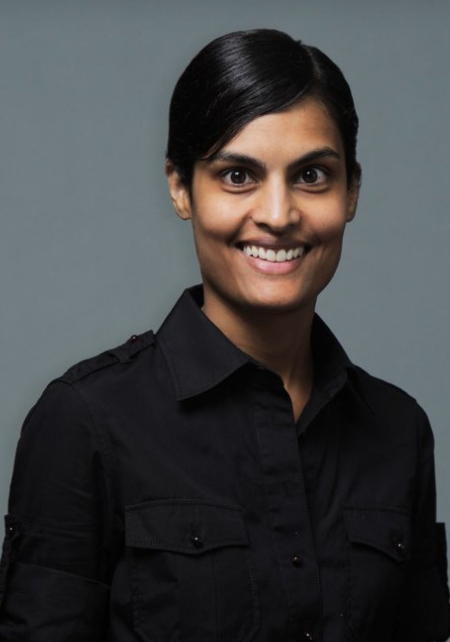 Shailee V. Lala合作论文数Division of Pediatric Radiology, Department of Radiology, New York University2
 Hersh Chandarana合作论文数Division of Abdominal Imaging, Department of Radiology, New York University2Sumant Inamdar合作论文数Department of Gastroenterology, Hofstra North Shore-LIJ School of Medicine2Williamson Alex K合作论文数2
 Daniel K. Sodickson合作论文数Department of Radiology, School of Medicine, NYU Grossman;Department of Neuroscience and Physiology, School of Medicine, NYU Grossman2Nancy R. Fefferman合作论文数2
 Kai Tobias Block合作论文数Department of Radiology, Grossman School of Medicine, New York University;Center for Advanced Imaging Innovation and Research, NYU Langone Health2Susan E. Sharp合作论文数1
 David Larson合作论文数Department of Radiology, Stanford University School of Medicine1Joshua Beiner合作论文数1
 Monica Sheth合作论文数1Igor Lobko合作论文数1Suchitra Acharya合作论文数1Kelsey Fawcett合作论文数1Rebecca Dennis合作论文数1
 Derek Hanson合作论文数1Pamela Singer合作论文数1Michelle J Vaz合作论文数1Stephen Dolgin合作论文数1Shari Gurusinghe合作论文数1Susmita N. Sarangi合作论文数1Deirdre Nolfi-Donegan合作论文数UPMC Childrens Hosp Pittsburgh1
 Monica Epelman合作论文数Nemours Childrens Hosp, Nemours Childrens Hlth Syst1Jan J W Lagendijk合作论文数1
 Nishith Khandwala合作论文数Department of Electrical Engineering, Stanford University1
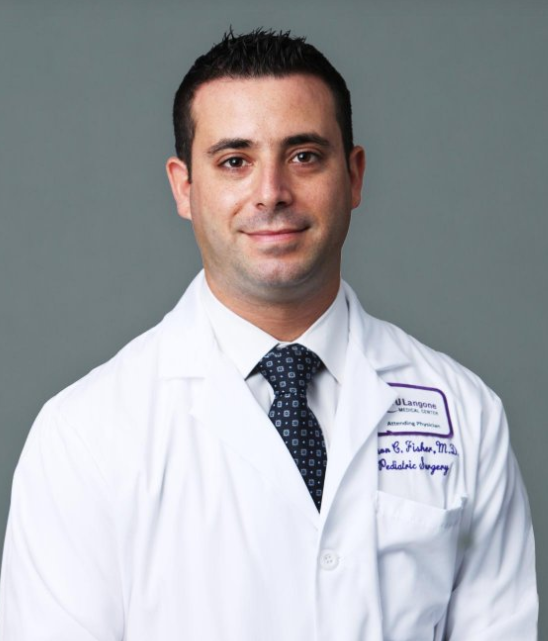 Jason C. Fisher合作论文数Division of Pediatric Surgery, Department of Surgery, Grossman School of Medicine, New York University1
 Jason C. Hoffmann合作论文数Department of Radiology, New York University1
 Sonia Mahajan合作论文数Weill Cornell Medicine;Department of Radiology, Memorial Sloan Kettering Cancer Center1
 Michael P. Recht合作论文数Division of Musculoskeletal Imaging, Department of Radiology, New York University1
 Jan Fritz合作论文数Division of Musculoskeletal Imaging, Department of Radiology, New York University1
 Cecilia L. Mercado合作论文数Division of Breast Imaging, Department of Radiology, New York University1
 Bari Dane合作论文数Division of Abdominal Imaging, Department of Radiology, New York University1
 Linda Granowetter合作论文数Grossman School of Medicine, New York University1




