Rationale and Objectives The COVID-19 pandemic necessitated a rapid transition to virtual interviews for Radiology residency programs, fundamentally altering the recruitment landscape. While virtual interviews have demonstrated cost-effectiveness and accessibility benefits, standardized etiquette guidelines remain underdeveloped. This study assesses Radiology residency program directors' perspectives on virtual interview etiquette and establishes evidence-based recommendations for professional conduct during virtual recruitment. Methods and Materials A comprehensive survey was distributed to all Radiology residency program directors in the United States to evaluate their experiences with virtual interviews and gather insights on appropriate virtual interview etiquette. Survey responses were analyzed using descriptive statistics and analysis to identify key themes and recommendations. Results Program directors identified several critical areas for virtual interview etiquette, including technical preparation, professional presentation, appropriate virtual environments, and effective communication strategies. Most respondents emphasized the importance of treating virtual interviews with the same professionalism as in-person encounters while adapting to the unique challenges of the digital format. Conclusion Virtual interview etiquette represents an essential component of professional residency recruitment. Establishing clear expectations for both applicants and programs should enhance the virtual interview experience.
RATIONALE AND OBJECTIVES:As part of promotion consideration, letters of evaluation are requested from external (to the candidate's institution) reviewers (referees). In some instances, internal letters are also requested. The process of responding to, reviewing the provided materials, writing the letter, and submitting it takes time. To our knowledge, there is no benchmark for the number of reference letters requested from senior faculty to or the time required to provide these letters. Our aim was to quantify this effort. MATERIALS AND METHODS:5 Professors at different institutions, in different parts of the United States, representing a range of subspecialties, were anonymously surveyed. The number and type of referee letters written, estimates of the time spent with each portion of letter writing, and the number of years writing letters were tabulated. RESULTS:In total, this group has written 218 letters over 49 total years of service (Table 1). This consisted of 179 external (average 4.7/year) and 39 internal (0.8/year). There were 28 instances of repeat letter requests (including consideration for different faculty ranks and changing institutions). We estimate it takes between 2.5 and 8 h to complete the necessary review of institutional guidelines and individual qualifications to complete one of these letters (Table 2). CONCLUSION:This work provides benchmarking information for the academic radiology community, including appointment and promotions committees, department chairs, and faculty members. The time, effort, energy, and attention that is required is significant but essential to academic career advancement. We recommend that departments and institutions recognize and assign value to this work.
Radiology medical education in the United States continues to face complex economic challenges. Training programs in this field are expanding at an insufficient rate relative to demand. Despite decades-old federal caps on radiology training positions, Medicare continues to be the largest provider of graduate medical education funding by a wide margin. The recent increase in radiology case volume is contributing to burnout and aggravating the pressures on academic radiology programs, further increasing the need for a larger and well-trained radiology workforce. Understanding the current radiology education funding structure is paramount for crafting potential solutions. Diversifying funding approaches, bolstering traditional funding sources, and developing new funding mechanisms for radiology education will enhance the pipeline for practicing radiologists. This review provides a summary of current radiology education funding, the impact of Medicare training positions caps on graduate medical education, and the impact of recent federal health care spending cuts on radiology training and proposes strategies to increase the number of radiology residency graduates.
RATIONALE AND OBJECTIVES:Radiology is a dynamic and ever-evolving field, necessitating research and innovation. However, the conventional medical training model falls short in fostering research skills, crucial for cultivating the upcoming cohort of physician-scientists. Our radiology residency research track (RT) was instituted to offer a dedicated research pathway, to foster the next generation of research-focused academic radiologists. The track provides an integrated 4-year longitudinal curriculum and academic time. This study assessed the impact and progress of our RT over 12 years. MATERIALS AND MATERIALS:Using publicly available online data from Doximity, PubMed, and Scopus we collected information on all graduates from our Diagnostic and Interventional Radiology residency program graduation classes between 2010 and 2022, including most recent job position, position type (academic vs. private), and publications. We compared RT and non-research track (NRT) residents. RESULTS:Out of 185 graduates, 179 profiles (97%) were retrievable, including all 13 RT residents. The average number of publications per resident during residency was 1.1 (186 total) for NRT graduates and 7.2 (93 total) for RT graduates (p < 0.001). Throughout their entire careers to date, NRT graduates averaged 7.3 publications per resident (1249 total), while RT graduates averaged 31.7 publications per resident (412 total) (p < 0.001). The average number of citations per graduate was 123 (21212 total) for NRT and 552 (7175 total) for RT (p < 0.001). Additionally, 36% of NRT graduates and 92% of RT graduates (p = 0.005) held academic job positions. CONCLUSION:Residents from the radiology residency research track were more likely to assume academic positions and had a higher number of publications and citations per resident compared to their non-research track counterparts, suggesting the track serves as an effective pipeline for cultivating academic radiologists.
Establishing a clinical education track as part of a radiology residency is essential in shaping future radiology educators. Many obstacles will be encountered while starting, maintaining, and improving these educational pathways. Hurdles may include recruiting suitable residents for the track, recruiting and supporting faculty advisors, sustaining long-term resident engagement, counteracting educational exclusivity, and providing adequate time and financial support. Although every program and institution may face individualized "mountains" to overcome, they are not insurmountable. The goal of this review is to address different conflicts we have encountered while maintaining the clinical education tract at our institution and to provide tips for overcoming them.
Many radiology departments have successfully increased trainee research involvement by providing protected academic time for research, offering travel funding for conferences, and developing research-focused curriculum via resident research tracks and other mechanisms. A departmental platform for trainees to share their scholarly projects can foster intradepartmental awareness and collaborations, supplement the existing resident research curriculum, encourage peer learning amongst trainees, and allow departmental celebration of their trainees' accomplishments. The authors describe the development of a departmental symposium for resident scholarly activity at their institution and detail a practical framework for implementation and lessons learned, which may serve as a guide for other radiology departments interested in establishing a similar event.
Rationale and Objectives Promotion is an important milestone in the career of academic radiologists. Appointments, Promotion and Tenure (APT) committees require multiple letters of support from both internal and external referees. Traditional narrative letters are highly subjective, have high inter-reader variability, are time-intensive, and vulnerable to gender and other biases. The Alliance of Directors and Vice Chairs of Education in Radiology (ADVICER) recognized the need for a standardized template to assist academic faculty, letter writers, and APT committees. Materials and Methods An ADVICER ad hoc committee of six educators with experience serving as external referees was convened to create a standardized template. Committee members performed a search of the relevant literature and internet sites, spoke with stakeholders such as APT chairs, and ultimately developed a template for faculty reviewer letters using the common clinician-educator pathway as a focal point. Results An open source, modifiable, standardized, template was produced. The template has been made available to ADVICER members and is available on the Association of University Radiologists (AUR) website at: https://www.aur.org/resources/Template-for-Faculty-Reviewer-Letters-for-Promotion-and-Appointment Conclusion This external referee template has the potential to reduce subjectivity, eliminate bias, and provide a flexible, modifiable, comprehensive faculty review letter template which will be useful for academic faculty, letter writers, and promotions committees.
The first year of radiology residency presents many unique challenges, from transitioning into a completely new, specialized field to preparing for call. Implementation of a longitudinal lecture series dedicated towards the clinical demands of being a first-year radiology resident may improve their knowledge and comfort level, as well as benefit the entire program. In this article, we outline our experience with the development of a resident-led dedicated first-year radiology resident lecture series providing targeted, high-yield instruction on rotation logistics, basic physics and artifacts, examination protocolling, and common and "don't miss" pathology.
Rationale and Objectives: Imaging Informatics is an emerging and fast-evolving field that encompasses the management of information during all steps of the imaging value chain. With many information technology tools being essential to the radiologists' day-to-day work, there is an increasing need for qualified professionals with clinical background, technology expertise, and leadership skills. To answer this, we describe our experience in the development and implementation of an Integrated Imaging Informatics Track (13T) for radiology residents at our institution. Materials and Methods: The 13T was created by a resident-driven initiative funded by an intradepartmental resident grant. Its curriculum is delivered through a combination of monthly small group discussions, operational meetings, recommended readings, lectures, and early exposure to the National Imaging Informatics Course. The track is steered and managed by the 13T Committee, including trainees and faculty advisors. Up to two first-year residents are selected annually based on their curriculum vitae and an interest application. Successful completion of the program requires submission of a capstone project and at least one academic deliverable (national meeting presentation, poster, exhibit, manuscript and/or grant). Results: In our three-year experience, the seven I3T radiology residents have reported a total of 58 scholarly activities related to Imaging Informatics. I3T residents have assumed leadership roles within our organization and nationally. All residents have successfully carried out their clinical responsibilities. Conclusion: We have developed and implemented an I3T for radiology residents at our institution. These residents have been successful in their clinical, scholarship and leadership pursuits.
Automated co-registration and subtraction techniques have been shown to be useful in the assessment of longitudinal changes in multiple sclerosis (MS) lesion burden, but the majority depend on T2-fluid-attenuated inversion recovery sequences. We aimed to investigate the use of a novel automated temporal color complement imaging (CCI) map overlapped on 3D double inversion recovery (DIR), and to assess its diagnostic performance for detecting disease progression in patients with multiple sclerosis (MS) as compared to standard review of serial 3D DIR images. We developed a fully automated system that co-registers and compares baseline to follow-up 3D DIR images and outputs a pseudo-color RGB map in which red pixels indicate increased intensity values in the follow-up image (i.e., progression; new/enlarging lesion), blue-green pixels represent decreased intensity values (i.e., disappearing/shrinking lesion), and gray-scale pixels reflect unchanged intensity values. Three neuroradiologists blinded to clinical information independently reviewed each patient using standard DIR images alone and using CCI maps based on DIR images at two separate exams. Seventy-six follow-up examinations from 60 consecutive MS patients who underwent standard 3 T MR brain MS protocol that included 3D DIR were included. Median cohort age was 38.5 years, with 46 women, 59 relapsing–remitting type MS, and median follow-up interval of 250 days (interquartile range: 196–394 days). Lesion progression was detected in 67.1
Magnetic resonance spectroscopy (MRS) is a valuable tool for imaging brain tumors, primarily as an adjunct to conventional imaging and clinical presentation. MRS is useful in initial diagnosis of brain tumors, helping differentiate tumors from possible mimics such as metastatic disease, lymphoma, demyelination, and infection, as well as in the subsequent follow-up of patients after resection and chemoradiation. Unfortunately, the spectroscopic appearance of many pathologies can overlap, and ultimately follow-up or biopsy may be required to make a definitive diagnosis. Future developments may continue to increase the value of MRS for initial diagnosis, treatment planning, and early detection of recurrence.
BACKGROUND AND PURPOSE:Multiple radiographic terms can be used to describe enlarged ventricles on noncontrast head computed tomography (CTs); however, precise terminology is important to determine etiology and clinical management. The purpose of this study was to characterize how ventricular size was described in radiology reports, especially in the setting of hydrocephalus.MATERIALS AND METHODS:A retrospective review of adult patients with at least 2 consecutive noncontrast head CTs from 2010 to 2016 was performed. Reports were grouped based on ventricular descriptions into 3 categories: "acceptable," "unclear," and "not acceptable." An additional subgroup was created for an examination indication of "hydrocephalus." Descriptive statistics and subgroup analysis were performed.RESULTS:A total of 270 patients with noncontrast head CTs were included, of which 53.3% (n = 144) used "acceptable" terms, 18.2% (n = 49) "unclear," and 28.5% (n = 77) "not acceptable." Ventricle size was reported as normal in 21.1% (n = 57) of cases. "Hydrocephalus" was given as an indication for 57 exams, of which 84.2% (n = 48) were categorized as "acceptable," 7.0% (n = 4) "unclear," and 8.8% (n = 5) "not acceptable." Chi-square test of independence revealed a significant relation between "acceptable" terminology and "hydrocephalus" indication (χ2 = 27.68, P< 0.001).CONCLUSION:Approximately half of radiology reports had an "acceptable" description of the ventricles. When "hydrocephalus" was in the indication, the report was more likely to have an "acceptable" description. Accurate clinical indications, and standardized terminology may improve the clinical utility of radiology reports for patients with hydrocephalus.
Consider the following scenario. You are the Chair of a small academic radiology department in the Midwest. It is Thursday morning, July 1, the start of a new academic year. You receive an email from Drs Smith and Hall, requesting to meet with you as soon as possible. Dr Smith is the Vice Chair of Education and the Residency Program Director, and Dr Hall is the Director of Medical Student Education. They are married to each other. During the meeting, they inform you that they have accepted positions at another academic center because of an urgent family situation and intend to start these new positions on August 1. You immediately go into crisis management mode because you do not have anyone in the leadership pipeline to assume their responsibilities. The radiology clerkship begins on August 1, residency applicants start submitting applications on September 1, and the residency program is scheduled for a reaccreditation site visit on September 10.
RATIONALE AND OBJECTIVES:An increase in the administrative work in our healthcare system has led to an increase in the number of administrative positions in radiology departments. Many of these are Vice Chair roles, including Vice Chair for Education (VCEd). The responsibility of this position has expanded, often far beyond the original definition. This article defines the role and expectations of the Vice Chair for Education and provides suggestions for success.MATERIALS AND METHODS:This article will review 12 vital roles that a Vice Chair for Education must play to be an effective advocate for radiology education within a department.RESULTS:Key attributes of an educational leader are delineated, divided into 12 areas or roles.CONCLUSION:This article summarizes key leadership skills needed by Vice Chairs for Education in order for them to be effective in their role.
A brief introduction is provided of the different imaging modalities encountered in the intensive care unit (ICU). The spectrum of intracranial pathology as well as potential postsurgical complications is reviewed, with a focus on pearls and pitfalls. A brief overview also is provided of imaging of the spine in an ICU patient.
Engaging and effectively educating learners is a long-standing issue in graduate medical education [1Sawatsky A.P. Zickmund S.L. Berlacher K. Lesky D. Granieri R. Understanding the challenges to facilitating active learning in the resident conferences.Med Educ Online. 2015; 20: 27289Crossref PubMed Scopus (24) Google Scholar]. We noted a decrease in resident attendance and focus during our daily traditional didactic curriculum. Although there is protected resident conference time, attendance is difficult to verify because of videoconferencing. Trainees are motivated to learn by a variety of factors, including intrinsic intellectual curiosity, desire to excel, examinations, and clinical rotations [1Sawatsky A.P. Zickmund S.L. Berlacher K. Lesky D. Granieri R. Understanding the challenges to facilitating active learning in the resident conferences.Med Educ Online. 2015; 20: 27289Crossref PubMed Scopus (24) Google Scholar]. However, these can be undermined by competing clinical or research demands, learners' variable interests in the subject, and perception of its relevance for clinical practice or examinations [1Sawatsky A.P. Zickmund S.L. Berlacher K. Lesky D. Granieri R. Understanding the challenges to facilitating active learning in the resident conferences.Med Educ Online. 2015; 20: 27289Crossref PubMed Scopus (24) Google Scholar]. Even if trainees recognize value in the curriculum, presentation styles may affect effective learning. Passive learning, in which knowledge is gained purely through listening and watching without instructor feedback, is limited in efficacy [2Pamarthi V. Grimm L. Johnson K. Maxfield C. Hybrid interactive and didactic teaching format improves resident retention and attention compared to traditional lectures.Acad Radiol. 2019; 26: 1269-1273Abstract Full Text Full Text PDF PubMed Scopus (12) Google Scholar]. Active learning, in which learners participate in the discovery process, can increase information retention [2Pamarthi V. Grimm L. Johnson K. Maxfield C. Hybrid interactive and didactic teaching format improves resident retention and attention compared to traditional lectures.Acad Radiol. 2019; 26: 1269-1273Abstract Full Text Full Text PDF PubMed Scopus (12) Google Scholar]. However, it may be difficult to implement active learning in residency curricula [1Sawatsky A.P. Zickmund S.L. Berlacher K. Lesky D. Granieri R. Understanding the challenges to facilitating active learning in the resident conferences.Med Educ Online. 2015; 20: 27289Crossref PubMed Scopus (24) Google Scholar]. A major barrier is lack of "buy-in" (ie, learners are not motivated by active learning methods and view them as pointless) [1Sawatsky A.P. Zickmund S.L. Berlacher K. Lesky D. Granieri R. Understanding the challenges to facilitating active learning in the resident conferences.Med Educ Online. 2015; 20: 27289Crossref PubMed Scopus (24) Google Scholar]. There is increasing interest in gamification and its effect on engagement and buy-in [3Nacke L.E. Deterding S. The maturing of gamification research.Comput Human Behav. 2017; 71: 450-454Crossref Scopus (332) Google Scholar]. Theoretically, gamification increases participant motivation by endowing them with "epic purpose" [3Nacke L.E. Deterding S. The maturing of gamification research.Comput Human Behav. 2017; 71: 450-454Crossref Scopus (332) Google Scholar]. By framing educational goals as a contest, gamification offers immediately attainable challenges and consistent rules against which accomplishments are rewarded through positive feedback [4Ortiz M, Chiluiza K, Valcke M. Gamification in higher education and STEM: a systematic review of literature. Proceedings of Edulearn 16: 8th International Conference on Education and New Learning Technologies, p6548-6558.Google Scholar]. Gamification encourages teamwork, because players have implicitly agreed to the same rules and goals [4Ortiz M, Chiluiza K, Valcke M. Gamification in higher education and STEM: a systematic review of literature. Proceedings of Edulearn 16: 8th International Conference on Education and New Learning Technologies, p6548-6558.Google Scholar]. For medical education, gamification has shown promising results in participant acceptance and knowledge retention [5Nevin C.R. Westfall A.O. Rodriguez J.M. et al.Gamification as a tool for enhancing graduate medical education.Postgrad Med J. 2014; 90: 685-693Crossref PubMed Scopus (175) Google Scholar]. Mindful of increasing engagement, we redesigned our resident neuroradiology curriculum by updating lecture contents and gamifying the delivery method. Our main objectives were to increase resident awareness, attendance, and participation by redefining high-yield didactic topics and encouraging healthy competition. A major determinant of participant buy-in of a gamified system is alignment of participant and game-designer goals [3Nacke L.E. Deterding S. The maturing of gamification research.Comput Human Behav. 2017; 71: 450-454Crossref Scopus (332) Google Scholar]. Given changes in the ABR Initial Certification, we redesigned our current curriculum based on neuroradiology topics within the ABR "Diagnostic Radiology CORE Examination Study Guide" [6ABRDiagnostic Radiology CORE Examination Study Guide.https://www.theabr.org/wp-content/uploads/2018/12/CORE_Exam_Study_Guide_2019.pdfDate accessed: December 19, 2019Google Scholar]. Main study guide topics were divided into conferences to be repeated on a 2-year cycle to ensure redundancy. Advanced topics, considered beneficial by faculty but not part of the study guide, were repeated every 4 years (Table 1). Each faculty member was asked to participate by teaching at least one conference yearly. We made the curriculum available to residents, highlighting correlations with the study guide [6ABRDiagnostic Radiology CORE Examination Study Guide.https://www.theabr.org/wp-content/uploads/2018/12/CORE_Exam_Study_Guide_2019.pdfDate accessed: December 19, 2019Google Scholar] to alleviate concerns that chosen topics were solely centered around faculty interest.Table 1Redesigned neuroradiology residency lecture curriculum developed based on the ABR CORE Examination Study Guide [6ABRDiagnostic Radiology CORE Examination Study Guide.https://www.theabr.org/wp-content/uploads/2018/12/CORE_Exam_Study_Guide_2019.pdfDate accessed: December 19, 2019Google Scholar]Main Topics (Every 2 y)Advanced Topics (Every 4 y)Brain Brain anatomyNeuroradiology and machine learning Intracranial infections or emergency neuroradiologyRadiogenomics White matter inflammatory or demyelinating diseaseSpine interventions Traumatic brain injuryFetal MRI Adult intracranial neoplasms Cerebrovascular disease or stroke Intracranial hemorrhage Increased and decreased intracranial pressure Aging and neurodegeneration Midline structures (including sella and pineal space) Neurovascular anatomy, vasculitis, aneurysms and AVMsSpine Spine anatomy and degenerative changes Spine trauma Neoplastic disease of the spine Inflammatory, infectious, and vascular disease of the spineHead and neck Overview of head and neck anatomy and emergencies Sinonasal cavities and orbits Temporal bone anatomy and pathology, include IAC or CPA Skull base, cranial nerves, and CSF leak Calvarium, facial bones, mandible, and TMJ Salivary glands and aerodigestive tract Cervical adenopthy and visceral spacePediatric neurology Epilepsy, metabolic and toxic brain injury Hydrocephalus, intracranial cysts Pediatric neuroradiologic emergencies and nonaccidental injury Pediatric headache and back pain Congenital brain and spine malformations Pediatric seizure disorder and phakomatoses Pediatric inherited metabolic and white matter disorders Pediatric brain and spine neoplasmsArtifacts in neuroradiologyAdvanced techniques in neuroimagingAVM = arteriovenous malformation; CPA = cerebellopontine angle; CSF = cerebrospinal fluid; IAC = internal auditory canal; TMJ = temporomandibular joint. Open table in a new tab AVM = arteriovenous malformation; CPA = cerebellopontine angle; CSF = cerebrospinal fluid; IAC = internal auditory canal; TMJ = temporomandibular joint. Curriculum gamification was based on the popular HBO television show Game of Thrones, in which powerful families vied for political and military dominance [7Benioff D. Weiss D.B. Game of Thrones. Home Box Office Home Entertainment, 2019Google Scholar]. Residents were divided into four houses (Lannisters, Targaryens, Starks, and the Night's Watch [7Benioff D. Weiss D.B. Game of Thrones. Home Box Office Home Entertainment, 2019Google Scholar]), balanced across different postgraduate-year levels, subspecialty interest, and gender (Table 2). House assignments were made by a Clinical Competency Committee faculty member familiar with residents' clinical interests. Each house elected a leader (king or queen), who encouraged team members to respond to questions and online campaigns and arbitrated differing responses. Teams sat together during each conference.Table 2Demographic breakdown of the houses in 2019 to 2020 based on gender, postgraduate year, and potential interest in neuroradiology from R2-R4 residentsDemographicStark (%)Lannister (%)Night Watch (%)Targaryen (%)Male68.868.868.866.7Female31.331.331.333.3R131.325.031.326.7R225.025.025.020.0R325.025.018.826.7R418.825.025.026.7Estimate interest in neuroradiology (R2-R4)50.036.440.033.3R1, residency year 1; R2, residency year 2; R3, residency year 3; R4, residency year 4. Open table in a new tab R1, residency year 1; R2, residency year 2; R3, residency year 3; R4, residency year 4. Each conference lecturer divided 20 points among the houses as they liked, based on attendance, multiple-choice responses, free response, anatomic drawing, and individual or group problem-solving. Faculty were encouraged to use creative challenges to maximize active learning and supported with resources including interactive slide deck templates (such as Jeopardy!), active learning ideas (including reversed-classroom teaching or drawing exercises), and polling software (including Poll Everywhere [8Poll Everywhere.https://www.polleverywhere.com/Date accessed: April 1, 2020Google Scholar], Kahoot! [9Kahoot!.https://kahoot.com/Date accessed: April 1, 2020Google Scholar], and RSNA Diagnosis Live [10RSNA Diagnosis Live.https://live.rsna.org/Date accessed: April 1, 2020Google Scholar]), which were accessible on a dedicated intranet site. Administrative support was provided for the translation of existing lectures into interactive formats. Faculty were familiarized with the gamified curriculum before implementation with collaborative discussions facilitated by the sectional education committee and received regular e-mail reminders throughout the year to sustain utilization of gamification techniques. Twice a year, residents participated in "campaigns" worth 50 points each. These were open-book, online case-based challenges coupled with questions regarding imaging findings, diagnosis, and management. Each king or queen solicited and submitted a consensus response for their house, and the 50 points were divided by the faculty organizer among the houses based on accuracy of their responses. Each house's score was periodically posted on a highly trafficked, secure resident website to remind learners of their progress. At graduation, the winning house was honored with a "crowning ceremony," and their banner was hung in the residency conference room as a visual challenge to motivate residents during the following academic year. Before and after the first year of the gamified curriculum's implementation, we performed an anonymized, voluntary, institutional review board–exempt online survey to assess resident attitudes and their attendance habits (Table 3). All 61 residents (residency years 1-4) were surveyed, and questions were predominantly phrased using 5-point Likert scale or true-or-false formats. Results were compared across the two time points with statistical significance assessed by the Mann-Whitney U test.Table 3Institutional review board–exempt, anonymized survey questions assessing self-reported resident attitudes toward the old, as well as redesigned neuroradiology curriculaQuestionPossible answersWhich year of residency are you in?R1, R2, R3, R4Are you aware that a predetermined neuroradiology lecture curriculum exists?Yes, noHow organized is the neuroradiology lecture curriculum?1: very disorganized, 2: disorganized, 3: neither organized or disorganized, 4: organized, 5: very organizedHow much does the neuroradiology lecture curriculum prepare you for your rotations, call, and clinical practice?1: not useful at all, 2: somewhat not useful, 3: neither useful or not useful, 4: useful, 5: very usefulHow well does the neuroradiology lecture curriculum prepare you for the CORE examination?1: not well at all, 2: not well, 3: neither well or not well, 4: well, 5: very wellAre you aware that there is an ABR outline for the topics tested on the CORE examination?Yes, noIf yes to the previous question, have you ever read through the outline?Yes, noIf yes to the previous questions, how well does the neuroradiology lecture curriculum match the topics shown on the outline?1: not well at all, 2: not well, 3: neither well or not well, 4: well, 5: very wellHow often do you attend the neuroradiology noon lectures in person?0%, 1%-25%, 26%-50%, 51%-75%, 76-99%, 100%When you do not attend a neuroradiology lecture in person, how often do you watch it remotely?0%, 1%-25%, 26%-50%, 51%-75%, 76%-99%, 100%Please rank your reasons for not attending neuroradiology lectures, where 1 = most frequent reason.•Too busy on a clinical rotation; have to finish dictating cases to leave on time•What's happening on rotation is more interesting than the lecture topic, even if I'm not required to stay (eg, rare procedure)•The lecture topic is not interesting•The lecturer is not interesting•Inconvenient location•Webcasting not working•I've heard this lecture before and do not feel like I need it again•OtherHow well do you retain information presented in neuroradiology lectures?1: not well at all, 2: not well, 3: neither well or not well, 4: well, 5: very wellHow much redundancy is built into the neuroradiology curriculum?•Not enough—I hear things once and do not remember it well enough•Barely enough —I vaguely remember hearing something before, but could probably use more repetition•Just right—I feel comfortable with the amount of reinforcement offered by repeated lectures•A little too much—I feel myself getting bored occasionally by materials I recognize•Way too much—I realized I've seen a lecture before and I completely zone outR1, residency year 1; R2, residency year 2; R3, residency year 3; R4, residency year 4. Open table in a new tab R1, residency year 1; R2, residency year 2; R3, residency year 3; R4, residency year 4. Before implementation, 22 of 61 (36%) residents responded to the survey and 25 of 61 (41%) responded afterward. Significant differences were found in awareness of the existence of a neuroradiology curriculum (increasing from 18% to 76%, P = .0007), and the number of residents who thought the curriculum was "organized" or "very well organized" (P = .005). No significant differences were found in how well residents believed the curriculum prepared them for rotations, call, or CORE examination. However, a greater percentage of learners believed that the redesigned curriculum prepared them "well" or "very well" as compared with before. None of the other questions yielded statistically different results. An informal evaluation of neuroradiology in-service examination results from 2016 to 2020 did not show significant differences in performance trends before and after curriculum gamification, although this assessment may be underpowered given high individual and postgraduate-year class variability. Our intervention altered both content and delivery method of the curriculum, making it difficult to distill the impact of each of these changes. However, because there was no change in residents' awareness of the ABR study guide [6ABRDiagnostic Radiology CORE Examination Study Guide.https://www.theabr.org/wp-content/uploads/2018/12/CORE_Exam_Study_Guide_2019.pdfDate accessed: December 19, 2019Google Scholar] or their perception of how closely the curriculum mimicked it, it is likely that these outcomes are at least partly attributable to gamification. We refrained from asking leading questions regarding self-perceived engagement and enjoyment of the curriculum. However, marketing research suggests that increasing awareness forms the baseline step toward increasing customer engagement. Thus, improved awareness of the gamified curriculum and perception of its organization may pave way for better engagement. A challenge of gamifying the curriculum is the ability and willingness of faculty members to alter lecture styles toward active learning. We were fortunate to have a faculty with high levels of interest in educational improvement, and we easily filled the schedule with teachers eager to give multiple conferences. Informal faculty feedback indicated that the centralized intranet resource with applications and templates encouraged creativity, and administrative support helped decrease associated technological challenges. Though regular reminders were necessary to encourage uniform utilization of gamification techniques (such as appropriate point assignments), most faculty successfully adopted active learning techniques. This questionnaire is limited by the subjectivity of self-reporting and self-selection, in which attendance and information retention can be overreported. Our sample size is limited by voluntary participation from a single year of residents. The nonindependent, unpaired sample population may have also undermined statistical power, limiting ability to detect significant changes in resident attitudes. In the future, we plan to improve objective tracking of resident engagement, including attendance. Because the recent coronavirus disease 2019 pandemic necessitated remote learning, it has become easier to monitor attendance and participation as residents now individually log in. The gamified curriculum can also be brought out of the lecture hall by allowing teams to earn points during the clinical workday. Through innovative approaches to educational strategy including gamification, we hope to continue improving the quality of radiology education.
Although education is one mission of institutions that sponsor graduate medical education, funding to support these activities has relatively diminished, while simultaneously, increasing clinical volumes have made it more challenging for faculty to teach trainees the necessary skills for independent practice. There are a few programs currently with strong alumni networks; however, all departments with training programs could benefit substantially by fostering strong relationships with their graduates. Alumni of residency programs offer a multitude of potential resources from actively teaching current trainees to mentorship or even sponsorship as graduating trainees seek to establish themselves in practice. Alumni might also desire to give back to their residency programs by providing financial support for new innovative educational programming.
There are few detailed investigations of neurologic complications in severe acute respiratory syndrome coronavirus 2 infection. We describe 3 patients with laboratory-confirmed coronavirus disease who had encephalopathy and encephalitis develop. Neuroimaging showed nonenhancing unilateral, bilateral, and midline changes not readily attributable to vascular causes. All 3 patients had increased cerebrospinal fluid (CSF) levels of anti-S1 IgM. One patient who died also had increased levels of anti-envelope protein IgM. CSF analysis also showed markedly increased levels of interleukin (IL)-6, IL-8, and IL-10, but severe acute respiratory syndrome coronavirus 2 was not identified in any CSF sample. These changes provide evidence of CSF periinfectious/postinfectious inflammatory changes during coronavirus disease with neurologic complications.
BACKGROUND AND PURPOSE Interview selection of candidates for academic radiology faculty positions is variable and subject to unconscious biases. The purpose of this study was to retrospectively apply a quantitative curriculum vitae (CV) rubric as a screening tool to identify qualified candidates for further consideration in the hiring process. MATERIALS AND METHODS Archived CVs submitted by applicants between 2012 and 2017 for neuroradiology faculty positions at our institution were anonymized. One blinded reviewer scored resumes based on categories that included education, work experience, extracurricular/teaching experience, and research. Logistic regression and receiver operating characteristics analysis were performed. This study was IRB exempted. RESULTS Of the total 102 applicants, 17 interviews were conducted and 10 candidates were offered a position. Maximum score of the model was 24 points. Mean score was 14 ± 4 (n = 102, range 5-22). Higher total CV score (P = 0.01), medical school ranking (P = 0.03), and number of published manuscripts (P = 0.03) were significantly associated with interview selection. The area under the curve in the ROC analysis for differentiating interview selection based on total CV scoring was 0.69 (95% confidence interval 0.56-0.82). At a cutoff of 14, the model is 82.4% sensitive, and 54.1% specific. CONCLUSION Standardized CV scoring is feasible with a cut-off score of 14 points providing high sensitivity in identifying candidates eligible for interview. This tool can potentially be applied in the future to the hiring process as it is neutral to factors such as gender and race and provides an opportunity to address diversity in academic medicine.
 Amit M. Saindane合作论文数8
 Lori Deitte合作论文数8
 Brent Weinberg合作论文数Department of Radiology and Imaging Sciences, School of Medicine, Emory University7
 Slanetz Priscilla J合作论文数Beth Israel Deaconess Medical Center, Harvard Medical School5Meltzer Carolyn C合作论文数5Richard Duszak合作论文数5Ryan B. Peterson合作论文数4Chokshi Falgun H合作论文数4
 Christopher P Ho合作论文数4
 Elizabeth Krupinski合作论文数Department of Radiology and Imaging Sciences, School of Medicine, Emory University;Winship Cancer Institute, Emory University4Theresa C. Mcloud合作论文数4A. Jayashankar合作论文数4S. M. Sabourin合作论文数4
 Petra J. Lewis合作论文数Dartmouth-Hitchcock Medical Center, Norris Cotton Cancer Center4Grant R Webber合作论文数3Dexter P Mendoza合作论文数Harvard Medical School, Massachusetts General Hospital3Maria Braileanu合作论文数3
 Judith Gadde合作论文数3
 Vijayasarathi Arvind合作论文数Department of Radiology and Imaging Sciences, Emory University Hospital3Hu Ranliang合作论文数3Harri Peter合作论文数3Adam Prater合作论文数3
 Robert A Novelline合作论文数Department of Radiology, Harvard Medical School;Massachusetts General Hospital3Anna Rozenshtein合作论文数3
 Zhengjia Nelson Chen合作论文数Division of Epidemiology and Biostatistics, School of Public Health, University of Illinois Chicago;Cancer Center, University of Illinois Chicago;Winship Cancer Institute, Emory University3
 Ramon Gonzalez合作论文数Division of Neuroradiology, Department of Radiology, Massachusetts General Hospital, Mass General Brigham;Mass General Cancer Center, Massachusetts General Hospital, Mass General Brigham;Harvard Medical School, Harvard University3Deborah A. Baumgarten合作论文数School of Medicine, Emory University3
 Leonie Gordon合作论文数3Jason W. Allen合作论文数2Sravanthi Reddy合作论文数2Pedro J Diaz-Marchan合作论文数2Hyunsuk Shim合作论文数2
 Patricia Balthazar合作论文数Sch Med, Emory Univ2
 Syed Ali Raza合作论文数Emory Univ2
 Seena Dehkharghani合作论文数Department of Neurology, Department of Medicine, New York University2
 Sadigh Gelareh合作论文数Dept Radiol & Imaging Sci, Emory Univ2
 Manohar Kuruva合作论文数Department of Nuclear Medicine and PET, Postgraduate Institute of Medical Education and Research, Chandigarh, India.2Jallow Ngoneh合作论文数2
 Kadom Nadja合作论文数Department of Diagnostic Imaging and Radiology, Children's National Medical Center2
 Pamela Schaefer合作论文数Massachusetts General Hospital;Harvard Medical School2
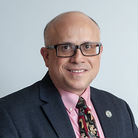 Michael Lev合作论文数Harvard Medical School;Massachusetts General Hospital2
 Chetlen Alison L合作论文数Department of Radiology, Penn State Milton S. Hershey Medical Center2
 Rondi Beth Gelbard合作论文数2
 C Matthew Hawkins合作论文数2
 Dharmadhikari Shalmali合作论文数Purdue University School of Health Sciences;Department of Radiology, Indiana University School of Medicine;Purdue University School of Health Sciences, Indiana University School of Medicine2
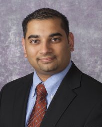 Vikas Agarwal合作论文数2Hoch Michael合作论文数2
 Danny R. Hughes合作论文数School of Economics, Ivan Allen College of Liberal Arts, Georgia Institute of Technology2Linda A. Deloney合作论文数University of Arkansas for Medical Sciences2




