Regiomethylation of the tetrazolato ligand in cyclometalated iridium complexes demonstrates control of photophysical properties for brain imaging lipid probes.
Zinc ions are highly abundant in pancreatic islet tissue, and multiple lines of evidence link loss of zinc homeostasis to poor glucose regulation in both type 1 and type 2 diabetes. Two major islet zinc-binding proteins, insulin and metallothionein, play crucial roles in beta cell function and glucose regulation. Here we used X-ray fluorescence microscopy (XFM) to map zinc and five additional elements (Cl, K, Ca, Fe, and Cu) to compare the metallome of exocrine, peri-islet and islet regions in young and old, non-diabetic control and diabetic (db/db) mice. We also determined the main forms of zinc found in pancreatic tissue using X-ray absorption near-edge structure (XANES) spectroscopic imaging. This allowed investigation of the relationship between zinc speciation and its protein ligands using correlative immunofluorescent imaging to assess whether zinc coordination may play a role in diabetes pathology. The anticipated depletion of zinc in young diabetic islets was accompanied by a significant decrease in insulin expression and increase in metallothionein expression. A parallel change in the contribution of cysteine vs histidine zinc speciation was also observed. Counter-intuitively, zinc abundance and speciation appeared to normalise in old diabetic animals with more advanced disease, despite large differences in labile zinc-binding protein content. These results are consistent with disrupted zinc coordination, where metallothionein-regulated muffling to minimise ionic activity is overwhelmed and zinc binds to unidentified ligands in histidine-like conformations. This opens future study questions focussed on the complex interplay between labile zinc, metallothionein, and oxidative mechanisms that may interfere with normal zinc homeostasis.
The quantity and bioavailability of iron (Fe) in commercial chicken eggs have been subject to ongoing debate. Understanding the chemical form of Fe in eggs, and how different laying conditions or cooking environments may alter chemical form is important to guide future studies of Fe bioavailability. To address these unanswered questions, this study aimed to accurately quantify and characterise Fe speciation (including haem and non-haem Fe) in egg yolk, albumen and whole eggs (mixed yolk and albumen), in both raw and cooked eggs. Eggs were obtained from four different hen housing systems: free-range, cage, barn and organic. Total Fe was measured using microwave plasma atomic emission spectrometry, and X-ray absorption near-edge structure spectroscopy was used to quantify the relative proportions of different chemical forms of Fe (Fe speciation). These analyses were conducted on raw albumen, yolk, and whole egg samples (combined yolk and albumen) from eggs produced across all housing systems, as well as on baked and boiled albumen, yolk, and whole egg samples from free-range eggs. Haem Fe was not detected by the analytical methods used, confirming that eggs are not a nutritionally relevant source of haem Fe. Mixing yolk and albumen alters Fe speciation, decreasing relative phosphate coordination of Fe and increasing Fe associated with protein carboxylate and chloride groups. Subsequent baking causes a significant reduction in carboxylate- and chloride-bound Fe, accompanied by an increase in sulfur-bound Fe. Boiling eggs was found to have minimal effects on Fe speciation. Despite contributing little Fe, albumen plays an important role in modulating Fe speciation, which may subsequently impact bioavailability. Cooking eggs changes the Fe speciation, particularly increasing the amount of Fe-S coordination, which should be taken into consideration for future study design when assessing Fe bioavailability.
This study demonstrates the thermal, structural, and rheological properties of flours and protein isolates of five Lupinus angustifolius L. varieties using soy flour and protein isolate as references. Fourier transform infrared (FTIR) analysis of lupin protein isolates in the amide I region revealed significantly higher beta-sheet content than alpha-helices, together comprising similar to 50 % of the secondary protein structures. In differential scanning calorimetry (DSC), lupin flour exhibited two distinct peak denaturation temperatures (Td), first between 90 and 91 degrees C, second between 103 and 105 degrees C. Similarly, for lupin protein isolates, the two Tds were at 84-86 degrees C and 96-98 degrees C, which represent beta-conglutin and alpha-conglutin, respectively. The microstructure of lupin proteins was less interconnected than soy, which had a more compact and continuous protein network. Lupin protein isolates form a weaker and easily deformed gel network compared to soy protein isolates due to their lower complex viscosity (& eng;& lowast;), storage (G ') and loss (G") moduli, and higher loss factor (tan delta) compared to soy protein isolates. Flours were characterised as more frequency-dependent than protein isolates, indicating weaker gel networks. Rapid viscosity analyser (RVA) profiles revealed that lupin flours exhibited higher peak (237.00-265.00 cP) and final viscosities (3.38-4.89 cP) than soy flour (166.33 cP and 1.11 cP, respectively), indicating the influence of non-starch components on enhanced pasting properties in lupin flours. Since lupin proteins form weaker gels than soy, process modulations are essential to improve their functionality in food products. Moreover, higher Td associated with lupin flour over soy flour demonstrate greater thermal stability, which is potentially beneficial in heat-processed, high-protein products.
During endochondral ossification, chondrocytes undergo maturation and biochemically modify the collagenous extracellular matrix of cartilage. Similar modifications to cartilage proteoglycans (PGs), which are predominantly chondroitin sulfate PGs, have not been characterized. Using synchrotron X-ray fluorescence imaging, we demonstrated that PG sulfation significantly decreased during cartilage maturation of chick embryos. Laser-capture microdissection and RNAseq revealed upregulation of Arylsulfatase I (Arsi) in mature cartilage of mouse. ARSI protein also increased in mature cartilage of mouse and chick in vivo and during maturation of ATDC5 chondrocytes in vitro, whereas expression of the two known chondroitin sulfate PG sulfatases (ARSB and GALNS) was not specific to mature cartilage. Colocalization studies suggested that ARSI is lysosomal, and lysosome homeostasis was altered in ARSI loss of function chondrocytes. Biochemical analyses of ARSI gain and loss of function cell lines and isolated cell-free systems revealed that ARSI is a novel chondroitin endosulfatase, specifically desulfating chondroitin-4-sulfate at pH 4.5. Finally, Arsi knockout in RCS chondrocytes caused increased expression of maturation genes, such as Col10a1 and Mmp13. In total, these data identify ARSI as a novel PG sulfatase regulating endochondral ossification.
Zinc ions (Zn2+) are the second most abundant trace metal ion in the brain of rodents and primates, often serving functions as a structure-stabilizing element or catalytic role. There is an additional pool of Zn2+, ∼15% of total brain Zn2+, which exists in a labile chemical form in a specific subset of glutamatergic neurons ('zinergic' or 'zincergic' neurons). The labile pool of Zn2+ is now well established to be critical for healthy memory function, with disturbance to the labile Zn2+ pool implicated in diminished memory performance during the ageing process or neurodegeneration. The chemical form of Zn2+ in the labile Zn2+ pool has however, remained unknown, largely due to the difficulty of imaging metal speciation for 'spectroscopically silent' metals such as Zn2+. In this study, we have developed X-ray absorption near edge structure (XANES) spectroscopic protocols to enable chemically specific imaging of Zn2+ speciation in murine brain (hippocampal) tissue. The protocols capitalise on the unique sensitivity of the XANES spectral region to metal ion coordination environment, enabling a direct in situ measurement of metal speciation. Key findings of our method development are characterisation of the effects of sample preparation on metal speciation, and revelation that Zn2+ coordination with histidine is likely to be the dominant coordination environment of the labile Zn2+ pool in the murine hippocampus.
Iron (Fe) is an essential mineral for poultry, playing a critical role in oxygen transport and energy metabolism. Consequently, it is routinely supplemented into commercial poultry diets to safeguard against deficiencies that can negatively impact productive performance. Conversely, excessive dietary Fe can compromise performance by inducing oxidative stress, reducing absorption of other nutrients and negatively disrupting the gastrointestinal microbiota. The bioavailability, and thus absorption, of Fe is dictated by the form it is present in the feed, with haem, the organic form of Fe, being preferentially absorbed. Modern poultry diets, depending on location or context, may be devoid of animal proteins, and therefore without haem. Inorganic Fe sulphates have been the primary source of supplemental Fe in poultry diets, via premixes. These sources may provide Fe in the ferric state (Fe3+), which must first be converted to a ferrous state (Fe2+) for uptake, though it is more commonly supplied as ferrous sulphate monohydrate (Fe2+). The poor bioavailability of inorganic Fe has led to increased interest in alternative organic Fe sources, such Fe-amino acid chelates and Fe proteinates, with higher absorption efficiency at lower active inclusion levels. Iron absorption is also dictated by interactions with anti-nutrients, notably phytic acid and mineral antagonists. There is considerable variation in recommended supplemental Fe levels for poultry across published guidelines, feeding standards and scientific articles. For example, in broilers and broiler breeders recommended total Fe ranges from 20 to 110 mg/kg and in laying hens from 30 to 80 mg/kg. Advances in feed formulation, including use of organic sources of Fe, reduced concentration of antagonistic trace minerals and phytase supplementation, has enhanced bioavailability of Fe, indicating that commercial poultry may now be over-supplemented with Fe. Updated and context-specific Fe recommendations are essential for optimising poultry productivity. This review examines how to assess Fe utilisation in poultry, including methods for determining Fe status, proposes Fe recommendations for broilers, broiler breeders and laying hens based on outputs from research trials, and explores how Fe interacts with other nutrients and the microbiota.
Background/Objectives: Traumatic brain injury (TBI) is a leading cause of morbidity and mortality worldwide. Electrolyte disturbances are common in this patient cohort, with serum chloride frequently elevated. Chloride dysregulation may be associated with poor neurological outcomes through mechanisms including paradoxical gamma amino butyric acid receptor excitation, cytotoxic edema, and ferroptosis. The aim of this review was to evaluate the relationship between serum chloride levels and outcomes in patients with TBI. Methods: A literature review was performed to identify all potential studies that reported on serum chloride levels and TBI. All study types and patient groups were included. Studies were included if they reported on serum chloride measurements as well as outcomes such as mortality, surgical intervention, intracranial pressure, and neurological/functional outcome scores in patients with TBI. References and citations were also reviewed. Results: A small number of mostly retrospective studies with modest patient numbers demonstrate an association between high chloride levels and increased mortality in patients with TBI, with this relationship persisting independent of hypernatremia. Recent large, randomized trials showed that balanced crystalloid solutions, despite lower chloride content, may be associated with worse outcomes in TBI patients compared to saline. No studies directly correlated chloride levels with intracranial pressure measurements. Chloride level rather than total chloride load appears more strongly associated with adverse outcomes, with non-hypertonic saline sources contributing substantially to chloride burden. Mechanistic evidence links chloride channel dysregulation to ferroptosis and cytotoxic edema, with sex-specific patterns of transporter expression. Conclusions: Limited available evidence suggests that hyperchloremia is independently associated with increased mortality in TBI though causality remains unestablished. The findings regarding balanced solutions challenge conventional fluid management assumptions and highlight the complexity of chloride's role in TBI pathophysiology. The absence of studies directly correlating chloride with intracranial pressure represents a critical evidence gap. Future studies with larger patient numbers, prospective designs, and multimodal neuromonitoring should further define these relationships to inform evidence-based chloride management strategies.
We introduce a powerful, integrated workflow that fuses cryo-optical fluorescence microscopy with cryogenic synchrotron radiation X-ray fluorescence nanoimaging to achieve nanoscale insights into cellular ultrastructure and composition. Our method delivers sharp 2D and 3D visualizations, enabling simultaneous elemental mapping, nanoparticle tracking, and imaging of mitochondrial features via a luminescent cyclometalated iridium complex. We further demonstrate that combining well-chosen molecular probes possessing different heavy elements (e.g., rhenium, iridium, bromine, and iodine) allows elemental multiplex "painting" of different organelles to provide X-ray fluorescent elemental contrast of some intracellular structures. By eliminating the need for separate sample preparations, this streamlined approach maximizes limited synchrotron beamtime and dramatically accelerates data acquisition, providing a practical reference workflow for advanced cryo-nanoscale imaging studies.
Understanding the role of metal ions in normal and abnormal cell function continues to emerge as a critical research area in the biological and biochemical sciences. This is especially true in the context of brain health and neurodegenerative diseases, as the brain is especially enriched in metal ions. A range of microscopy and bioanalytical techniques are available to assist in characterizing and observing changes to the brain metallome. As is the case in many other scientific fields, the integration of multiple analytical methods often yields a more complete chemical picture and deeper biological understanding. Herein, we present a case study applying 4 different analytical methods to provide spatially resolved characterization of chemical and biochemical parameters relating to the iron (Fe) metallome within a specific brain region, cornu ammonis sector 1 (CA1) of the hippocampus. The CA1 hippocampal sector was chosen for investigation due to its known endogenous enrichment in Fe and its selective vulnerability to neurodegeneration. The 4 analytical techniques applied were X-ray fluorescence microscopy (to quantify Fe distribution); X-ray absorption near-edge structure (XANES) spectroscopy to reveal information on Fe oxidation state and coordination environment; immuno-fluorescence to reveal relative abundance of Fe storage proteins (heavy chain ferritin and mitochondrial ferritin); and spatial transcriptomics to reveal gene expression pathways relevant to Fe homeostasis. Collectively, the results highlight that although pyramidal neurons in lateral and medial regions of the hippocampal CA1 sector are morphologically similar, key differences in the Fe metallome are evident. The observed differences within the hippocampal CA1 sector potentially indicate a higher oxidative environment and higher metabolic turnover in medial CA1 neurons relative to lateral CA1 neurons, which may account for the heightened vulnerability to neurodegeneration that is observed in the medial CA1 sector.
The liver is essential for numerous metabolic functions and is the primary site of iron storage and regulation in addition to maintaining critical functions in lipid metabolism. Both iron deficiency and overload have been demonstrated as being involved with metabolic dysfunction; hence, tight regulation of iron absorption is essential to maintain health. Approximately one-third of individuals suffering from non-alcoholic fatty liver disease have elevated hepatic iron concentrations, with increased iron associated with increased disease severity, suggesting a convergence in dysregulation between lipid and iron metabolism. Increasingly, the literature is demonstrating, using a myriad of model organisms and iron-loading methods, that iron loading induces dysregulation in multiple aspects of hepatic lipid metabolism. However, the molecular mechanisms involved, and their subsequent effects on human diseases, are unclear. As iron is a fundamental component of many enzymes and proteins involved in lipid metabolism and is involved in the production of free radicals and oxidative stress, the mechanisms are numerous. In this review, we examine and summarise the dysregulation that iron loading elicits on hepatic lipid availability, de novo synthesis, catabolism, and export. We propose that understanding the interplay between iron and lipid metabolism holds the key to unlocking the complexities of disease development and progression, ultimately leading to improved therapeutic avenues.
Sample preparation is a key consideration for FTIR spectroscopic analysis of biological cells and tissue and the effects of paraffin embedding and formalin fixation have been well studied. More recently, the effect of DNA and RNA hydration and its effect on nucleic acid absorbance bands has been studied and characterised. Surprisingly, although the effects of lipid hydration have been characterised with FTIR spectroscopy in pure lipid or model lipid bilayer systems, there has not yet been a study on the effects of lipid hydration on FTIR spectra collected from biological tissues. The X-ray fluorescence microscopy and X-ray absorption spectroscopy communities have commenced studies on the effect of tissue dehydration on the distribution and speciation of metal ions (and nonmetal elements such as sulfur) in tissue samples. Therefore, the aim of this study was to investigate differences in FTIR spectra that exist when comparing frozen-hydrated tissues and air-dried dehydrated tissues, with a specific focus on lipid absorbance bands. The results highlight that not surprisingly, lipid dehydration is a key event that occurs when air-drying tissue sections, potentially removing valuable biochemical information. Through use of a temperature-controlled sample stage we demonstrate the tissues can be analysed with lipids still hydrated, in a frozen-hydrated state, which represents as close as possible to the in vivo condition currently achievable for organs such as brain tissue.
Brain metal homeostasis is essential for healthy neurological function, and disturbed brain metal homeostasis has deleterious consequences for neurodevelopment or cognitive outcome following injury or during disease. Specific regions of the brain (e.g. the hippocampus and subregions within) are known to be enriched with transition metals (i.e. ions of iron, copper, and zinc). Neither the physiological need for localized enrichment, nor the mechanisms driving the enrichment, however, are well understood. In this study we have applied a multimodal template, incorporating elemental mapping using X-ray fluorescence microscopy with spatial transcriptomics, to help reveal a molecular basis for metallomic heterogeneity across key subregions of the hippocampus. Our results reveal that significant differences in iron, zinc, and copper enrichment are associated with regional enrichment of specific transcripts related to metal transport, metal storage, and metal regulatory proteins. In addition to providing novel biological insight into the neurometallomic profile of the hippocampus, this study also provides an important template for others to integrate transcriptomics into multimodal workflows investigating the neurometallome.
Natural aging is associated with mild memory loss and cognitive decline, and age is the greatest risk factor for neurodegenerative diseases, such as Alzheimer's disease. There is substantial evidence that oxidative stress is a major contributor to both natural aging and neurodegenerative disease, and coincidently, levels of redox active metals such as Fe and Cu are known to be elevated later in life. Recently, a pronounced age-related increase in Cu content has been reported to occur in mice and rats around a vital regulatory brain region, the subventricular zone of lateral ventricles. In our study herein, we have characterized lateral ventricle Cu content in a unique murine model of accelerated aging, senescence accelerated mouse-prone 8 (SAMP8) mice. Our results confirm an age-related increase in ventricle Cu content, consistent with the studies by others in wild-type mice and rats. Specifically, we observed Cu content to increase over the time frame 1 to 5 months and 5 to 9 months, but interestingly, no significant increase occurred between 9 and 12 months (although brain Cu content at 12 months was significantly elevated relative to 1 and 5 month-old animals). Despite the magnitude of Cu increase observed within the cells that comprise the subventricular zone of lateral ventricles (average 3 mM Cu, with isolated subcellular concentrations of 17 mM), we did not detect spectroscopic markers of thiol oxidation, protein aggregation, or lipid oxidation. The lack of evidence for oxidative stress in ex vivo animal tissue is in contrast to in vitro studies demonstrating that thiol, protein, and lipid oxidation is pronounced at these Cu concentrations. We suggest that our findings most likely indicate that the Cu ions in this brain region are sequestered in an unreactive form, possibly extended chains of Cu-thiolate complexes, which do not readily redox cycle in the aqueous cytosol. These results also appear to partially challenge the long-held view that age-related increases in brain metal content drive oxidative stress as we did not observe a concomitant association between age-related Cu increase and markers of oxidative stress, nor did we observe a net increase in Cu content between mice aged 9 and 12 months.
This early career research highlight provides a review of my own research program over the last decade, a time frame that encompasses my transition from postdoctoral fellowships to independent researcher. As an analytical chemist and applied spectroscopist, the central theme of my research program over this time has been protocol development at synchrotron facilities, with the main objective to investigate brain metal homeostasis during both brain health and brain disease. I will begin my review with an overview of brain metal homeostasis, before introducing analytical challenges associated with its study. I will then provide a brief summary of the two main X-ray techniques I have used to study brain metal homeostasis, X-ray fluorescence microscopy (XFM) and X-ray absorption near edge structure spectroscopy (XANES). The review then finishes with a summary of my main research contributions using these two techniques, put in the context of the results from others in the field.
Brain iron content is widely reported to increase during “ageing”, across multiple species from nematodes, rodents (mice and rats) and humans. Given the redox-active properties of iron, there has been a large research focus on iron-mediated oxidative stress as a contributor to tissue damage during natural ageing, and also as a risk factor for neurodegenerative disease. Surprisingly, however, the majority of published studies have not investigated brain iron homeostasis during the biological time period of senescence, and thus knowledge of how brain homeostasis changes during this critical stage of life largely remains unknown. This commentary examines the literature published on the topic of brain iron homeostasis during ageing, providing a critique on limitations of currently used experimental designs. The commentary also aims to highlight that although much research attention has been given to iron accumulation or iron overload as a pathological feature of ageing, there is evidence to support functional iron deficiency may exist, and this should not be overlooked in studies of ageing or neurodegenerative disease.
Neutral Re(I) morpholine complexes exhibiting long emission lifetimes, high photo- and pH stability and low cytotoxicity for imaging endosome-lysosome compartments.
Neurobiological research relies heavily on imaging techniques, such as fluorescence microscopy, to understand neurological function and disease processes. However, the number and variety of fluorescent probes available for ex vivo tissue section imaging limits the advance of research in the field. In this review, we outline the current range of fluorescent probes that are available to researchers for ex vivo brain section imaging, including their physical and chemical characteristics, staining targets, and examples of discoveries for which they have been used. This review is organised into sections based on the biological target of the probe, including subcellular organelles, chemical species (e.g., labile metal ions), and pathological phenomenon (e.g., degenerating cells, aggregated proteins). We hope to inspire further development in this field, given the considerable benefits to be gained by the greater availability of suitably sensitive probes that have specificity for important brain tissue targets.
Imaging with multiple modalities can maximise the information gained from the analysis of a single sample. probes for optical fluorescence and X-ray fluorescence microscopy based on brominated 4-amino-1,8-naphthalimide and BODIPY scaffolds have been successfully designed and synthesised. Herein we show that these prototype probes, based on each of these scaffolds, can be imaged in two different cancer cell lines, and that the respective optical fluorescence and X-ray fluorescence signals are well correlated in these images.
 Ingrid Pickering合作论文数Department of Geological Sciences, College of Arts and Science, University of Saskatchewan21
 Graham George合作论文数Department of Geological Sciences, University of Saskatchewan21
 Virginie Lam合作论文数Curtin Health Innovation Research Institute, Curtin University21Ashley Hollings合作论文数Curtin University20
 M. Jake Pushie合作论文数Structural Biology Research Group, University of Calgary20
 Michael E Kelly合作论文数17
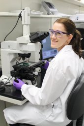 Nicole J Sylvain合作论文数Department of Biological Sciences, University of Alberta17
 Phyllis G. Paterson合作论文数University of Saskatchewan College of Pharmacy and Nutrition16
 John Mamo合作论文数School of Population Health, Faculty of Health Sciences, Curtin University;Curtin Medical Research Institute, Curtin University;Perron Institute14Helen Nichol合作论文数Anatomy and Cell Biology, University of Saskatchewan13Massimiliano Massi合作论文数12Huishu Hou合作论文数11
 Jitraporn Vongsvivut合作论文数School of Life and Environmental Sciences, Deakin University11Sally Caine合作论文数11
 Daryl L. Howard合作论文数Department of Chemistry, University of Otago11Ryu Takechi合作论文数10Frederick Colbourne合作论文数Department of Psychology;Centre for Neuroscience;University of Alberta;Centre for Neuroscience, University of Alberta10
 Mark J Tobin合作论文数Australian Synchrotron10Ryusuke Takechi合作论文数9
 Peter Lay合作论文数School of Chemistry, Faculty of Science, The University of Sydney9
 David Hartnell合作论文数School of Molecular and Life Sciences, Curtin University8
 Simon W Lewis合作论文数7Elena Dallerba合作论文数7Meg Willans合作论文数7Rhiannon E Boseley合作论文数7
 David Paterson合作论文数Division of Infectious Diseases, University of Pittsburgh Medical Center7
 Elizabeth Carter合作论文数School of Chemistry, The University of Sydney7Martin D de Jonge合作论文数School of Physics;University of Melbourne;School of Physics, University of Melbourne7Juliane Reinhardt合作论文数Australian Nucl Sci & Technol Org, Australian Synchrotron6Michael Nesbit合作论文数6
 Georges Grau合作论文数Department of Pathology, School of Medical Sciences, Faculty of Medicine & Health, The University of Sydney6Rebecca J Tidy合作论文数5Nicholas Fimognari合作论文数5H. Hou合作论文数5
 Gaewyn Ellison合作论文数Curtin Health Innovation Research Institute, Curtin University5
 Nicholas John Hunt合作论文数5
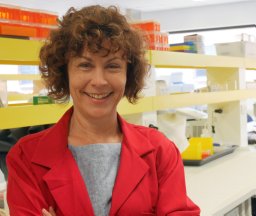 Melinda Fitzgerald合作论文数School of Animal Biology, University of Western Australia5
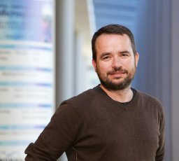 Hugh H. Harris合作论文数School of Chemistry, The University of Sydney5
 Mariam Alaverdashvili合作论文数Canadian Centre for Behavioural Neuroscience, University of Lethbridge5S. Caine合作论文数4
 Andrew Crawford合作论文数4Bradley J Schwehr合作论文数4Karina Khambatta合作论文数4
 Georgina Sauzier合作论文数School of Molecular and Life Sciences, Curtin University4Aitken Jade B合作论文数4
 El-Assaad Fatima合作论文数Vasc Immunol Unit, Univ Sydney4
 Lins Brittney R合作论文数Department of Physiology, University of Saskatchewan4D. Cohen合作论文数4
 Rainer Siegele合作论文数4




