BACKGROUND:Isocitrate dehydrogenase (IDH) is a commonly mutated gene in gliomas. Although IDH-mutant WHO grade 4 astrocytomas tend to occur in younger patients and show an improved survival compared to IDH-wildtype, prognosis remains poor and treatment options are limited. Ivosidenib is a small-molecule inhibitor of IDH-1 that has shown promise for treating low-grade IDH-mutant glioma. However, little is known about its efficacy in IDH-mutant WHO grade 4 astrocytoma. OBSERVATIONS:The authors present the case of a 36-year-old male with a IDH-mutant WHO grade 4 astrocytoma. He was originally treated with resection, followed by radiation therapy and temozolomide. At the second recurrence, ivosidenib was started, after lomustine therapy had failed to control the disease. Radiological surveillance showed interval decrease in tumor size starting 2 months after initiation of ivosidenib. The patient remained neurologically intact, and no adverse effects were observed. LESSONS:Here the authors report the effects of the off-label use of ivosidenib. The INDIGO trial published evidence of efficacy in using an IDH inhibitor for low-grade gliomas. The role of these drugs in high-grade IDH-mutant gliomas is currently unknown. Further studies are needed to assess their impact on overall and progression-free survival. https://thejns.org/doi/10.3171/CASE25572.
Background Aggressive meningiomas often display chromosomal abnormalities disrupting more than 20% of the genome, but the mechanisms that allow meningioma cells to thrive nevertheless are unknown.Methods This study used orthogonal small-molecule screens, gene expression analyses, DNA-binding analyses, epigenetic histone mark datasets, and orthotopic animal models to identify pathways essential for meningioma cell survival.Results Epigenetic buffering of proteotoxic stress by euchromatic histone methyltransferase 2 (EHMT2) enables meningioma cell survival. A high-throughput small-molecule screen and orthogonal gene expression analysis identified EHMT2 as important for meningioma growth. EHMT2 preferentially binds cis-regulatory elements affecting genes involved in protein processing in the endoplasmic reticulum (ER) in meningioma cells, and EHMT2 inhibition activates the ER stress apoptotic pathway. EHMT2 inhibition also decreases Sirtuin 1 (SirT1)-mediated activation of heat shock factor 1 (HSF1) transcriptional activity, thereby causing collapse of heat shock protein expression. The molecular chaperone and ER stress inhibitor, 4-phenylbutyric acid, abrogates meningioma cell death occurring after EHMT2 inhibition. Importantly, EHMT2 inhibition decreases meningioma growth in mice.Conclusions EHMT2/SirT1/HSF1-dependent mitigation of proteotoxic stress represents a promising therapeutic target in meningioma.
Animal studies and human tissue experiments have demonstrated that traumatic brain injury (TBI) causes damage to the extracellular matrix (ECM). To test the hypothesis that TBI causes disruption of sulfated glycosaminoglycan (sGAG) in the ECM, we measured levels of sGAG in the cerebrospinal fluid (CSF), blood, and urine, in patients with severe TBI in the acute postinjury period. Samples of CSF, blood, and urine were obtained within 72 h of injury in patients who received external ventricular drains as part of their treatment of severe TBI. Levels of chondroitin and heparan sGAGs were measured, along with their disaccharide constituents. Demographic information, presence of polytrauma, brain injury load, and distance of radiologically visible parenchymal injury from the ventricle were analyzed for correlation with total subtype sGAG levels. Levels were measured in 14 patients ranging in age from 17 to 90 years. CSF sGAG levels were variable among patients, with higher sGAG levels in plasma compared with CSF. Patients with polytrauma had nonsignificantly higher blood sGAG compared with patients with isolated head injury. Subcategories of CSF sGAG levels correlated with distance from the ventricle of parenchymal injury but not with brain injury load. This study is the first to measure sGAG levels in ventricular CSF and the first to analyze levels in TBI. These data demonstrate the elevation locally of intracranial sGAGs after severe TBI and suggest rapid local metabolism of these breakdown products. The consequences of ECM breakdown may provide unique therapeutic and preventive avenues to mitigate postinjury sequelae.
BACKGROUND:Human herpesvirus 6 (HHV-6) is a neurotropic virus known to induce encephalitis in immunocompromised patients as well as a subset of pediatric patients. However, ventriculitis secondary to HHV-6 is a rare occurrence among immunocompetent adults. Although acquired hydrocephalus has been described to infrequently co-occur with HHV-6 encephalitis in pediatric and immunocompromised adult patients, it has not been described in an immunocompetent adult. OBSERVATIONS:The authors present a rare case of HHV-6 encephalitis and obstructive hydrocephalus in an immunocompetent adult patient. Despite CSF diversion, the emergence and proliferation of multiple septations throughout the ventricular system resulted in the progression of severe hydrocephalus. LESSONS:Previous studies have linked HHV-6 to inflammatory states, occurring predominantly among children and immunocompromised adults, that manifest primarily as meningitis and encephalitis. This case highlights the fact that HHV-6 can cause life-threatening ventriculitis and hydrocephalus in immunocompetent adults. Pathophysiological mechanisms of virus-induced neuroinflammation yielding intraventricular septation formation are reviewed. https://thejns.org/doi/10.3171/CASE25273.
Improvements in single-cell whole-genome sequencing (scWGS) assays have enabled detailed characterization of somatic copy number alterations (CNAs) at the single-cell level. Yet, current computational methods are mostly designed for detecting chromosome-scale changes in cancer samples with low sequencing coverage. Here, we introduce HiScanner (High-resolution Single-Cell Allelic copy Number callER), which combines read depth, B-allele frequency, and haplotype phasing to identify CNAs with high resolution. In simulated data, HiScanner consistently outperforms state-of-the-art methods across various CNA types and sizes. When applied to high-coverage scWGS data from 65 cells across 11 neurotypical human brains, HiScanner shows a superior ability to detect smaller CNAs, uncovering distinct CNA patterns between neurons and oligodendrocytes. We also generated low-coverage scWGS data from 179 cells sampled from the same meningioma patient at two time points. For this serial dataset, integration of CNAs with point mutations revealed evolutionary trajectories of tumor cells. These findings show that HiScanner enables accurate characterization of frequency, clonality, and distribution of CNAs at the single-cell level in both non-neoplastic and neoplastic cells.
BACKGROUND AND IMPORTANCE:Trephination is a procedure in which a small hole is made in the skull. Rare cases of self-trephination by individuals seeking medical benefit have been reported. Excoriation disorder is a compulsive skin-picking condition in which an individual self-inflicts cutaneous lesions. Left untreated, severe excoriation disorder can pose significant health risks. CLINICAL PRESENTATION:Here, we describe 5 patients who presented with self-trephination due to a severe form of compulsive cranial excoriation at 2 neighboring academic medical centers over a 4-year period. We review the clinical presentation of self-trephination in cranial excoriation disorder and associated risk factors, surgical and nonsurgical interventions, complications of the disease, treatments, and mortality. Defining clinical characteristics include repetitive self-induced destruction of the scalp and skull with entry into the intracranial compartment, frequent psychiatric comorbidities, infection or injury of the brain with consequent neurological morbidity or mortality, and frequent treatment failures because of poor adherence. CONCLUSION:Self-trephination in cranial excoriation disorder is a severe neuropsychological disorder and neurosurgical emergency that exposes the brain and is often life-threatening. Appropriate therapy requires antibiotics, surgical debridement and repair of the wound, and concomitant effective psychiatric management of the underlying compulsion, including the use of antidepressants and behavioral therapy.
Background Meningiomas frequently recur after surgery. Existing guidelines for postoperative surveillance are based on customary practices or limited data. This may result in excessive or inadequate surveillance.Methods We compared 8 studies involving 1519 resected meningiomas with postoperative follow-up ranging from 7 to 23 years. Meningiomas were stratified using the World Health Organization and Simpson grading systems, and progression-free survival data were compared. Recurrence patterns were validated using 2 additional studies involving 2463 meningiomas.Results Incompletely resected meningiomas of all grades displayed recurrences throughout the observation period. The 5-year and 10-year cumulative incidence of recurrence for completely resected Grade 1 meningiomas was 10% and 20%, with no recurrences beyond 11 years. For completely resected Grade 2 meningiomas, the 5-year and 10-year cumulative incidence of recurrence was 24% and 50%, with ongoing recurrences throughout the observation period. Elevated recurrence rates for Grade 1/2 meningiomas persisted beyond 5 years. For completely resected Grade 3 meningiomas, the 5-year cumulative incidence of recurrence was 63%, and all recurred before 10 years.Conclusions Postoperative magnetic resonance imaging (MRI) at 48 h to determine the extent of resection and at 4 months to detect rapid regrowth is recommended. For completely resected Grade 1 meningiomas, annual MRI followed by discontinuation of surveillance if there is no recurrence after 11 years is reasonable. For completely resected Grade 2 meningiomas, annual MRI indefinitely is recommended. For Grade 3 meningiomas, MRI every 3-4 months for 2 years, followed by every 6 months indefinitely, is recommended. Incompletely resected meningiomas should be followed indefinitely.
Supplementary Figures 1-5 from A Genome-Wide Screen Reveals Functional Gene Clusters in the Cancer Genome and Identifies EphA2 as a Mitogen in Glioblastoma
Idiopathic normal pressure hydrocephalus (iNPH) is an enigmatic neurological disorder that develops after age 60 and is characterized by gait difficulty, dementia, and incontinence. Recently, we reported that heterozygous CWH43 deletions may cause iNPH. Here, we identify mutations affecting nine additional genes (AK9, RXFP2, PRKD1, HAVCR1, OTOG, MYO7A, NOTCH1, SPG11, and MYH13) that are statistically enriched among iNPH patients. The encoded proteins are all highly expressed in choroid plexus and ependymal cells, and most have been associated with cilia. Damaging mutations in AK9, which encodes an adenylate kinase, were detected in 9.6% of iNPH patients. Mice homozygous for an iNPH-associated AK9 mutation displayed normal cilia structure and number, but decreased cilia motility and beat frequency, communicating hydrocephalus, and balance impairment. AK9+/- mice displayed normal brain development and behavior until early adulthood, but subsequently developed communicating hydrocephalus. Together, our findings suggest that heterozygous mutations that impair ventricular epithelial function may contribute to iNPH.
Supplementary Table 1 from A Genome-Wide Screen Reveals Functional Gene Clusters in the Cancer Genome and Identifies EphA2 as a Mitogen in Glioblastoma
Supplementary Information from A Genome-Wide Screen Reveals Functional Gene Clusters in the Cancer Genome and Identifies EphA2 as a Mitogen in Glioblastoma
Heterozygous mutations affecting FOXJ1, a transcription factor governing multiciliated cell development, have been associated with obstructive hydrocephalus in humans. However, factors that disrupt multiciliated ependymal cell function often cause communicating hydrocephalus, raising questions about whether FOXJ1 mutations cause hydrocephalus primarily by blocking cerebrospinal fluid (CSF) flow or by different mechanisms. Here, we show that heterozygous FOXJ1 mutations are also associated with communicating hydrocephalus in humans and cause communicating hydrocephalus in mice. Disruption of one Foxj1 allele in mice leads to incomplete ependymal cell differentiation and communicating hydrocephalus. Mature ependymal cell number and motile cilia number are decreased, and 12% of motile cilia display abnormal axonemes. We observed decreased microtubule attachment to basal bodies, random localization and orientation of basal body patches, loss of planar cell polarity, and a disruption of unidirectional CSF flow. Thus, heterozygous FOXJ1 mutations impair ventricular multiciliated cell differentiation, thereby causing communicating hydrocephalus. CSF flow obstruction may develop secondarily in some patients harboring FOXJ1 mutations. Heterozygous FOXJ1 mutations impair motile cilia structure and basal body alignment, thereby disrupting CSF flow dynamics and causing communicating hydrocephalus.
Supplementary Tables 1-4 from A Developmental Taxonomy of Glioblastoma Defined and Maintained by MicroRNAs
Supplementary Table 2 from A Genome-Wide Screen Reveals Functional Gene Clusters in the Cancer Genome and Identifies EphA2 as a Mitogen in Glioblastoma
Most research on the evolution of damage after traumatic brain injury (TBI) focuses on cellular effects, but the analysis of human tissue slices and animal research have shown that TBI causes concomitant damage in the extracellular matrix, which can play a significant role in both short-term consequences such as edema, and late effects such as post-traumatic epilepsy (PTE). To test the hypothesis that traumatic brain injury (TBI) in human patients causes disruption of sulfated glycosaminoglycan (sGAG) in the extracellular matrix, we measured levels of these substances in the ventricular cerebrospinal fluid (CSF) in patients with severe TBI in the acute post-injury period, along with concomitant levels in blood and urine. We assessed whether levels corresponded to parenchymal injury load, distance of traumatic brain lesions from the ventricle, presence of polytrauma, or host demographic factors. Methods Samples of CSF, blood, and urine were obtained within 72 hours of injury in patients who received external ventricular drains as part of their treatment of severe TBI, and levels of chondroitin and heparan sGAGs were measured, along with their disaccharide constituents. Basic demographic information, presence and severity of polytrauma, brain injury load based on imaging findings, and distance of radiologically visible parenchymal injury from the ventricle were analyzed for correlation with total subtype sGAG levels in each patient. Results Levels were measured in 14 patients ranging in age from 17-90 years. CSF sGAG levels were variable among patients, and sGAG levels were higher in plasma than in CSF and variable in urine. Patients with polytrauma had non-significantly higher blood sGAG compared to patients with isolated head injury. Subcategories of CSF sGAG levels correlated with distance from the ventricle of parenchymal injury but not with brain injury load, which may reflect rapid metabolism in the parenchyma, contamination by blood, or bulk directional CSF flow from the ventricle to the subarachnoid space. Conclusion This study is the first to measure sGAG levels in ventricular CSF and also provides the first measurements in patients with TBI. Damage to the extracellular matrix may play a major role in acute and chronic injury sequelae, and these data demonstrate elevation locally of intracranial sGAGS after severe TBI and also suggest rapid local metabolism of these breakdown products. The consequences of extracellular matrix breakdown may provide unique therapeutic and preventive avenues to mitigate post-injury sequelae.
 Hong Wei Yang (杨宏伟)合作论文数南京理工大学理学院25
 Peter J Park合作论文数Department of Biomedical Informatics, Harvard Medical School;Division of Genetics, Brigham and Women’s Hospital23
 Jay Roberts合作论文数Clinical Research and Development, Smith Kline and French Laboratories18
 Hakan Gurdal合作论文数Dept Med Pharmacol, Univ Ankara17Rona S Carroll合作论文数16
 Alexandra J. Golby合作论文数Brigham and Women's Hospital;Image Guided Neurosurgery Laboratory, Department of Neurosurgery, Harvard Medical School, Harvard University13
 Ian Dunn合作论文数The Roslin Institute, The University of Edinburgh/The Royal (Dick) School of Veterinary Studies, The University of Edinburgh12
 Nils D. Arvold合作论文数Harvard Radiation Oncology Program, Harvard Medical School11
 E. Antonio Chiocca合作论文数Department of Neurosurgery, Brigham and Women's Hospital;Institute for the Neurosciences, Brigham and Women's Hospital;Harvard Medical School;Center for Neuro-oncology, Dana-Farber Cancer Institute11
 Brian M. Alexander合作论文数Dana-Farber Cancer Institute;Harvard Medical School;GCAR;Roche/Genentech Pharma R&D11
 Elizabeth Claus合作论文数Department of Biostatistics, School of Public Health, Yale University;Department of Neurosurgery, Brigham and Women’s Hospital;Harvard Medical School11Xiuli Jiang合作论文数10JB Parker合作论文数9E Friedman合作论文数9
 Eudocia Q. Lee合作论文数Division of Cancer Neurology, Department of Neurology, Brigham and Women's Hospital, Harvard Medical School;Center for Neuro-Oncology, Dana-Farber Cancer Institute9Dl Snyder合作论文数8Yi Yu合作论文数8
 David Reardon合作论文数Center for Neuro-Oncology, Dana-Farber Cancer Institute;Harvard Medical School7
 Margaret Horvath合作论文数Dana-Farber/Brigham and Women's Cancer Center, Harvard Medical School7Lakshmi Nayak合作论文数7
 Patrick Y. Wen合作论文数Department of Neurology, Harvard Medical School;Center For Neuro-Oncology, Dana-Farber Cancer Institute7M McMillian合作论文数6Wei Huang合作论文数6S Bryant合作论文数6
 Bradley D. Anderson合作论文数College of Pharmacy, University of Kentucky6
 Xin Wang合作论文数Brigham and Women's Hospital6Weil Lai合作论文数6
 Ayal A. Aizer合作论文数Harvard Medical School;Department of Radiation Oncology, Brigham and Women's Hospital6David R. Tribble合作论文数6Amanda Brosius合作论文数5Meredith Thomas合作论文数5Tm Seasholtz合作论文数5Guoping Cai合作论文数5J Wan合作论文数5
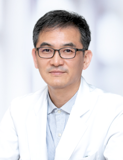 Tae Min Kim合作论文数Department of Hemato Oncology, Seoul National University Hospital;Department of Internal Medicine, Seoul National University Hospital5
 Elizabeth A Maher合作论文数5Lalani Tahaniyat合作论文数5
 Jamie L. Fraser合作论文数5
 Laura E. Durso合作论文数Center for American Progress5Wang Zhen合作论文数5Park Richard合作论文数5
 Lynda Chin合作论文数Apricity Health5Xing Hongyan合作论文数5
 Ronald DePinho合作论文数Department of Cancer Biology, Division of Basic Science Research, The University of Texas MD Anderson Cancer Center5
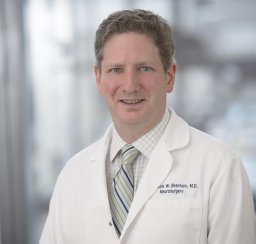 Cameron Walter Brennan合作论文数Department of Neurosurgery, Memorial Sloan-Kettering Cancer Center5
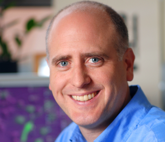 Keith L. Ligon合作论文数Center for Patient Derived Models, Dana-Farber Cancer Institute;Division of Neuropathology, Brigham and Women's Hospital;Medical School, Harvard University;Travera;Broad Institute, Massachusetts Institute of Technology;Boston Children's Hospital5
 Yanyang Tu (涂艳阳)合作论文数5Anton Bittner合作论文数5
 Rona Carroll合作论文数5




