De novo membranous nephropathy (dnMN) is an uncommon immune complex-mediated late complication of human kidney allografts that causes proteinuria. We report here the first case of dnMN in a pig-to-baboon kidney xenograft. The donor was a double knockout (GGTA1 and β4GalNT1) genetically engineered pig with a knockout of the growth hormone receptor and addition of 6 human transgenes (hCD46, hCD55, hTBM, hEPCR, hHO1, and hCD47). The recipient developed proteinuria at 42 days posttransplant, which progressively rose to the nephrotic-range at 106 days, associated with an increase in serum antidonor IgG. Kidney biopsies showed antibody-mediated rejection (AMR) with C4d and thrombotic microangiopathy that eventually led to graft failure at 120 days. In addition to AMR, the xenograft had diffuse, global granular deposition of C4d and IgG along the glomerular basement membrane on days 111 and 120. Electron microscopy showed extensive amorphous subepithelial electron-dense deposits with intervening spikes along the glomerular basement membrane. These findings, in analogy to human renal allografts, are interpreted as dnMN in the xenograft superimposed on AMR. The target was not identified but is hypothesized to be a pig xenoantigen expressed on podocytes. Whether dnMN will be a significant problem in other longer-term xenokidneys remains to be determined.
Dedifferentiated chondrosarcoma is rare, aggressive, and microscopically bimorphic. How pathologic features such as the amounts of dedifferentiation affect prognosis remains unclear. We evaluated the percentages and sizes of dedifferentiation in a consecutive institutional series of dedifferentiated chondrosarcomas from 1999 to 2021. The statistical analysis included cox proportional hazard models and log-rank tests. Of the 67 patients (26 women, 41 men; age, 39 to >89 [median 61] years; 2 with Ollier disease), 58 presented de novo; 9 were identified with conventional chondrosarcomas 0.6-13.2 years (median, 5.5 years) prior. Pathologic fracture and distant metastases were noted in 27 and 7 patients at presentation. The tumors involved the femur (n = 27), pelvis (n = 22), humerus (n = 7), tibia (n = 4), scapula/ribs (n = 4), spine (n = 2), and clivus (n = 1). In the 56 resections, the tumors ranged in size from 3.5 to 46.0 cm (median, 11.5 cm) and contained 1%-99.5% (median, 70%) dedifferentiated components that ranged in size from 0.6 to 24.0 cm (median, 7.3 cm). No correlation was noted between total size and percentage of dedifferentiation. The dedifferentiated components were typically fibrosarcomatous or osteosarcomatous, whereas the associated cartilaginous components were predominantly grade 1-2, rarely enchondromas or grade 3. The entire cohort's median overall survival and progression-free survival were 11.8 and 5.4 months, respectively. In the resected cohort, although the total size was not prognostic, the percentage of dedifferentiation ≥20% and size of dedifferentiation >3.0 cm each predicted worse overall survival (9.9 vs 72.5 months; HR, 3.76; 95% CI, 1.27-11.14; P = .02; 8.7 vs 58.9 months; HR, 3.03; 95% CI, 1.21-7.57; P = .02, respectively) and progression-free survival (5.3 vs 62.1 months; HR, 3.05; 95% CI, 1.13-8.28; P = .03; 5.3 vs 56.6 months; HR, 2.50; 95% CI, 1.06-5.88; P = .04, respectively). In conclusion, both the percentages and sizes of dedifferentiation were better prognostic predictors than total tumor sizes in dedifferentiated chondrosarcomas, highlighting the utility of their pathologic evaluations.
Deoxysphingolipids (1-deoxySLs) are neurotoxic sphingolipids associated with obesity and diabetic neuropathy (DN) and have been linked to severity of functional peripheral neuropathies. While l-serine supplementation can reduce 1-deoxySL accumulation and improve insulin sensitivity and sensory nerve velocity, long-term outcomes have not yet been examined. To assess this, we treated 2 month old db/db mice, a model of DN, with 5-20 % oral l-serine for 6 months and longitudinally quantified the extent of functional neuropathy progression. We examined putative biomarkers of neuropathy in blood and tissue and quantified levels of small fiber neuropathy, looking for associations between lowered 1-deoxySL and phenotypes. Toxic 1-deoxySLs were suppressed long-term in plasma and various tissue including the sciatic nerve, which is particularly targeted in DN. Functional neuropathy and sensory modalities were significantly improved in the treatment group well into advanced stages of disease. However, structural assessments revealed prominent axonal degeneration, apoptosis and Schwann cell pathology, suggesting that neuropathy was ongoing. Hyperglycemia and dyslipidemia persisted during our study, and high levels of glutathione were seen in the spinal cord. Our results demonstrate that despite significant functional improvements, l-serine does not prevent chronic degenerative changes specifically at the structural level, pointing to other processes such as oxidative damage and hyperglycemia, that persist despite 1-deoxySL reduction.
Megakaryocytes (MKs) are precursors to platelets, the second most abundant cells in the peripheral circulation. However, while platelets are known to participate in immune responses and play significant functions during infections, the role of MKs within the immune system remains largely unexplored. Histological studies of sepsis patients identified increased nucleated CD61 + cells (MKs) in the lungs, and CD61 + staining (likely platelets within microthrombi) in the kidneys, which correlated with the development of organ dysfunction. Detailed imaging cytometry of peripheral blood from patients with sepsis found significantly higher MK counts, which we predict would likely be misclassified by automated hematology analyzers as leukocytes. Utilizing in vitro techniques, we show that both stem cell derived MKs (SC MKs) and cells from the human megakaryoblastic leukemia cell line, Meg-01, undergo chemotaxis, interact with bacteria, and are capable of releasing chromatin webs in response to various pathogenic stimuli. Together, our observations suggest that MK cells display some basic innate immune cell behaviors and may actively respond and play functional roles in the pathophysiology of sepsis.
Peripheral neuropathy is a dose-limiting side effect of chemotherapy with paclitaxel. Paclitaxel-induced peripheral neuropathy (PIPN) is typically characterized by a predominantly sensory neuropathy presenting with allodynia, hyperalgesia and spontaneous pain. Oxidative mitochondrial damage in peripheral sensory neurons is implicated in the pathogenesis of PIPN. Reactive sulfur species, including persulfides (RSSH) and polysulfides (RSnH), are strong nucleophilic and electrophilic compounds that exert antioxidant effects and protect mitochondria. Here, we examined the potential neuroprotective effects of glutathione trisulfide (GSSSG) in a mouse model of PIPN. Intraperitoneal administration of paclitaxel at 4 mg/kg/day for 4 days induced mechanical allodynia and thermal hyperalgesia in mice. Oral administration of GSSSG at 50 mg/kg/day for 28 days ameliorated mechanical allodynia, but not thermal hyperalgesia. Two hours after oral administration, 34S-labeled GSSSG was detected in lumber dorsal root ganglia (DRG) and in the lumber spinal cord. In mice treated with paclitaxel, GSSSG upregulated expression of genes encoding antioxidant proteins in lumber DRG, prevented loss of unmyelinated axons and inhibited degeneration of mitochondria in the sciatic nerve. In cultured primary neurons from cortex and DRG, GSSSG mitigated paclitaxel-induced superoxide production, loss of axonal mitochondria, and axonal degeneration. These results indicate that oral administration of GSSSG mitigates PIPN by preventing axonal degeneration and mitochondria damage in peripheral sensory nerves. The findings suggest that administration of GSSSG may be an approach to the treatment or prevention of PIPN and other peripheral neuropathies.
Mutations in the peroxisomal half-transporter ABCD1 cause X-linked adrenoleukodystrophy, resulting in elevated very long-chain fatty acids (VLCFA), progressive neurodegeneration and an associated pain syndrome that is poorly understood. In the nervous system of mice, we found ABCD1 expression to be highest in dorsal root ganglia (DRG), with satellite glial cells (SGCs) displaying higher expression than neurons. We subsequently examined sensory behavior and DRG pathophysiology in mice deficient in ABCD1 compared to wild-type mice. Beginning at 8 months of age, Abcd1−/y mice developed persistent mechanical allodynia. DRG had a greater number of IB4-positive nociceptive neurons expressing PIEZO2, the mechanosensitive ion channel. Blocking PIEZO2 partially rescued the mechanical allodynia. Beyond affecting neurons, ABCD1 deficiency impacted SGCs, as demonstrated by high levels of VLCFA, increased glial fibrillary acidic protein (GFAP), as well as genes disrupting neuron-SGC connectivity. These findings suggest that lack of the peroxisomal half-transporter ABCD1 leads to PIEZO2-mediated mechanical allodynia as well as SGC dysfunction. Given the known supportive role of SGCs to neurons, this elucidates a novel mechanism underlying pain in X-linked adrenoleukodystrophy.
Clear cell sarcoma (CCS) is an uncommon malignant mesenchymal neoplasm of young adults with a predilection for tendons and aponeuroses of distal extremities, a distinctive nested growth pattern, melanocytic differentiation, and usually an EWSR1::ATF1 fusion. Distinction from melanoma can be challenging but is critical for clinical management. Rare cases of primary bone CCS have been reported. The purpose of this study was to evaluate the clinicopathologic features of a series of primary bone CCS. Three cases of primary bone CCS were identified out of 140 CCS diagnosed between 2010 and 2021. Two patients were female, and 1 patient was male; ages were 19, 47, and 61 years. All tumors arose in the long bones of the extremities (femur, humerus, fibula). Two tumors also involved regional lymph nodes at presentation. Two showed characteristic histologic features, in the form of nests and fascicles of uniform epithelioid to spindle cells with prominent nucleoli and pale eosinophilic to clear cytoplasm; 1 tumor showed sheet-like growth, unusual focal pleomorphism, and more notable nuclear atypia. By immunohistochemistry, S100 protein was positive in 2/3 cases, SOX10 in 3/3, HMB-45 in 2/3, MiTF in 2/2, and melan A in 1/3. All cases were confirmed to harbor EWSR1 rearrangement and EWSR1::ATF1 fusion or t(12;22). On follow-up, all 3 patients developed metastases and died of disease, 5, 18, and 21 months after diagnosis. In summary, CCS rarely presents in the skeleton. At such locations, distinction from metastatic melanoma is particularly challenging. Clinical and pathologic features are similar to conventional CCS of soft tissue. Primary bone CCS may pursue an aggressive clinical course.
Moran, Jakob M.T. MD; Hung, Yin P. MD, PhD; Selig, Martin K. BA; Nielsen, G.P. MD Author Information
MEGD(H)EL syndrome is a rare autosomal recessive disorder caused by mutations in SERAC1, a protein necessary for phosphatidylglycerol remodeling. It is characterized by 3-methylglutaconic aciduria, deafness-dystonia, (hepatopathy), encephalopathy, and Leigh-like syndrome, but has a wide spectrum of severity. Here, we present a case of a child with MEGD(H)EL syndrome with infantile hepatopathy, neurodevelopmental delays, characteristic biochemical abnormalities, and biallelic novel SERAC1 mutations: (1) deletion of (at least) exons 2-4, pathogenic; and (2) c.1601A>T (p.H534L), likely pathogenic. Her initial clinical presentation was notable for persistently elevated transaminases, speech delay, delayed motor milestones, and sensorineural hearing loss. However, her verbal and motor development has progressively improved and now, at 4 years of age, she has only speech and mild gross motor delays as compared to her unaffected peers and is exceeding clinical expectations. The histologic features of a liver biopsy are described, which has not previously been published in detail for this syndrome. Hepatocytes showed granular cytoplasm and fine intracytoplasmic lipid droplets. The ultrastructural findings included abnormal circular mitochondrial cristae. These findings are consistent with a mitochondrial disorder.
Introduction: Thyroblastoma, a primary thyroid neoplasm with histological features of primitive thyroid tissue has recently been described and is included as a distinct entity in the most recent edition of the World Health Organization (WHO) Classification of Tumors (5th edition). In this study, we expand the clinical, morphological, and molecular profile of this aggressive neoplasm. Patient Findings: The patients are females, 19 and 45 years of age, referred for large thyroid nodules. Tumor morphology is biphasic, composed of nests and follicles of epithelial cells, some with colloid-like secretions reminiscent of fetal thyroid follicles intertwined with a primitive stromal spindle cell component. By immunohistochemistry, the epithelial component is diffusely positive for PAX8 and TTF1 markers. Molecular studies showed DICER1 aberrations. Conclusion: A primary primitive thyroid malignancy reminiscent of early fetal embryology with no teratoid element, recently reported as thyroblastoma represents a unique entity, novel in its description, and is likely underdiagnosed.
Background Histiocytic and dendritic cell neoplasms are a diverse group of tumors arising from monocytic or dendritic cell lineage. Whereas the genomic features for Langerhans cell histiocytosis and Erdheim-Chester disease have been well described, other less common and often aggressive tumors in this broad category remain poorly characterized, and comparison studies across the World Health Organization diagnostic categories are lacking. Methods Tumor samples from a total of 102 patient cases within four major subtypes of malignant histiocytic and dendritic cell neoplasms, including 44 follicular dendritic cell sarcomas (FDCSs), 41 histiocytic sarcomas (HSs), 7 interdigitating dendritic cell sarcomas (IDCSs), and 10 Langerhans cell sarcomas (LCSs), underwent hybridization capture with analysis of up to 406 cancer-related genes. Results Among the entire cohort of 102 patients, CDKN2A mutations were most frequent across subtypes and made up 32% of cases, followed by TP53 mutations (22%). Mitogen-activated protein kinase (MAPK) pathway mutations were present and enriched among the malignant histiocytosis (M) group (HS, IDCS, and LCS) but absent in FDCS (72% vs. 0%; p < .0001). In contrast, NF-kappa B pathway mutations were frequent in FDCSs but rare in M group histiocytoses (61% vs. 12%; p < .0001). Tumor mutational burden was significantly higher in M group histiocytoses as compared with FDCSs (median 4.0/Mb vs. 2.4/Mb; p = .012). We also describe a pediatric patient with recurrent secondary histiocytic sarcoma treated with targeted therapy and interrogated by molecular analysis to identify mechanisms of therapeutic resistance. Conclusion A total of 42 patient tumors (41%) harbored pathogenic mutations that were potentially targetable by approved and/or investigative therapies. Our findings highlight the potential value of molecular testing to enable precise tumor classification, identify candidate oncogenic drivers, and define personalized therapeutic options for patients with these aggressive tumors. Implications for Practice This study presents comprehensive genomic profiling results on 102 patient cases within four major subtypes of malignant histiocytic and dendritic cell neoplasms, including 44 follicular dendritic cell sarcomas (FDCSs), 41 histiocytic sarcomas (HSs), 7 interdigitating dendritic cell sarcomas (IDCSs), and 10 Langerhans cell sarcomas (LCSs). MAPK pathway mutations were present and enriched among the malignant histiocytosis (M) group (HS, IDCS, and LCS) but absent in FDCSs. In contrast, NF-kappa B pathway mutations were frequent in FDCSs but rare in M group histiocytosis. A total of 42 patient tumors (41%) harbored pathogenic mutations that were potentially targetable by approved and/or investigative therapies.
"Pulmonary Hemosiderosis with Calcification Associated with IgA Nephropathy." American Journal of Respiratory and Critical Care Medicine, 204(2), pp. e24–e25
Nuclear protein in testis (NUT) carcinoma, an aggressive tumour driven by NUTM1 rearrangements, often involves the lung/mediastinum and shows squamous differentiation. We encountered an index patient with a thoracic NUT carcinoma diagnosed by molecular testing, showing extensive pleural involvement and diffuse thyroid transcription factor‐1 (TTF‐1) expression, initially suggestive of lung adenocarcinoma with pseudomesotheliomatous growth. We thus gathered an institutional series of thoracic NUT carcinomas to examine their pathological spectrum.
Amyloid arthropathy is a joint disease associated with systemic amyloidosis. Herein, we present a model case and review the clinicopathologic features and pathophysiology of this disorder. Amyloid arthropathy results from elevation of serum amyloidogenic proteins and their deposition as aggregates in synovial fluid and articular tissues. The most common proteins are beta-2-microglobulin in the context of long-term hemodialysis therapy and immunoglobulin light chains associated with plasma cell proliferations. We provide a comprehensive update on the pathogenesis, clinical manifestations, and pathologic features of amyloid arthropathy. We provide detailed insights on amyloid protein deposition and aggregation in joints and proper details for diagnosis.
An 88-year-old man presented with diplopia, limitation of extraocular movements, and a firm palpable mass in the superolateral orbit. Biopsy revealed a sclerosing signet ring cell carcinoma with histopathologic features mimicking those of a primary signet ring cell (histiocytoid) carcinoma of the eyelid of eccrine or apocrine gland origin, a metastasis from an invasive lobular breast carcinoma or a metastatic diffuse-type gastric carcinoma. An extensive panel of immunohistochemical stains and molecular genetic analyses unequivocally failed to establish a precise diagnosis. Electron microscopy demonstrated features of a primary lacrimal gland lesion with intracytoplasmic lumens and zymogen granules typical of lacrimal secretory pyramidal cells. A thorough initial systemic work-up failed to reveal a primary visceral malignancy. Fifteen months of follow-up have failed to detect the emergence of another primary malignancy. To the best of our knowledge, a tumor with the morphology of the current lesion has not been previously described in the major or accessory lacrimal glands.
 G. Petur Nielsen合作论文数Division of Bone and Soft Tissue Pathology, Massachusetts General Hospital;Harvard Medical School26
 Vikram Deshpande合作论文数Department of Pathology, Beth Israel Deaconess Medical Center, Harvard Medical School12
 Lawrence R. Zukerberg合作论文数10
 Rex Hung合作论文数Department of Pathology, Massachusetts General Hospital8
 Mari Mino-Kenudson合作论文数Harvard Medical School;Massachusetts General Hospital6
 Henry Kronenberg合作论文数Endocrine Division, Mass General Research Institute;Massachusetts General Hospital;Harvard Medical School;Harvard Stem Cell Institute5Richard L. Kradin合作论文数5Ivan Chebib合作论文数5Aleksandr Vasilyev合作论文数4
 Jason L. Hornick合作论文数Department of Pathology, Harvard Medical School;Immunohistochemistry Laboratory, Department of Pathology, Brigham and Women's Hospital;Dana-Farber Cancer Institute4
 Robert Colvin合作论文数Harvard Medical School;The Immunopathology Research Laboratory, Center for Transplantation Sciences, Massachusetts General Hospital;Transplantation Biology Research Center, Massachusetts General Hospital4
 Anat Stemmer-Rachamimov合作论文数C. S. Kubik Laboratory for Neuropathology, Massachusetts General Hospital;Department of Pathology, Massachusetts General Hospital;Harvard Medical School4Erik A. Williams合作论文数3
 Anika L Marand合作论文数Dept Surg, Massachusetts Gen Hosp3Lucas R Massoth合作论文数Dept Pathol, Massachusetts Gen Hosp3
 Cristina Ferrone合作论文数Department of Surgery, Cedars-Sinai Medical Center;Division of General and GI Surgery, Department of Surgery, Massachusetts General Hospital3
 Joe Lennerz合作论文数BostonGene3
 Jorgensen, J.A.合作论文数3
 Ivy A Rosales合作论文数Histopathology Research Core Laboratory, Department of Pathology, Massachusetts General Hospital;Harvard University3
 Daniel Irimia合作论文数Center for Engineering in Medicine & Surgery, Massachusetts General Hospital3
 Galit H Frydman合作论文数3Olaleye David合作论文数3
 Esther Oliva合作论文数Department of Pathology, Massachusetts General Hospital, Harvard Medical School3
 Miriam A. Bredella合作论文数Department of Radiology, Harvard Medical School, Massachusetts General Hospital3William C. Faquin合作论文数3
 Felix Ellett合作论文数Department of Medical Biology, University of Melbourne3
 Charles Vanderburg合作论文数Stanley Center for Psychiatric Research, Broad Institute of MIT and Harvard;National Aeronautics and Space Administration3
 James G. Fox合作论文数Department of Biological Engineering, Massachusetts Institute of Technology3
 Keith H. K. Wong合作论文数Department of Biomedical Engineering, Boston University3
 Iain Drummond合作论文数Harvard Medical School and Renal Unit, Massachusetts General Hospital3Florian Eichler合作论文数3
 Shannon N. Tessier合作论文数Harvard Medical School;Center for Engineering in Medicine & Surgery, Massachusetts General Hospital;Shriners Hospitals for Children3
 Valentina Nardi合作论文数Harvard Medical School and Division of Pediatric Hematology/Oncology, Dana Farber Cancer Institute3Martin S Taylor合作论文数2Emily Berner合作论文数2Quinn Taylor-Black合作论文数2Julie M. Guilmette合作论文数2Svetlana Stoykova合作论文数2Hugh Auchincloss合作论文数2Partha Sinha合作论文数2
 Brandon Nicolay合作论文数Massachusetts General Hospital Cancer Center, Harvard Medical School2
 Alexa Burger合作论文数Univ Zurich, Inst Mol Life Sci, CH-8057 Zurich, Switzerland2Yi Gong合作论文数Harvard Med Sch, Massachusetts Gen Hosp, Dept Neurol, 55 Fruit St,ACC 708, Boston, MA 02114 USA2Joseph H Schwab合作论文数2
 Tatsuya Kobayashi合作论文数Massachusetts Gen Hosp, Endocrine Unit, Boston, MA 02114 USA2
 Rushika M Perera合作论文数Center for Vector-Borne Infectious Diseases, Colorado State University2
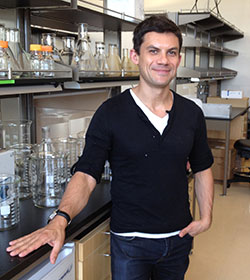 Roberto Zoncu合作论文数Biochemistry, Biophysics and Structural Biology Faculty, Department of Molecular and Cell Biology, University of California, Berkeley2
 Dudley William Lamming合作论文数University of Wisconsin-Madison2Fulzele Keertik合作论文数Harvard Medical School, Massachusetts General Hospital2




