The remarkable efficiency of double fertilization in flowering plants depends on the precise delivery of immotile sperm cells to two female gametes. Although sperm cells are rapidly released from the pollen tube, how they are accurately directed to their fertilization site has remained unknown. Here we identify a transient extracellular structure, termed the gamete pit, that forms at the egg cell–central cell (EC–CC) interface immediately after pollen tube discharge and serves as the landing site for sperm cells. Gamete pit formation is enabled by localized dissociation of the EC–CC interface, mediated by extracellular protein assemblies designated EC1 bodies. These structures contain extracellular deposits enriched in EGG CELL 1 (EC1) peptides, including amyloid-like assemblies. Loss of EC1 bodies prevented gamete pit formation, causing sperm-cell backflow or catastrophic over-penetration of pollen tube contents into the central cell. Surprisingly, ectopic deposition of a heterologous amyloidogenic protein alleviated over-penetration and partially restored fertility. Our findings reveal a mechanism by which extracellular protein assemblies remodel the gamete interface for sperm-cell reception, uncover an unexpected structural role for the conserved gamete-activating peptide EC1, and establish protein-mediated cell-interface remodeling as a fundamental principle underlying double fertilization in flowering plants.
Volume electron microscopy based on serial sectioning allows for three-dimensional (3D) visualization and analysis of the internal structures of tissues, cells, and organelles. One such technique, focused ion beam (FIB) scanning electron microscopy (SEM), has the advantages of nanoscale sectioning and high z-resolution, but the disadvantage of limited volume processing. Because of this limitation, targeting localized objects by FIB-SEM is difficult. Here, we developed a FIB-SEM observation workflow that enables the analysis of the filiform apparatus of synergid cells enclosed in the Arabidopsis ovule. In this protocol, plant samples are stained, embedded, trimmed, and carbon-coated while maintaining their orientation within the tissue. Then, sequential observations are performed using Cut & See function of FIB-SEM, followed by image processing for 3D reconstruction. Utilization of multi-scanning and image cropping from high-resolution data helps to identify localized targets within plant tissue. The filiform apparatus, which is an invaginated cell wall structure of the synergid cells, shows distinct contrast in each image, allowing for segmentation using brightness-based binarization. Such segmentation avoids the need to manually trace complex structures and facilitates 3D reconstruction by volume electron microscopy.
The pollen tube plays a pivotal role in double fertilization by delivering sperm cells (SCs) to the ovule. In Arabidopsis thaliana , a pair of SCs tightly connects with the vegetative nucleus (VN) to form the male germ unit (MGU), which is located in the apical region during pollen-tube growth, keeping the VN ahead of the SCs. MGU transport relies on independent motility of VN and SC pairs. However, the complexity of this dual motive force has hindered our understanding of MGU behavior, including its positioning and nuclear order. We used Arabidopsis mutants or transgenic plants that produced semi-motile MGUs with aberrant VNs or SCs to analyze the positioning of VN or SCs after stochastically disconnecting the MGU. In pollen tubes with an immotile SC pair, the VN was ∼70 μm away from the tip, whereas in pollen tubes with an immotile VN, the SC pair was ∼100 μm away from the tip, implying that the VN and SCs have independent home positions. The position of MGU moved forward owing to the loss of the microtubule-destabilizing kinesin KINESIN-13A . Conversely, microtubule depolymerization by oryzalin treatment or introducing mutations in TUBULIN BETA 4 ( TUB4 ) deregulated the position of the MGU and shifted its position backward. In addition, tub4 plants exhibited reduced fertility. These data indicate a significant role of microtubules in stable MGU positioning to ensure reproductive success. ### Competing Interest Statement The authors have declared no competing interest.
Pollen tubes exhibit one of the fastest apical growth rates among plant cells. Maintaining the proper balance between turgor pressure and cell wall synthesis at the pollen tube tip is crucial for this rapid growth, and any disruption can result in pollen tube rupture. In our study, we reveal that exposure to short-wavelength visible light, specifically blue light, induces pollen tube rupture. The frequency of pollen tube rupture increases in an intensity-dependent manner. Additionally, we observed Ca2+ influx after blue light irradiation, accompanying with either pollen tube rupture or a temporary halt in elongation. These findings offer insights into the interplay between pollen tube integrity maintenance and Ca2+ influx at the pollen tube tip, presenting a novel and efficient method to control pollen tube burst. Subject Areas (1) growth and development (11) new methodology ### Competing Interest Statement The authors have declared no competing interest.
In the anthers and ovaries of flowers, pollen grains and embryo sacs are produced with uniform cell compositions. This stable gametogenesis enables elaborate interactions between male and female gametophytes after pollination, forming the highly successful sexual reproduction system in flowering plants. As most ovules are fertilized with a single pollen tube, the resulting genome set in the embryo and endosperm is determined in a single pattern by independent fertilization of the egg cell and central cell by two sperm cells. However, if ovules receive four sperm cells from two pollen tubes, the expected options for genome sets in the developing seeds would more than double. In wild-type Arabidopsis thaliana plants, around 5% of ovules receive two pollen tubes. Recent studies have elucidated the abnormal fertilization in supernumerary pollen tubes and sperm cells related to polytubey, polyspermy, heterofertilization and fertilization recovery. Analyses of model plants have begun to uncover the mechanisms underlying this new pollen tube biology. Here, we review unusual fertilization phenomena and propose several breeding applications for flowering plants. These arguments contribute to the remodeling of plant reproduction, a challenging concept that alters typical plant fertilization by utilizing the current genetic toolbox.
Endosperm-embryo development in flowering plants is regulated coordinately by signal exchange during seed development. However, such a reciprocal control mechanism has not been clearly identified. In this study, we identified an endosperm-specific gene, LBD35, expressed in an embryonic development-dependent manner, by a comparative transcriptome and cytological analyses of double-fertilized and single-fertilized seeds prepared by using the kokopelli mutant, which frequently induces single fertilization events. Transcriptome analysis using LBD35 as a marker of the central cell fertilization event identified that 141 genes, including 31 genes for small cysteine-rich peptides, are expressed in a double fertilization-dependent manner. Our results reveal possible embryonic signals that regulate endosperm gene expression and provide a practicable method to identify genes involved in the communication during endosperm-embryo development.
Apomixis, defined as the transfer of maternal germplasm to offspring without fertilization, enables the fixation of F1-useful traits, providing advantages in crop breeding. However, most apomictic plants require pollination to produce the endosperm. The endosperm is essential for embryogenesis, and its development is suppressed until fertilization. We show that the expression of a chimeric repressor of the Elongation of Siliques without Pollination 3 (ESP3) gene (Pro35S:ESP3-SRDX) induces ovule enlargement without fertilization in Arabidopsis thaliana. The ESP3 gene encodes a protein similar to the flowering Wageningen homeodomain transcription factor containing a StAR-related lipid transfer domain. However, ESP3 lacks the homeobox-encoding region. Genes related to the cell cycle and sugar metabolism were upregulated in unfertilized Pro35S:ESP3-SRDX ovules similar to those in fertilized seeds, while those related to autophagy were downregulated similar to those in fertilized seeds. Unfertilized Pro35S:ESP3-SRDX ovules partially nourished embryos when only the egg was fertilized, accumulating hexoses without central cell proliferation. ESP3 may regulate nutrient flow during seed development, and ESP3-SRDX could be a useful tool for complete apomixis that does not require pseudo-fertilization.
In the synergid cells, F-actin controls secretion of pollen tube attractants toward the outside of the ovule, and its temporal destruction may terminate further attractant supply after fertilization. Pollen tube attraction is a key event of sexual reproduction in flowering plants. In the ovule, two synergid cells neighboring the egg cell control pollen tube arrival via the active secretion of attractant peptides such as AtLURE1 and XIUQIU from the filiform apparatus (FA) facing toward the micropyle. Distinctive cell polarity together with longitudinal F-actin and microtubules are hallmarks of the synergid cell in various species, though the functions of these cellular structures are unclear. In this study, we used genetic and pharmacological approaches to indicate the roles of cytoskeletal components in FA formation and pollen tube guidance in Arabidopsis thaliana. Genetic inhibition of microtubule formation reduced invaginations of the plasma membrane but did not abolish micropylar AtLURE1.2 accumulation. By contrast, the expression of a dominant-negative form of ACTIN8 induced disorganization of the FA and loss of polar AtLURE1.2 distribution toward the FA. Interestingly, after pollen tube reception, F-actin became unclear for a few hours in the persistent synergid cell, which may be involved in pausing and resuming pollen tube attraction during early polytubey block. Our data suggest that F-actin plays a central role in maintaining cell polarity and in mediating male-female communication in the synergid cell.
In pollen and pollen tubes, immotile sperm cells are enclosed by an inner vegetative plasma membrane (IVPM), a single endomembrane originating from the vegetative-cell plasma membrane. It is widely believed that sperm cells must be removed from the IVPM prior to gamete associations and fusions; however, details of the timing and morphological changes upon IVPM dissociation remain elusive. Here, we report a rapid IVPM breakdown immediately before double fertilization in Arabidopsis thaliana. The IVPM was stably observed in coiling pollen tubes when pollen tube discharge was prevented using lorelei mutant ovules. In contrast, a semi-in vivo fertilization assay in wild-type ovules demonstrated fragmented IVPM around sperm nuclei 1 min after pollen tube discharge. These observations revealed the dynamic alteration of released sperm cells and provided new insights into double fertilization in flowering plants. With a summary of recent findings on IVPM lipid composition, we discussed the possible physiological signals controlling IVPM breakdown.
The vegetative cell nucleus proceeds ahead of a pair of sperm cells located beneath the pollen tube tip during germination. The tip-localized vegetative nucleus had been considered to play a pivotal role in the control of directional pollen tube growth and double fertilization. However, we recently reported the female-targeting behavior of pollen tubes from mutant plants, of which the vegetative nucleus and sperm nuclei were artificially immotile. We showed that the apical region of the mutant pollen tubes became physiologically enucleated after the first callose plug formation, indicating the autonomously growing nature of pollen tubes without the vegetative nucleus and sperm cells. Thus, in this study, we further analyzed another Arabidopsis thaliana mutant producing physiologically enucleated pollen tubes and discussed the mechanism by which a pollen tube can grow without de novo transcription from the vegetative nucleus. We propose several possible molecular mechanisms for persistent pollen tube growth, such as the contribution of transcripts before and immediately after germination and the use of persistent transcripts, which may be important for a competitive race among pollen tubes.
The pollen tube is a male reproductive tissue in angiosperms that transports sperm cells to the ovule where double fertilization takes place. It was believed that the nuclei, always present at the apical region of the pollen tube, were responsible for the pollen-tube growth and directional control during its long journey to the ovule. However, we recently showed that pollen tubes could reach the ovule even when de novo transcripts are not supplied to a tip during pollen-tube elongation. This review focuses on recent topics about the directional control of pollen tubes including our research and discusses the importance of these studies for plant reproduction.
During the double fertilization process, pollen tubes deliver two sperm cells to an ovule containing the female gametes. In the pollen tube, the vegetative nucleus and sperm cells move together to the apical region where the vegetative nucleus is thought to play a crucial role in controlling the direction and growth of the pollen tube. Here, we report the generation of pollen tubes in Arabidopsis thaliana whose vegetative nucleus and sperm cells are isolated and sealed by callose plugs in the basal region due to apical transport defects induced by mutations in the WPP domain-interacting tail-anchored proteins (WITs) and sperm cell-specific expression of a dominant mutant of the CALLOSE SYNTHASE 3 protein. Through pollen-tube guidance assays, we show that the physiologically anuclear mutant pollen tubes maintain the ability to grow and enter ovules. Our findings provide insight into the sperm cell delivery mechanism and illustrate the independence of the tip-localized vegetative nucleus from directional growth control of the pollen tube.
The female gametophytes of angiosperms contain cells with distinct functions, such as those that enable reproduction via pollen tube attraction and fertilization. Although the female gametophyte undergoes unique developmental processes, such as several rounds of nuclear division without cell plate formation and final cellularization, it remains unknown when and how the cell fate is determined during development. Here, we visualized the living dynamics of female gametophyte development and performed transcriptome analysis of individual cell types to assess the cell fate specifications in Arabidopsis thaliana. We recorded time lapses of the nuclear dynamics and cell plate formation from the 1-nucleate stage to the 7-cell stage after cellularization using an in vitro ovule culture system. The movies showed that the nuclear division occurred along the micropylar-chalazal (distal-proximal) axis. During cellularization, the polar nuclei migrated while associating with the forming edge of the cell plate, and then, migrated toward each other to fuse linearly. We also tracked the gene expression dynamics and identified that the expression of MYB98pro::GFP-MYB98, a synergid-specific marker, was initiated just after cellularization in the synergid, egg, and central cells and was then restricted to the synergid cells. This indicated that cell fates are determined immediately after cellularization. Transcriptome analysis of the female gametophyte cells of the wild-type and myb98 mutant revealed that the myb98 synergid cells had egg cell-like gene expression profiles. Although in myb98, egg cell-specific gene expression was properly initiated in the egg cells only after cellularization, but subsequently expressed ectopically in one of the 2 synergid cells. These results, together with the various initiation timings of the egg cell-specific genes, suggest complex regulation of the individual gametophyte cells, such as cellularization-triggered fate initiation, MYB98-dependent fate maintenance, cell morphogenesis, and organelle positioning. Our system of live-cell imaging and cell type-specific gene expression analysis provides insights into the dynamics and mechanisms of cell fate specifications in the development of female gametophytes in plants.
A putative component protein of the nuclear lamina, KAKU4, modulates nuclear morphology in Arabidopsis thaliana seedlings, but its physiological significance is unknown. KAKU4 was highly expressed in mature pollen grains, each of which has a vegetative cell and two sperm cells. KAKU4 protein was highly abundant on the envelopes of vegetative nuclei and less abundant on the envelopes of sperm cell nuclei in pollen grains and elongating pollen tubes. Vegetative nuclei are irregularly shaped in wild-type pollen. However, KAKU4 deficiency caused them to become more spherical. After a pollen grain germinates, the vegetative nuclei and sperm cells enter and move along the pollen tube. In the wild type, the vegetative nucleus preceded the sperm cell nuclei in >90% of the pollen tubes, whereas, in kaku4 mutants, the vegetative nucleus preceded the sperm cell nuclei in only about half of the pollen tubes. kaku4 pollen was less competitive for fertilization than wild-type pollen after pollination. These results led us to hypothesize that the nuclear shape in vegetative cells of pollen grains affects the orderly migration of the vegetative nucleus and sperm cells in pollen tubes.
After eukaryotic fertilization, gamete nuclei migrate to fuse parental genomes in order to initiate development of the next generation. In most animals, microtubules control female and male pronuclear migration in the zygote. Flowering plants, on the other hand, have evolved actin filament (F-actin)-based sperm nuclear migration systems for karyogamy. Flowering plants have also evolved a unique double-fertilization process: two female gametophytic cells, the egg and central cells, are each fertilized by a sperm cell. The molecular and cellular mechanisms of how flowering plants utilize and control F-actin for double-fertilization events are largely unknown. Using confocal microscopy live-cell imaging with a combination of pharmacological and genetic approaches, we identified factors involved in F-actin dynamics and sperm nuclear migration in Arabidopsis thaliana (Arabidopsis) and Nicotiana tabacum (tobacco). We demonstrate that the F-actin regulator, SCAR2, but not the ARP2/3 protein complex, controls the coordinated active F-actin movement. These results imply that an ARP2/3-independent WAVE/SCAR-signaling pathway regulates F-actin dynamics in female gametophytic cells for fertilization. We also identify that the class XI myosin XI-G controls active F-actin movement in the Arabidopsis central cell. XI-G is not a simple transporter, moving cargos along F-actin, but can generate forces that control the dynamic movement of F-actin for fertilization. Our results provide insights into the mechanisms that control gamete nuclear migration and reveal regulatory pathways for dynamic F-actin movement in flowering plants.
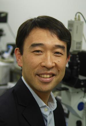 Tetsuya Higashiyama (東山哲也)合作论文数Institute of Transformative Bio-Molecules, Nagoya University25
 Shuh-Ichi Nishikawa合作论文数Faculty of Science, University of Tokyo12
 Susaki Daichi合作论文数Graduate School of Science, Nagoya University11
 Toshiya Endo合作论文数Dept Mat & Biol Chem, Yamagata Univ9
 Tomokazu Kawashima合作论文数Austrian Academy of Sciences, Gregor Mendel Institute9
 Kazuki Motomura合作论文数Graduate School of Arts and Sciences, The University of Tokyo9
 Tetsu Kinoshita合作论文数Faculty of Science, Kobe University8Naoya Sugi合作论文数7
 Daisuke Kurihara合作论文数Graduate School of Science, Nagoya University;Higashiyama Live-Holonics Project, Japan Science and Technology Exploratory Research for Advanced Technology;Graduate School of Science, Nagoya University7
 Hidenori Takeuchi合作论文数Nagoya Univ, Grad Sch Sci, Div Biol Sci, Chikusa Ku, Aichi 4648602, Japan4
 Frédéric Berger合作论文数Gregor Mendel Institute, Austrian Academy of Sciences, Vienna BioCenter4Ryushiro D. Kasahara合作论文数4Yoshikatsu Sato合作论文数Graduate School of Science, Tokyo Metropolitan University4
 Masaya Yamamoto合作论文数3
 Atsushi Takeda合作论文数3Yuki Hamamura合作论文数3Mina Ohtsu合作论文数3Haruna Tsuchi合作论文数2Satsuki Nishimaki合作论文数2Yilin Zhang合作论文数2Takamasa Suzuki合作论文数Chubu Univ, Coll Biosci & Biotechnol, Kasugai, Aichi, Japan2Kentaro Tamura合作论文数Kyoto Univ, Grad Sch Sci, Dept Bot, Kyoto 6068502, Japan2
 Ueli Grossniklaus合作论文数Department of Plant and Microbial Biology, University of Zurich;Zurich-Basel Plant Science Center, University of Zurich2
 Masaru Ohme Takagi合作论文数National Institute of Advanced Industrial Science and Technology;Institute for Environmental Science and Technology, Saitama University2
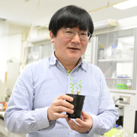 Masayoshi Kawaguchi (川口正代司)合作论文数Department of Basic Biology, School of Life Science, The Graduate University for Advanced Studies;National Institute for Basic Biology2
 Yelagandula Ramesh合作论文数Austrian Acad Sci, Vienna Bioctr VBC2Nagahara Shiori合作论文数2
 David Twell合作论文数UiT The Arctic University of Norway;Department of Biological Sciences, Korea Advanced Institute of Science and Technology;Department of Experimental Botany, Radboud University;Plant Gene Expression Center, University of California, Berkeley;French National Centre for Scientific Research;Universitat Zurich;Department of Genetics, University of Leicester;Charles University in Prague;Cardiff University;Temasek Life Sciences Laboratory2
 Minako Ueda合作论文数2Chieko Goto合作论文数2
 Takao Oi (大井崇生)合作论文数Department of Plant Production Sciences, Graduate School of Bioagricultural Sciences, Nagoya University2
 Mengxiang Sun (孙蒙祥)合作论文数College of Life Sciences, Wuhan University2
 Nobutaka Mitsuda合作论文数CREST, Japan Science and Technology Agency2Hironori Takasaki合作论文数National Institute of Crop Science2
 Michitaka Notaguchi合作论文数Institute for Advanced Research, Nagoya University2
 Takuya Suzaki合作论文数Heidelberg Institute of Zoology, University of Heidelberg2Tomoko Igawa合作论文数2
 Arp Schnittger合作论文数Zentrum für Molekularbiologie der Pflanzen;Universität Tübingen;Zentrum für Molekularbiologie der Pflanzen, Universität Tübingen2
 Ikuko Hara-Nishimura合作论文数Kyoto University;Konan University;Nara National Institute of Higher Education and Research;Nara Institute of Science and Technology;Kanagawa University2Xin'Ai Zhao合作论文数1yu jin kim合作论文数1John E. Fowler合作论文数1Yuki Yamaguchi合作论文数1Yonsheng Guan合作论文数1Reika Hasegawa合作论文数1Gaëtan Pochon合作论文数1Andrea R M Calhau合作论文数1Takashi Sakakibara合作论文数1Chihiro Sato合作论文数1




