Abstract INTRODUCTION: Liver disease occurs on a continuum from steatosis to fibrosis, cirrhosis and ultimately hepatocellular carcinoma (HCC), with a 30% lifetime risk of HCC among those with cirrhosis (LCr). If identified early, steatosis and fibrosis are potentially reversible, and in LCr, surveillance can reduce cancer morbidity and mortality. Despite these benefits, conventional LCr detection modalities are invasive or have limited performance. We previously demonstrated that cost-effective liquid biopsies of genome-wide cell-free DNA (cfDNA) fragmentomes enable early detection of HCC. Here, we use these technologies to detect liver steatosis, fibrosis, and cirrhosis towards improved pre-cancer intervention and HCC surveillance. METHODS: We performed low-coverage, whole genome sequencing of plasma cfDNA from separate Discovery (n=423) and Validation (n=221) cohorts including individuals with no known liver disease (n=397), chronic liver disease and early fibrosis (n=91) including viral hepatitis and metabolic associated steatotic liver disease, or advanced fibrosis/cirrhosis (n=156). We computed genome-wide fragment length, coverage, and repeat element features (DELFI and ARTEMIS), cross-validated a machine learning classifier for fibrosis and LCr detection in the Discovery Cohort and evaluated the locked model in the Validation Cohort. We then performed whole methylome sequencing (n=28) and cell-type deconvolution to reveal mechanisms of change to cfDNA fragmentomes in LCr. RESULTS: Individuals with early liver disease/fibrosis and advanced fibrosis/cirrhosis were detected with high performance (AUC=0.90, 95% CI=0.86-0.95 and AUC=0.95, 95% CI=0.93-0.98, respectively) in the Discovery Cohort. At an 80% specificity locked cutpoint, Validation Cohort sensitivity was 70.8% (90% CI=52.3%-87.5%) for early liver disease/fibrosis and 90.1% (90% CI=84.4%-94.4%) for advanced fibrosis/cirrhosis. The model displayed low cross-reactivity for other fibrotic origin conditions including benign lung nodules or chronic pancreatitis (median scores 0.087 and 0.068 respectively vs. 0.55 for LCr, p<0.0002). The approach outperformed the existing fibrosis index FIB-4, detecting 5.07x (95% CI=3.03-17.35) and 1.2x (95% CI=1.18-1.32) more cases of early liver disease/fibrosis and advanced fibrosis/cirrhosis in simulations. cfDNA methylome deconvolution revealed increased contributions of liver endothelium (p=0.00016) and blood monocytes (p=5.2x10⁻⁵) and decreased contribution of hepatocytes (p=0.00035) with shorter fragment lengths in LCr. CONCLUSIONS: A cfDNA fragmentome biomarker enabled early detection of liver disease including LCr and reflected both liver-derived and immune-cell related changes. These analyses may enable accessible early detection of pre-cancer conditions with potential to improve liver disease management and early detection of HCC. Citation Format: Akshaya Vijaya Annapragada, Zachariah Foda, Hope Orjuela, Carter Norton, Shashi Koul, Noushin Niknafs, Sarah Short, Keerti Boyapati, Adrianna Bartolomucci, Dimitrios Mathios, Michael Noe, Chris Cherry, Jacob Carey, Alessandro Leal, Bryan Chesnick, Nic Dracopoli, Jamie Medina, Nicholas Vulpescu, Daniel Bruhm, Sarah Bacus, Vilmos Adleff, Amy Kim, Steve Baylin, Greg Kirk, Andrei Sorop, Razvan Iacob, Speranta Iacob, Liana Gheorghe, Simona Dima, Katherine McGlynn, Manuel Ramirez-Zea, Claus Feltoft, Julia Johansen, John Groopman, Jillian Phallen, Rob Scharpf, Victor Velculescu. Non-invasive early detection of cancer-predisposing liver diseases using genome-wide cfDNA fragmentomes [abstract]. In: Proceedings of the American Association for Cancer Research Annual Meeting 2026; Part 1 (Regular Abstracts); 2026 Apr 17-22; San Diego, CA. Philadelphia (PA): AACR; Cancer Res 2026;86(7 Suppl):Abstract nr 4074.
Supplementary Table S1. Summary of clinical information. Supplementary Table S2. Summary of sample characteristics. Supplementary Table S3. Summary of whole-genome sequencing and fragment analyses. Supplementary Table S4. Summary of protein analyses. Supplementary Table S5. Summary of machine learning models and scores. Supplementary Table S6. Summary of performance for ovarian cancer detection.
Supplementary Figure S1. Evaluation of screening model DELFI-Pro scores and comorbidities in individuals without cancer. Supplementary Figure S2. DELFI-Pro score evaluation in available clinical characteristics of patients with cancer. Supplementary Figure S3. Stability analysis across fold repeats and collection source. Supplementary Figure S4. Detection of ovarian cancer subtypes using DELFI-Pro screening model. Supplementary Figure S5. ROC analyses of asymptomatic individuals in the using screening or diagnostic models in the Discovery Cohort. Supplementary Figure S6. Performance of ichorCNA and median cfDNA fragment lengths in the Discovery Cohorts. Supplementary Figure S7. Detection of ovarian cancer subtypes using DELFI-Pro at high specificity. Supplementary Figure S8. Genome-wide fragmentation profiles are altered in patients in the Validation Cohort with ovarian cancer. Supplementary Figure S9. Analyses of chromosomal changes in Discovery and Validation cohorts. Supplementary Figure S10. Performance of DELFI-Pro for detection of ovarian cancer in the Validation Cohort. Supplementary Figure S11. Correlation of rank ordered DELFI-Pro scores for the Screening and Diagnostic models. Supplementary Figure S12. Assessment of DELFI-Pro in women with benign lesions. Supplementary Figure S13. Performance of DELFI-Pro for distinguishing ovarian cancer from benign masses. Supplementary Figure S14. Performance of DELFI-Pro for distinguishing ovarian cancer subtypes from benign masses. Supplementary Figure S15. Performance of DELFI-Pro for distinguishing ovarian cancer subtypes from benign masses in Validation Cohort. Supplementary Figure S16. Evaluation of overall tumor burden using the sum of reported lesion diameters. Supplementary Figure S17. Evaluation of CA1-25 and HE4 blood concentrations measured at different centers.
Survival analyses of patients with molecularly defined untreated GBM using ARTEMIS–DELFI model components. A, The horizontal axis indicates the classifier score cutoff used to dichotomize the GBM patient cohort to low- and high-score categories. The vertical axis indicates the P value of the univariate survival analysis of the Cox proportional hazard ratios for each ARTEMIS–DELFI submodel at all evaluated score cutoffs. Each submodel of the ARTEMIS–DELFI model is represented with a different color. Kaplan–Meier survival analyses of the GBM cohort for the chromosomal arm model (B) and the epigenetic model (C) both at a score cutoff of 0.75. P values are calculated based on the log-rank test, and the estimated HR and 95% CI are shown. TE, transposable element, SINE, short interspersed nuclear elements, LINE, long interspersed nuclear elements.
Extracranial metastasis of IDH-wildtype glioblastoma is very rare and poorly understood at the molecular level. We report a case of FGFR3::TACC3 fusion IDH-wildtype glioblastoma in a 61-year-old male, whose preoperative blood sample showed highly aberrant cfDNA fragmentation patterns, which could be suggestive of early systemic dissemination, undetected by standard-of-care imaging of his body. Eleven months post-resection and adjuvant therapy, he developed widespread extracranial metastases. Comprehensive molecular profiling of matched primary and metastatic tumors revealed broadly conserved genomic, transcriptomic, and copy number landscapes, with the metastasis harboring an additional ERCC6 deletion and enriched expression of receptor tyrosine kinase signaling genes. These findings provide rare insight into the genetic continuity and evolution underlying IDH-wildtype glioblastoma metastasis.
80% of adults in the US have metabolic risk factors for Liver Cirrhosis (LCr), but LCr diagnosis is challenging. Elastography and blood-based fibrosis indices have limited performance, and biopsies are invasive. The lifetime risk of hepatocellular carcinoma (HCC) in individuals with LCr is ∼30%, yet <20% of individuals undergo any HCC surveillance. We previously demonstrated that genome-wide cell-free DNA (cfDNA) fragmentomes can detect HCC in the blood. Here, we expand these approaches to pre-neoplasia, for LCr detection to facilitate management and HCC surveillance. We evaluated cfDNA fragmentomes in separate Discovery (n=465) and External Validation (n=279) Cohorts. These cohorts comprised individuals with LCr (n=132), at high-risk for LCr with viral hepatitis (n=26), metabolic associated steatotic liver disease (MASLD) and/or non-cirrhotic fibrosis (n=44), or aflatoxin exposure (n=10), or from healthy screening populations (n=532, including 126 with metabolic risk factors). For all individuals, we extracted cfDNA from plasma, performed low coverage (1-2x) whole genome sequencing, and computed genome-wide fragment length, coverage and repeat element features (DELFI and ARTEMIS). We cross-validated a machine learning model with these features for detection of LCr in the Discovery Cohort and evaluated the locked model in the Validation Cohort. In the Discovery Cohort, individuals with LCr were detected with high performance (AUC=0.97, 95% CI 0.94-1.0 and AUC=0.95, 95% CI=0.92-0.98, for individuals with and without metabolic risk factors). Scores were higher in LCr than in healthy populations and increased with cirrhosis severity (p<3.2x10⁻⁶ for Child-Pugh A, B and C). In the Validation Cohort, the locked model achieved 78% sensitivity and 92% specificity when locked at a threshold of 90% specificity and 90% sensitivity in the Discovery Cohort (AUC=0.95, 95% CI=0.91-0.99, and AUC=0.93, 95% CI=0.89-0.97, for individuals with and without metabolic risk factors), outperforming common fibrosis indices APRI and FIB-4. Among high-risk individuals without LCr but with aflatoxin exposure, MASLD, or fibrosis, scores were higher than in healthy individuals (p<6.0x10⁻¹⁶), but remained lower than for individuals with LCr (p<2.2x10⁻¹⁶). Fragmentomic analyses of transcription factor binding sites and single nucleotide variants revealed molecular alterations linked to both liver-tissue derived and inflammatory changes of cirrhosis. cfDNA fragmentomes enable detection of LCr, a pre-cancer condition that increases HCC risk. HCC surveillance in high-risk populations is critical, but accessibility and adherence remain low. A facile, effective screening approach for LCr may enable early identification towards improved management and initiation of HCC surveillance. Akshaya V. Annapragada, Zachariah H. Foda, Noushin Niknafs, Sarah Short, Dimitrios Mathios, Shashikant Koul, Keerti Boyapati, Adrianna Bartolomucci, Jamie E. Medina, Nicholas A. Vulpescu, Chris Cherry, Daniel C. Bruhm, Vilmos Adleff, Amy Kim, Andrei Sorop, Razvan Iacob, Speranta Iacob, Liana Gheorghe, Simona Dima, Katherine A. McGlynn, Manuel Ramirez-Zea, John Groopman, Jillian Phallen, Robert B. Scharpf, Victor E. Velculescu. Cell-free DNA fragmentomes enable early identification of liver cirrhosis to facilitate cancer surveillance [abstract]. In: Proceedings of the American Association for Cancer Research Annual Meeting 2025; Part 1 (Regular Abstracts); 2025 Apr 25-30; Chicago, IL. Philadelphia (PA): AACR; Cancer Res 2025;85(8_Suppl_1):Abstract nr 6426.
Abstract Ovarian cancer is a leading cause of death for women worldwide, in part due to ineffective screening methods. In this study, we used whole-genome cell-free DNA (cfDNA) fragmentome and protein biomarker [cancer antigen 125 (CA-125) and human epididymis protein 4 (HE4)] analyses to evaluate 591 women with ovarian cancer, with benign adnexal masses, or without ovarian lesions. Using a machine learning model with the combined features, we detected ovarian cancer with specificity >99% and sensitivities of 72%, 69%, 87%, and 100% for stages I to IV, respectively. At the same specificity, CA-125 alone detected 34%, 62%, 63%, and 100%, and HE4 alone detected 28%, 27%, 67%, and 100% of ovarian cancers for stages I to IV, respectively. Our approach differentiated benign masses from ovarian cancers with high accuracy (AUC = 0.88, 95% confidence interval, 0.83–0.92). These results were validated in an independent population. These findings show that integrated cfDNA fragmentome and protein analyses detect ovarian cancers with high performance, enabling a new accessible approach for noninvasive ovarian cancer screening and diagnostic evaluation. Significance: There is an unmet need for effective ovarian cancer screening and diagnostic approaches that enable earlier-stage cancer detection and increased overall survival. We have developed a high-performing accessible approach that evaluates cfDNA fragmentomes and protein biomarkers to detect ovarian cancer.
Delayed diagnosis of brain tumors is common and negatively impacts patient outcomes. A noninvasive, blood-based assay for early detection may accelerate clinical intervention and improve prognosis. This study explores genome-wide cell-free DNA (cfDNA) fragmentome profiling for the detection of brain tumors, focusing on gliomas. Genome-wide cfDNA fragmentation profiles and repeat element landscapes were analyzed from plasma samples of individuals with brain tumors (n = 148) and controls without cancer (n = 357). A machine learning classifier was trained on cfDNA fragmentome features to distinguish tumor-bearing individuals and validated in an independent, prospectively collected cohort. To elucidate the cellular origins of cfDNA signals, we developed a statistical framework that integrates cfDNA coverage at transcription factor binding sites (TFBS) with pan-tissue and pan-cancer transcription factor expression profiles. The classifier demonstrated robust performance in detecting brain tumors across all glioma grades, achieving an area under the ROC curve (AUC) of 0.90 (95% CI, 0.87–0.93). Fragmentation signatures reflected contributions from both tumor-derived cfDNA and immunologically relevant leukocyte populations, consistent with the immunosuppressive environment of brain tumors. These cfDNA features were reproducible and stable across cohorts. Plasma cfDNA fragmentome analysis enables accurate, noninvasive detection of gliomas and reveals reproducible fragmentation changes associated with brain malignancy. These findings support the potential clinical utility of cfDNA fragmentomics as a liquid biopsy platform for earlier diagnosis and disease monitoring in neuro-oncology.
Diagnostic delays in patients with brain cancer are common and can impact patient outcome. Development of a blood-based assay for detection of brain cancers could accelerate brain cancer diagnosis. In this study, we analyzed genome-wide cell-free (cfDNA) fragmentomes, including fragmentation profiles and repeat landscapes, from the plasma of individuals with (n = 148) or without (n = 357) brain cancer. Machine learning analyses of cfDNA fragmentome features detected brain cancer across all-grade gliomas (AUC = 0.90; 95% confidence interval, 0.87-0.93), and these results were validated in an independent prospectively collected cohort. cfDNA fragmentome changes in patients with gliomas represented a combination of fragmentation profiles from glioma cells and altered white blood cell populations in the circulation. These analyses reveal the properties of cfDNA in patients with brain cancer and open new avenues for noninvasive detection of these individuals. SIGNIFICANCE:Brain cancer is one of the deadliest and most challenging cancers to detect with liquid biopsy approaches in blood, hampering efforts for earlier noninvasive diagnosis. We have developed a machine learning genome-wide cfDNA fragmentation method that provides a sensitive and accessible approach for brain cancer detection.
Supplementary Table S1 shows Clinical information of US/EU patients analyzed. Supplementary Table S2 is a summary of whole genome cfDNA analyses. Supplementary Table S3 shows clinical information of Hong Kong patients analyzed. Supplementary Table S4 shows the lowest scoring transcription factors in US/EU cohort samples. Supplementary Table S5 lists the plasma volume and cfDNA amounts used in genomic libraries.
Supplementary Figure S1 shows contributions of individual genomic regions to the final trained DELFI surveillance model. Supplementary Figure S2 shows DELFI scores in individuals without cancer by age and sex. Supplementary Figure S3 shows DELFI scores in high-risk individuals without cancer across racial or ethnic groups. Supplementary Figure S4 shows DELFI scores in cirrhotic patients in relation to severity. Supplementary Figure S5 shows DELFI scores in individuals in relation to BMI. Supplementary Figure S6 Shows Performance of alternative DELFI models. Supplementary Figure S7 shows Correlation between rank ordered DELFI. Supplementary Figure S8 shows DELFI scores in patients with HCC were related to tumor characteristics. Supplementary Figure S9 shows DELFI scores in by etiology of underlying liver disease. Supplementary Figure S10 shows the DELFI score in patients with HCC is correlated to AFP levels. Supplementary Figure S11 shows the correlation of fragmentation profiles. Supplementary Figure S12 shows Genome wide fragmentation profiles in the Hong Kong validation cohort Supplementary Figure S13 shows chromosomal copy number in Hong Kong cohort. Supplementary Figure S14 shows the implementation of DELFI in liver cancer screening.
Meningiomas are tumors of the central nervous system that vary in their presentation, ranging from benign and slow-growing to highly aggressive. The standard method for diagnosing and classifying meningiomas involves invasive surgery and can fail to provide accurate prognostic information. Liquid biopsy methods, which exploit circulating tumor biomarkers such as DNA, extracellular vesicles, micro-RNA, proteins, and more, offer a non-invasive and dynamic approach for tumor classification, prognostication, and evaluating treatment response. Currently, a clinically approved liquid biopsy test for meningiomas does not exist. This review provides a discussion of current research and the challenges of implementing liquid biopsy techniques for advancing meningioma patient care.
Circulating cell-free DNA (cfDNA) is emerging as an avenue for cancer detection, but the characteristics of cfDNA fragmentation in the blood are poorly understood. We evaluate the effect of DNA methylation and gene expression on genome-wide cfDNA fragmentation through analysis of 969 individuals. cfDNA fragment ends more frequently contained CCs or CGs, and fragments ending with CGs or CCGs are enriched or depleted, respectively, at methylated CpG positions. Higher levels and larger sizes of cfDNA fragments are associated with CpG methylation and reduced gene expression. These effects are validated in mice with isogenic tumors with or without the mutant IDH1, and are associated with genome-wide changes in cfDNA fragmentation in patients with cancer. Tumor-related hypomethylation and increased gene expression are associated with decrease in cfDNA fragment size that may explain smaller cfDNA fragments in human cancers. These results provide a connection between epigenetic changes and cfDNA fragmentation with implications for disease detection. Cell free DNA fragmentation is a promising biomarker for disease, but its epigenetic regulation is incompletely understood. Here, the authors investigated the effects of DNA methylation in the production of cfDNA fragmentation, and corelate these changes with gene expression in human cancer.
Abstract Introduction: Liquid biopsies are promising noninvasive tools for cancer detection. Variation in cell-free DNA concentrations (cfDNA-conc) has been observed between individuals with and without cancer. We assessed cfDNA-conc in 2287 treatment-naive individuals including those without cancer, with benign or high-risk conditions, or with one of eight cancer types. Methods: Plasma (0.3 to 9.8 mL) was separated from whole blood collected in Streck or EDTA tubes from individuals with bile duct, breast, colorectal, gastric, ovarian, liver, lung, pancreatic, or metastatic cancer (n=23, 54, 28, 26, 264, 75, 180, 31, 22, respectively) as well as benign or high-risk conditions (n=550, 133, respectively) such as cirrhosis or hepatitis B or C virus infection (HBV/HCV), and without cancer (n=901). cfDNA was extracted using the Qiagen QIAamp Circulating Nucleic Acid Kit. Quality and quantity were assessed using the High Sensitivity DNA assay on the Agilent Bioanalyzer. Total amount (ng) of cfDNA from all nucleosomal peaks was evaluated per volume (mL) of plasma to assess significance across cohorts using unpaired two-sample Wilcoxon tests. Results: We observed that in individuals without cancer, cfDNA-conc increased with age (Pearson R2=0.59; 18-40yrs vs 41-55yrs, vs 56-65yrs, vs 66-85yrs; p=2.31E-09, 1.50E-12, 8.31E-17, respectively). No difference was observed between cfDNA-conc from EDTA and Streck plasma from the same individual (n=9, p=0.359, Wilcoxon signed-rank test two-tailed). Individuals with benign adnexal masses or cirrhosis had similar cfDNA-conc compared to those without cancer while individuals with benign lung lesions or HBV/HCV had significantly different cfDNA-conc (p=0.70, 0.35, 6.96E-04, 1.88E-09, respectively). These observations were not affected by age. Individuals with cancer had significantly higher cfDNA-conc compared to those without cancer (bile duct p=2.08E-14, breast p=4.44E-16, colorectal p=6.04E-10, gastric p=5.93E-04, ovarian p=9.99E-16, liver p=1.22E-15, lung p=0, pancreatic p=8.57E-03, metastatic p=1.18E-06). Ovarian cancer exhibited higher cfDNA-conc than benign adnexal masses which was more pronounced in later stage disease (I/II p=3.83E-06, III/IV p=8.18E-08). Compared to cirrhosis or HBV/HCV we observed a stepwise increase in cfDNA-conc in liver cancer with progressing stage (0/A p= 6.50E-04, B p= 5.69E-10, C p= 1.47E-08). Higher cfDNA-conc were also observed with higher stage of lung cancer compared to benign lung lesions (I/II p=4.99E-03, III/IV p=3.89E-06). Using cfDNA-conc (ng/mL) as a single feature, we distinguished individuals with and without cancer with an AUC of 0.72 (95% CI=0.70-0.75). Conclusions: In a cohort of 2287 individuals, we observed cfDNA-conc increases with age and cancer stage that varied across cancer types. As a classifier, cfDNA-conc can be predictive of cancer status, yet further work is needed to understand the contribution of other comorbidities. Citation Format: Sarah Short, Akshaya V. Annapragada, Jamie E. Medina, Zachariah H. Foda, Dimitrios Mathios, Carolyn Hruban, Elaine J. Chiao, Kavya Boyapati, Adrianna Bartolomucci, Keerti Boyapati, Vilmos Adleff, Robert B. Scharpf, Victor E. Velculescu, Jillian Phallen. Variation of cell-free DNA concentrations in liquid biopsies [abstract]. In: Proceedings of the American Association for Cancer Research Annual Meeting 2024; Part 1 (Regular Abstracts); 2024 Apr 5-10; San Diego, CA. Philadelphia (PA): AACR; Cancer Res 2024;84(6_Suppl):Abstract nr 982.
Abstract Introduction: Repeat sequences comprise >50% of the human genome and structural and epigenetic changes in these regions are implicated in cancer. However, no systematic analysis of the compendium of repeat sequences has ever been performed in human cancer or cell-free DNA (cfDNA), largely due to the inability to identify and quantify repeat sequences genome-wide. We describe here the first comprehensive analysis of genome-wide repeat landscapes in cancer and demonstrate their utility in cfDNA liquid biopsies. Methods: We developed ARTEMIS (Analysis of RepeaT EleMents in dISease) an alignment-free, genome-wide approach for analyzing repeat landscapes in short read sequencing. This approach uses a de novo search of short sequences (kmers) in the telomere to telomere (chm13) reference genome to identify 1.2 billion 24-mers uniquely defining 1280 individual repeat types occurring genome-wide across 57 subfamilies and 6 families. We analyzed ARTEMIS kmers in whole genome sequences of 525 matched tumor/normal pairs from breast, colorectal, liver, lung, ovarian, cervical, prostate, thyroid, head and neck, gastric, and bladder cancers in the Pan Cancer Analysis of Whole Genomes (PCAWG), and in low coverage (1-2x) whole genome sequences of 1450 cfDNA samples from individuals with and without 8 types of cancer. Results: Analysis of ARTEMIS kmer repeat landscapes in 525 PCAWG tumors identified changes in all 1280 repeat element types, including 820 novel elements not previously known to be altered in cancer. A median of 807 repeat elements (range 246-1280) were altered in each tumor compared to its matched normal. The majority of changes were in repeat elements not previously described as altered in tumorigenesis and were most frequently found within Satellites, LINEs and SINEs, though changes were also observed in LTRs, Transposable Elements, and RNA Elements. A cross-validated cfDNA model using repeat landscapes (ARTEMIS) and fragmentation features (DELFI) detected individuals in a diagnostic cohort (n=287) across all stages of lung cancer with high performance (AUC 0.91, 95% CI 0.88-0.95) and was externally validated in a separate population (n=513). The locked model generated scores that correlated with circulating tumor mutant allele fractions for patients (n=19) undergoing targeted lung cancer therapy (r=0.80, p<2.2e-16), and stratified progression-free survival (p<0.001). ARTEMIS repeat landscape analyses of cfDNA also detected liver cancer in a high-risk cohort (n=208) of individuals with cirrhosis or viral hepatitis (AUC 0.91, 95% CI 0.87-0.95), and identified tissue of origin among seven tumor types (n=423). Conclusions: ARTEMIS reveals genome-wide repeat landscapes in human cancer, including in 820 novel elements not previously known to be altered in tumorigenesis. These repeat landscapes that can now be described are evaluable in the circulation and provide an avenue for noninvasive detection and characterization of cancer. Citation Format: Akshaya Annapragada, Noushin Niknafs, James R. White, Daniel C. Bruhm, Christopher Cherry, Jamie E. Medina, Vilmos Adleff, Carolyn Hruban, Dimitrios Mathios, Zachariah H. Foda, Jillian Phallen, Robert B. Scharpf, Victor E. Velculescu. Genome-wide repeat landscapes in cancer and cell-free DNA [abstract]. In: Proceedings of the American Association for Cancer Research Annual Meeting 2024; Part 1 (Regular Abstracts); 2024 Apr 5-10; San Diego, CA. Philadelphia (PA): AACR; Cancer Res 2024;84(6_Suppl):Abstract nr 988.
Genetic changes in repetitive sequences are a hallmark of cancer and other diseases, but characterizing these has been challenging using standard sequencing approaches. We developed a de novo kmer finding approach, called ARTEMIS (Analysis of RepeaT EleMents in dISease), to identify repeat elements from whole-genome sequencing. Using this method, we analyzed 1.2 billion kmers in 2837 tissue and plasma samples from 1975 patients, including those with lung, breast, colorectal, ovarian, liver, gastric, head and neck, bladder, cervical, thyroid, or prostate cancer. We identified tumor-specific changes in these patients in 1280 repeat element types from the LINE, SINE, LTR, transposable element, and human satellite families. These included changes to known repeats and 820 elements that were not previously known to be altered in human cancer. Repeat elements were enriched in regions of driver genes, and their representation was altered by structural changes and epigenetic states. Machine learning analyses of genome-wide repeat landscapes and fragmentation profiles in cfDNA detected patients with early-stage lung or liver cancer in cross-validated and externally validated cohorts. In addition, these repeat landscapes could be used to noninvasively identify the tissue of origin of tumors. These analyses reveal widespread changes in repeat landscapes of human cancers and provide an approach for their detection and characterization that could benefit early detection and disease monitoring of patients with cancer.
Purpose/Objective(s) Biomarkers to assess response to standard chemoradiation following resection for high grade glioma (HGG) are imprecise. Assessments often require multiple MRIs months apart and fail to differentiate treatment response from progression, in a disease with median progression-free survival (PFS) around 7 months. We previously developed DELFI (DNA Evaluation of Fragments for Early Interception) to use cost-effective, low-coverage, whole-genome sequencing (WGS) and machine learning to evaluate millions of cfDNA fragments in the blood that can reflect cancer-related genomic and epigenomic changes. Here we analyze fragmentation in patients with HGG to identify noninvasive biomarkers of RT response and disease progression. Materials/Methods We enrolled 39 patients with primary HGG with 116 plasma liquid biopsies collected pre-surgery, pre-RT, 2, 4 and 6 weeks on-RT, and 1-month post-RT. We extracted cfDNA from 58 liquid biopsies (n = 17 patients, with n = 5, 16, 13, 8, 14 and 2 draws at the pre-surgery, pre-RT, 2, 4 and 6 weeks on-RT, and 1-month post-RT time points, respectively) and performed WGS of cfDNA fragments. We analyzed cfDNA fragmentation profiles by summarizing the ratio of short (100-150 bp) to long (151-220 bp) fragments in 5Mb bins genome-wide, and correlated profiles between timepoints for each patient. We estimated PFS as time between diagnosis and first indication of new treatment post-RT. Results Pre-surgery cfDNA concentrations trended higher than at pre-RT (mean 18.9 ng/mL vs. 9.7 ng/mL) and further decreased on-RT and post-RT (mean 5.9 and 5.6 ng/mL, respectively). Fragmentation profiles pre-RT were more correlated to on-RT profiles than to pre-surgery profiles (median correlation 0.94 vs. 0.88), possibly reflecting high pre-surgery tumor burden that falls after resection and adjuvant treatment. At four weeks on-RT, a higher correlation to the pre-RT fragmentation profile was associated with longer PFS (p = 0.01, median PFS 15.6 vs. 4.8 months for patients with correlations above and below the mean, respectively), suggesting that patients with fragmentomes resembling their lowest tumor burden profile survive longer before progression. Conclusion We provide early evidence that genome-wide cfDNA fragmentation profiles reflect relative HGG tumor burden, and that changes to fragmentation may capture early molecular signs of progression. These changes are detectable on-RT and post-RT and are associated with PFS in as few as four weeks on-RT. This interim analysis demonstrates the utility of cfDNA fragmentomes for noninvasive monitoring of treatment response in patients with HGG. We are continuing patient accrual, sample collection and sequencing (goal n = 100 patients enrolled) with the objective of identifying response biomarkers to support real time treatment modification.
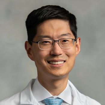 Michael Lim合作论文数School of Medicine, Johns Hopkins University;School of Medicine, Stanford University45
 Jillian Phallen合作论文数36
 Christina Jackson合作论文数Dept Neurosurg, Johns Hopkins Univ28
 Henry Brem合作论文数Department of Neurosurgery, School of Medicine, The Johns Hopkins University24
 Drew Mark Pardoll合作论文数Department of Oncology, School of Medicine, Johns Hopkins University;Bloomberg~Kimmel Institute for Cancer Immunotherapy, School of Medicine, Johns Hopkins University;Sidney Kimmel Comprehensive Cancer Center, School of Medicine, Johns Hopkins University23
 Victor E. Velculescu合作论文数Department of Genetic Medicine, School of Medicine, Johns Hopkins University;Delfi Diagnostics21
 Noushin Niknafs合作论文数Johns Hopkins Univ, Dept Biomed Engn, Baltimore, MD 21218 USA19
 Jamie Medina合作论文数18
 Betty Tyler合作论文数Departments of Neurology and Neurosurgery, School of Medicine, Biomedical Engineering18Vilmos Adleff合作论文数18
 Debebe Theodros合作论文数Department of Neurosurgery, School of Medicine, Stanford University17Daniel C Bruhm合作论文数Johns Hopkins Univ, Sch Med, Sidney Kimmel Comprehens Canc Ctr, Baltimore, MD 21205 USA16
 Annapragada Akshaya合作论文数16
 Xiaobu Ye合作论文数Departments of Neurology and Neurosurgery, School of Medicine, Biomedical Engineering15Stephen Cristiano合作论文数Johns Hopkins Univ, Sch Med, Sidney Kimmel Comprehens Canc Ctr, Baltimore, MD 21205 USA13Foda Zachariah H合作论文数Department of Pharmacological Sciences, Stony Brook University13Eileen Kim合作论文数12Robert Scharpf合作论文数Biostatistics, Bloomberg School of Public Health, Johns Hopkins University12Garzon-Muvdi Tomas合作论文数Department of †Neurosurgery;‡Oncology Center-Chemical Therapeutics, The Johns Hopkins University School of Medicine;§Technion Faculty of Medicine, Technion-Israel Institute of Technology;Technion-Israel Institute of Technology, The Johns Hopkins University School of Medicine12Zineb Belcaid合作论文数Department of Neurosurgery, Brain Tumor Center, Erasmus University Medical Center12
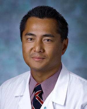 Phuoc T. Tran合作论文数Department of Radiation Oncology, School of Medicine, University of Maryland, Baltimore11
 Peter Burger合作论文数Division of Neuropathology, Department of Pathology, School of Medicine, The Johns Hopkins University11
 Charles Drake合作论文数Herbert Irving Comprehensive Cancer Center, Columbia University;The Janssen Pharmaceutical Companies of Johnson & Johnson11
 Jennifer Kim合作论文数Department of Neurology, Yale School of Medicine, Yale University10
 Chul-Kee Park合作论文数Department of Neurosurgery, College of Medicine, Seoul National University;Brain Tumor Center, Seoul National University Hospital10
 Bettegowda Chetan合作论文数The Johns Hopkins University9Antonella Mangraviti合作论文数Sant'Andrea University Hospital;Department of Neuroscience, Mental Health and Sense Organs, Sapienza University of Rome9Alessandro Olivi合作论文数8
 Michael Noe合作论文数Johns Hopkins Sch Med, Baltimore, MD USA8Carolyn Hruban合作论文数8
 Ernest Jan Bobeff合作论文数8Robert B. Scharpf合作论文数Department of Otolaryngology-Head and Neck Surgery, School of Medicine, Johns Hopkins University8Taube Janis M合作论文数8
 Patel Mira A合作论文数Johns Hopkins Hospital, Johns Hopkins University School of Medicine8Esteban Velarde合作论文数School of Medicine, Johns Hopkins University8
 Theodore H. Schwartz合作论文数Minimally Invasive Surgery, Weill Cornell Medicine, Cornell University;Neurological Surgery, Weill Cornell Medicine, Cornell University8Haiying Xu合作论文数8Allison M. Martin合作论文数7Sarah Harris-Bookman合作论文数7
 Nicholas A Vulpescu合作论文数Sch Med, Johns Hopkins Univ7
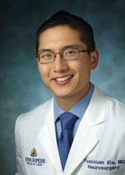 Yuanxuan Xia合作论文数Dept Neurosurg, Johns Hopkins Univ7
 Stephen B. Baylin合作论文数Johns Hopkins School of Medicine;Cancer Genetics and Epigenetics, Johns Hopkins University7
 Amy K Kim合作论文数Gastroenterology and Hepatology, Department of Medicine, Johns Hopkins University7
 Vijay K. Anand合作论文数Department of Otorhinolaryngology, Weill Cornell Medical College7
 Eric Sankey合作论文数Department of Neurosurgery, The Johns Hopkins University School of Medicine6Liu Ann合作论文数6Tam Ada合作论文数6Sarah Nicholas合作论文数5
 Pavan P Shah合作论文数Sch Med, Johns Hopkins Univ5




