Supplemental Figure S1. Comparison of clonogenic survival fractions of various cancer cell lines after treatment with SAR302503 and Ruxolitinib. Supplemental Figure S2. KRAS mutation by treatment sensitivities in NSCLC cell lines. Supplementary Figure S3. The response of NCIH1944 xenografts to ionizing radiation. Supplemental Figure S4. Top-ranked pathways in SAR sensitive NSCLC cell lines. Supplemental Figure S5. Cancer growth and proliferation gene network in SAR-sensitive NSCLC. Supplemental Figure S6. ROC analysis of SAR sensitivity by TSP-IRDS scores. Supplemental Figure S7. Distribution of TSP-IRDS scores in clinical NSCLCs. Supplemental Figure S8. TSP-IRDS predicts survival in clinical NSCLC. Supplemental Figure S9. TSP-IRDS scores predict the benefit of adjuvant cisplatin-based chemotherapy in clinical NSCLC. Supplementary Figure and Table Legends
Supplementary Table 1. Clonogenic survival of NSCLC cell lines after cytotoxic therapy.
Metastases are the cause of the vast majority of cancer deaths. In the metastatic process, cells migrate to the vasculature, intravasate, extravasate, and establish metastatic colonies. This pattern of spread requires the cancer cells to change shape and to navigate tissue barriers. Approaches that block this mechanical program represent new therapeutic avenues. We show that 4-hydroxyacetophenone (4-HAP) inhibits colon cancer cell adhesion, invasion, and migration in vitro and reduces the metastatic burden in an in vivo model of colon cancer metastasis to the liver. Treatment with 4-HAP activates nonmuscle myosin-2C (NM2C) (MYH14) to alter actin organization, inhibiting the mechanical program of metastasis. We identify NM2C as a specific therapeutic target. Pharmacological control of myosin isoforms is a promising approach to address metastatic disease, one that may be readily combined with other therapeutic strategies.
Metastases are the cause of the vast majority of cancer deaths. In the metastatic process, cells migrate to the vasculature, intravasate, extravasate, and establish new metastatic colonies. This pattern of spread requires the cancer cells to change shape and to navigate tissue barriers. Approaches that block this mechanical program represent new therapeutic avenues. We show that 4-hydroxyacetophenone (4-HAP) inhibits colon cancer cell adhesion, invasion, and migration in vitro, and reduces the metastatic burden in an in vivo model of colon cancer metastasis to the liver. Treatment with 4-HAP activates nonmuscle myosin-2C, increasing cortical tension and generating contractile, mixed-polarity arrangements of actin. These mechanical alterations overcome the cells ability to polarize, disseminate, and invade. Our results identify nonmuscle myosin-2C (MYH14) for the first time as a therapeutic target. Targeting actomyosin contractility is a promising approach to address metastatic disease, one that may be readily combined with other therapeutic strategies.
Abstract Lung cancer is the leading cause of cancer-related deaths worldwide. Approximately 85% of all lung cancers are non–small cell histology [non–small cell lung cancer (NSCLC)]. Modern treatment strategies for NSCLC target driver oncogenes and immune checkpoints. However, less than 15% of patients survive beyond 5 years. Here, we investigated the effects of SAR302503 (SAR), a selective JAK2 inhibitor, on NSCLC cell lines and tumors. We show that SAR is cytotoxic to NSCLC cells, which exhibit resistance to genotoxic therapies, such as ionizing radiation, cisplatin, and etoposide. We demonstrate that constitutive IFN-stimulated gene expression, including an IFN-related DNA damage resistance signature, predicts for sensitivity to SAR. Importantly, tumor cell–intrinsic expression of PD-L1 is IFN-inducible and abrogated by SAR. Taken together, these findings suggest potential dual roles for JAK2 inhibitors, both as a novel monotherapy in NSCLCs resistant to genotoxic therapies, and in tandem with immune checkpoint inhibition. Mol Cancer Ther; 17(4); 732–9. ©2018 AACR.
In the originally published version of this Article, the affiliation details for Kevin P. White inadvertently omitted 'Tempus Labs, Chicago, IL, 60654, USA'. This has now been corrected in both the PDF and HTML versions of the Article.
Multiple therapeutic agents are typically used in concert to effectively control metastatic tumors. Recently, we described microRNAs that are associated with the oligometastatic state, in which a limited number of metastatic tumors progress to more favorable outcomes. Here, we report the effective delivery of an oligometastatic microRNA (miR-655-3p) to colorectal liver metastases using nanoscale coordination polymers (NCPs). The NCPs demonstrated a targeted and prolonged distribution of microRNAs to metastatic liver tumors. Tumor-targeted microRNA miR-655-3p suppressed tumor growth when co-delivered with oxaliplatin, suggesting additive or synergistic interactions between microRNAs and platinum drugs. This is the first known example of systemically administered nanopartides delivering an oligometastatic microRNA to advanced metastatic liver tumors and demonstrating tumor-suppressive effects. Our results suggest a potential therapeutic strategy for metastatic liver disease by the co-delivery of microRNAs and conventional cytotoxic agents using tumor-specific NCPs.
Patients with a limited number of hepatic metastases and slow rates of progression can be successfully treated with local treatment approaches1,2. However, little is known about the heterogeneity of liver metastases, and animal models capable of evaluating the development of individual metastatic colonies are needed. Here, we present an advanced model of hepatic metastases that provides the ability to quantitatively visualize the development of individual tumor clones in the liver and estimate their growth kinetics and colonization efficiency. We generated a panel of monoclonal derivatives of HCT116 human colorectal cancer cells stably labeled with luciferase and tdTomato and possessing different growth properties. With a splenic injection followed by a splenectomy, the majority of these clones are able to generate hepatic metastases, but with different frequencies of colonization and varying growth rates. Using the In Vivo Imaging System (IVIS), it is possible to visualize and quantify metastasis development with in vivo luminescent and ex vivo fluorescent imaging. In addition, Diffuse Luminescent Imaging Tomography (DLIT) provides a 3D distribution of liver metastases in vivo. Ex vivo fluorescent imaging of harvested livers provides quantitative measurements of individual hepatic metastatic colonies, allowing for the evaluation of the frequency of liver colonization and the growth kinetics of metastases. Since the model is similar to clinically observed liver metastases, it can serve as a modality for detecting genes associated with liver metastasis and for testing potential ablative or adjuvant treatments for liver metastatic disease.
BACKGROUNDThe selection of patients for oligometastasis‐directed ablative therapy remains a challenge. The authors report on clinical and molecular predictors of survival from a stereotactic body radiotherapy (SBRT) dose‐escalation trial for oligometastases.METHODSPatients who had from 1 to 5 metastases, a life expectancy of >3 months, and a Karnofsky performance status of >60 received escalating SBRT doses to all known cancer sites. Time to progression, progression‐free survival, and overall survival (OS) were calculated at the completion of SBRT, and clinical predictors of OS were modeled. Primary tumor microRNA expression was analyzed to identify molecular predictors of OS.RESULTSSixty‐one evaluable patients were enrolled from 2004 to 2009. The median follow‐up was 2.3 years for all patients (range, 0.2‐9.3 years) and 6.8 years for survivors (range, 2.0‐9.3 years). The median, 2‐year, and 5‐year estimated OS were 2.4 years, 57%, and 32%, respectively. The rate of progression after SBRT was associated with an increased risk of death (hazard ratio [HR], 1.44; 95% confidence interval [CI], 1.24‐1.82). The time from initial cancer diagnosis to metastasis (HR, 0.98; 95% CI, 0.98‐0.99), the time from metastasis to SBRT (HR, 0.98; 95% CI, 0.98‐0.99), and breast cancer histology (HR, 0.12; 95% CI, 0.07‐0.37) were significant predictors of OS. In an exploratory analysis, a candidate classifier using expression levels of 3 microRNAs (miR‐23b, miR‐449a, and miR‐449b) predicted survival among 17 patients who had primary tumor microRNA expression data available.CONCLUSIONSA subset of oligometastatic patients achieves long‐term survival after metastasis‐directed SBRT. Clinical features and primary tumor microRNA expression profiling, if validated in an independent dataset, may help select oligometastatic patients most likely to benefit from metastasis‐directed therapy. Cancer 2016;122:2242–50. © 2016 American Cancer Society.
We present a model of hepatic colorectal metastases which represents monoclonal cell lines double-labeled by luciferase and tdTomato. These cells form liver metastasis in varying numbers and patterns similar to those observed in patients. Using in vivo and ex vivo luminescent and fluorescent imaging we determine the growth kinetics and clonogenic frequency of tumor cells colonizing liver. Molecular profiling detected stable expressional differences between clones consistent with their phenotypes. The data indicate that clinically relevant phenotypes of liver metastases can be modeled in vivo.
Colopericardial fistula after colonic interposition is a rare complication, with few prior reported cases. Management of such cases has usually consisted of resection of the colonic segment with cervical diversion. Here we present a case of successful primary repair of a colopericardial fistula in a 73-year-old woman who had initially undergone a colonic interposition graft 30 years before presentation. (C) 2015 by The Society of Thoracic Surgeons
 Ralph R. Weichselbaum合作论文数Department of Radiation and Cellular Oncology, The University of Chicago11
 Nikolai N. Khodarev合作论文数10
 Mitchell C. Posner合作论文数Department of Radiation Oncology, The University of Chicago9
 Sean Pitroda合作论文数Division of the Biological Sciences, The University of Chicago;PersonaDx8Xiaona Huang合作论文数6Abhineet Uppal合作论文数5
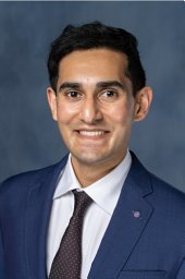 Akash D. Parekh合作论文数Department of Radiation and Cellular Oncology and Ludwig Center for Metastasis Research,5Suisui Song合作论文数4Lucy Chen合作论文数4
 Wightman Sean C合作论文数Division of Thoracic Surgery, Rush University Medical Center4Oshima Go合作论文数4
 Hua Liang合作论文数Department of Statistics, Columbian College of Arts & Sciences, The George Washington University4
 Paul Roach合作论文数School of Environmental & Life Sciences, University of Newcastle4Gene-Fu Liu合作论文数3Lai Xue合作论文数Univ Chicago Med, Dept Surg, Chicago, IL 60637 USA3
 Samuel Hellman合作论文数MacLean Center for Clinical Medical Ethics, University of Chicago;Bucksbuam Institute for Clinical Excellence;Department of Radiation and Cellular Oncology, University of Chicago3
 Darren Bryan合作论文数Department of Surgery, University of Chicago3Urszula Cichoń合作论文数2Katarzyna Krysztofiak合作论文数2
 Elizabeth Carla Poli合作论文数Univ Texas MD Anderson Canc Ctr2
 Douglas Robinson合作论文数Department of Cell Biology, School of Medicine, Johns Hopkins University2
 Dustin Thomas合作论文数2
 Schiffhauer Eric合作论文数2
 Skowron Kinga B合作论文数2
 Ronald S. Rock合作论文数Institute for Biophysical Dynamics, The University of Chicago2
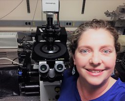 Alexandra Surcel合作论文数Department of Cell Biology, Johns Hopkins University School of Medicine2Jonathan J Oskvarek合作论文数1Lauren C Das合作论文数1
 Jorge Andrade合作论文数1
 Christopher Poon合作论文数University of Southern California1Nining Guo合作论文数Univ Chicago, Dept Chem, 929 E 57th St, Chicago, IL 60637 USA1Michael Beckett合作论文数Univ Chicago, Dept Radiat & Cellular Oncol, Ludwig Ctr Metastasis Res, Chicago, IL 60637 USA1Martin Forde合作论文数Univ Chicago, Inst Genom & Syst Biol, Chicago, IL 60637 USA1
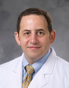 Joseph Kamel Salama合作论文数Department of Radiation Oncology, Duke University School of Medicine1
 Sajid A Khan合作论文数Yale Department of Surgery, Yale School of Medicine, Yale University1Jeremy Segal合作论文数Biological Sciences Division, The University of Chicago1
 Wickii T Vigneswaran合作论文数1Brown Miguel合作论文数1Wong Anthony合作论文数1
 Kadri Sabah合作论文数Lane Center for Computational Biology, Carnegie Mellon University1Son Christina H合作论文数1Watson Sydeaka合作论文数1
 Lei Huang (黄雷)合作论文数Zhiyuan College, Shanghai Jiao Tong University;Shanghai Jiao Tong University School of Medicine1Joseph Nora E合作论文数1
 Wenbin Lin (林文斌)合作论文数School of Science, Westlake University1
 Kevin White合作论文数Department of Biochemistry, Yong Loo Lin School of Medicine, National University of Singapore;A*STAR1
 Mark S. Talamonti合作论文数Department of Surgery, NorthShore University HealthSystem1
 Philip B. Paty合作论文数Memorial Sloan-Kettering Cancer Center1
 Karen L. Kaul合作论文数Evanston Hospital, Northwestern University Medical School1




