Regulatory T cells (Tregs), characterized by FOXP3 expression, are essential for maintaining immune homeostasis by controlling inflammation. However, in autoimmune diseases such as rheumatoid arthritis (RA), impaired Treg function contributes to immune dysregulation and disease pathology. While most studies of human Tregs have focused on blood, here we analyzed Tregs in synovial tissues from RA patients using single cell RNA sequencing (scRNAseq). We identified two predominant Treg states, CD25hiCXCR6pos Tregs with strong suppressive function, and CD25loAREGpos Tregs, a dysfunctional state exclusively enriched in synovial tissues but not in blood. Computational and in vitro analyses revealed that cortisol induced AREG expression, suppressed glycolysis, and impaired the suppressive function of CD25loAREGpos Tregs. In turn, AREG promoted an IL-33+ inflammatory phenotype in synovial fibroblasts. Importantly, we found that TNFR2 engagement can prevent or reverse this dysfunctional Treg state. In contrast to CD25loAREGpos Tregs, CD25hiCXCR6pos Tregs were highly suppressive, showed coordinated abundance with macrophages in synovial tissue, and functionally interacted with membrane-bound TNFα expressed by macrophages, which promoted their functional suppressive state. These two Treg subsets were similarly found in the synovial tissue in Juvenile Idiopathic Arthritis (JIA), another inflammatory arthritic disorder, indicating conserved mechanisms across arthritic diseases. Together, our findings define distinct pathways driving divergent functional and dysfunctional Treg states in inflamed tissues and point to interventions that may prevent or reverse the development of the dysfunctional state.
Treatment-refractory rheumatoid arthritis (RA) is a major unmet need, and the underlying mechanisms are poorly understood. To identify molecular determinants of refractory RA, we performed spatial transcriptomic profiling on synovial tissue biopsy samples taken 6 months before and after treatment. In the baseline biopsy samples of non-remitting patients, we identified increased fibrogenic signaling within vascular tissue niches, marked by high fibroblast COMP expression. We uncovered a role of endothelial-derived Notch signaling as an upstream regulator of fibroblast transforming growth factor beta (TGFβ) signaling via its opposing ability to induce TGFβ isoform expression while suppressing TGFβ receptors, generating a proximal-to-distal gradient of TGFβ sensitivity that can be altered with disruption of steady-state Notch signaling. In posttreatment biopsy samples, we observed significant immune depletion with expansion of fibrogenic niches, a process that can be reversed by inhibition of Notch and TGFβ signaling in RA patient-derived organoids. Collectively, our data implicate targeting of TGFβ signaling to prevent exuberant synovial tissue fibrosis as a potential therapeutic strategy for refractory RA. Wei et al. perform spatial transcriptomic profiling on synovial tissue biopsy samples from individuals with recent-onset rheumatoid arthritis, finding TGFβ signaling and fibrogenic fibroblast activation drive a treatment-refractory tissue phenotype.
Rheumatoid arthritis (RA) is a systemic autoimmune disease currently with no universally highly effective prevention strategies. Identifying pathogenic immune phenotypes in at-risk populations prior to clinical onset is crucial to establishing effective prevention strategies. Here, we applied multimodal single-cell technologies (mass cytometry and CITE-Seq) to characterize the immunophenotypes in blood from at-risk individuals (ARIs) identified through the presence of serum antibodies against citrullinated protein antigens (ACPAs) and/or first-degree relative (FDR) status, as compared with patients with established RA and people in a healthy control group. We identified significant cell expansions in ARIs compared with controls, including CCR2+CD4+ T cells, T peripheral helper (Tph) cells, type 1 T helper cells, and CXCR5+CD8+ T cells. We also found that CD15+ classical monocytes were specifically expanded in ACPA-negative FDRs, and an activated PAX5lo naive B cell population was expanded in ACPA-positive FDRs. Further, we uncovered the molecular phenotype of the CCR2+CD4+ T cells, expressing high levels of Th17- and Th22-related signature transcripts including CCR6, IL23R, KLRB1, CD96, and IL22. Our integrated study provides a promising approach to identify targets to improve prevention strategy development for RA.
Obesity worsens inflammatory arthritis severity, even in non-load-bearing joints, but the mechanism is unknown. Here, we show that there is an immunological mechanism mediated by T cells in adipose tissue. Using an antigen-induced arthritis model with trackable, arthritis-inducing CD8+ OT-I T cells, we found that OT-I T cells home to visceral adipose tissue (VAT) and expand there in the obese high-fat diet (HFD) context. Transplant of VAT from arthritic mice increased arthritis severity in naïve recipient mice and was ameliorated by CD8 T cell depletion. Bulk RNA sequencing identified pro-inflammatory changes to OT-I T cells in VAT characterized by increased IFN α and γ signaling after HFD. Intraperitoneal injection of IFNα, but not IFNγ, expanded CD8 T cell numbers in VAT. HFD-induced expansion of VAT CD8 T cells was ameliorated with global Ifnar1 deletion, and importantly, genetic deletion of Ifnar1 in T cells decreased arthritis severity in obese mice. These results provide a mechanistic explanation of how obesity worsens autoimmunity.
Granzymes are a family of serine proteases that are mainly expressed by CD8+ T cells, natural killer cells and innate-like lymphocytes1. Although their primary function is thought to be the induction of cell death in virally infected cells and tumours, accumulating evidence indicates that some granzymes can elicit inflammation by acting on extracellular substrates1. We previously found that most tissue CD8+ T cells in rheumatoid arthritis synovium, and in inflamed organs for some other diseases, express granzyme K (GZMK)2, a tryptase-like protease with poorly defined function. Here, we show that GZMK can activate the complement cascade by cleaving the C2 and C4 proteins. The nascent C4b and C2b fragments form a C3 convertase that cleaves C3, enabling the assembly of a C5 convertase that cleaves C5. The resulting convertases generate all the effector molecules of the complement cascade: the anaphylatoxins C3a and C5a, the opsonins C4b and C3b, and the membrane attack complex. In rheumatoid arthritis synovium, GZMK is enriched in regions with abundant complement activation, and fibroblasts are the main producers of complement proteins that serve as substrates for GZMK-mediated complement activation. Furthermore, Gzmk-deficient mice are significantly protected from inflammatory disease, exhibiting reduced arthritis and dermatitis, with concomitant decreases in complement activation. Our findings describe the discovery of a previously unidentified mechanism of complement activation that is driven entirely by lymphocyte-derived GZMK. Given the widespread abundance of GZMK-expressing T cells in tissues in chronic inflammatory diseases, GZMK-mediated complement activation is likely to be an important contributor to tissue inflammation in multiple disease contexts.
Rheumatoid arthritis (RA) is a chronic inflammatory disease where the synovial lining membrane undergoes pathological changes resulting in joint destruction. In healthy joints, the synovial lining is essential for joint homeostasis, forming a selective barrier and secreting lubricating molecules, yet the mechanisms that restores homeostatic synovial lining during RA remission remains poorly understood. Here, we applied spatial transcriptomics to examine biopsies of RA patients in remission to identify a mechanism that orchestrates a phenotypic switch specifying synovial quiescent lining fibroblast differentiation. Spatial transcriptomics revealed a proximity-sensing program where at low cell-density, fibroblasts adopt proliferative and fibrotic transcriptional state characterized by expression of MKI67 , COL1A2 and COL6A2 , whereas at high cell-density, fibroblasts induce a quiescent lining fibroblast transcriptional program characterized by PRG4 , CLU and PDPN . Mechanistically, fibroblasts sense spatial proximity through HB-EGF-EGFR signaling, which leads to phosphorylation of transcription factor CREB5. Perturbation of the EGFR-CREB5 axis abolishes fibroblast proximity-sensing and blocks synovial lining fibroblast differentiation. Conversely, EGFR activation by the ligand HB-EGF or pharmacologic activation of CREB5 is sufficient to induce synovial lining fibroblast differentiation. Together, our findings define a novel spatial proximity-sensing pathway underlying a return to homoeostatic fibroblast function during RA remission. By sensing their spatial proximity to neighboring fibroblasts, synovial fibroblasts translate these positional cues into signals that lead to restoration of normal, steady-state synovial lining membrane.
Once regarded as passive bystander cells of the tissue stroma, fibroblasts have emerged as active orchestrators of tissue homeostasis and disease. From regulating immunity and controlling tissue remodelling to governing cell growth and differentiation, fibroblasts assume myriad roles in guiding normal tissue development, maintenance and repair. By comparison, in chronic inflammatory diseases such as rheumatoid arthritis, fibroblasts recruit and sustain inflammatory leukocytes, become dominant producers of pro-inflammatory factors and catalyse tissue destruction. In other disease contexts, fibroblasts promote fibrosis and impair host control of cancer. Single-cell studies have uncovered striking transcriptional and functional heterogeneity exhibited by fibroblasts in both normal tissues and diseased tissues. In particular, advances in the understanding of fibroblast pathology in rheumatoid arthritis have shed light on pathogenic fibroblast states in other chronic diseases. The differentiation and activation of these fibroblast states is driven by diverse physical and chemical cues within the tissue microenvironment and by cell-intrinsic signalling and epigenetic mechanisms. These insights into fibroblast behaviour and regulation have illuminated therapeutic opportunities for the targeted deletion or modulation of pathogenic fibroblasts across many diseases. This Review provides a comprehensive overview of fibroblast biology in rheumatoid arthritis and other chronic inflammatory diseases. The authors discuss insights into fibroblast behaviour and pathogenicity from single-cell and functional studies and describe how these findings have informed efforts to therapeutically target fibroblasts.
Neurological injury drives most deaths and morbidity among patients hospitalized for out-of-hospital cardiac arrest (OHCA). Despite its clinical importance, there are no effective pharmacological therapies targeting post-cardiac arrest (CA) neurological injury. Here, we analyzed circulating immune cells from a large cohort of patients with OHCA, finding that lymphopenia independently associated with poor neurological outcomes. Single-cell RNA sequencing of immune cells showed that T cells with features of both innate T cells and natural killer (NK) cells were increased in patients with favorable neurological outcomes. We more specifically identified an early increase in circulating diverse NKT (dNKT) cells in a separate cohort of patients with OHCA who had good neurological outcomes. These cells harbored a diverse T cell receptor repertoire but were consistently specific for sulfatide antigen. In mice, we found that sulfatide-specific dNKT cells trafficked to the brain after CA and resuscitation. In the brains of mice lacking NKT cells (Cd1d-/-), we observed increased inflammatory chemokine and cytokine expression and accumulation of macrophages when compared with wild-type mice. Cd1d-/- mice also had increased neuronal injury, neurological dysfunction, and worse mortality after CA. To therapeutically enhance dNKT cell activity, we treated mice with sulfatide lipid after CA, showing that it improved neurological function. Together, these data show that sulfatide-specific dNKT cells are associated with good neurological outcomes after clinical OHCA and are neuroprotective in mice after CA. Strategies to enhance the number or function of dNKT cells may thus represent a treatment approach for CA.
Synovial tissue inflammation is the hallmark of rheumatoid arthritis (RA). Recent work has identified prominent pathogenic cell states in inflamed RA synovial tissue, such as T peripheral helper cells; however, the epigenetic regulation of these states has yet to be defined. We measured genome-wide open chromatin at single cell resolution from 30 synovial tissue samples, including 12 samples with transcriptional data in multimodal experiments. We identified 24 chromatin classes and predicted their associated transcription factors, including a CD8+ GZMK+ class associated with EOMES and a lining fibroblast class associated with AP-1. By integrating an RA tissue transcriptional atlas, we found that the chromatin classes represented 'superstates' corresponding to multiple transcriptional cell states. Finally, we demonstrated the utility of this RA tissue chromatin atlas through the associations between disease phenotypes and chromatin class abundance as well as the nomination of classes mediating the effects of putatively causal RA genetic variants.
Precision medicine in immune-mediated inflammatory diseases (IMIDs) requires an understanding of how cellular networks change following therapy. We describe a therapeutic atlas for Crohn’s disease (CD) and ulcerative colitis (UC) following anti-tumour necrosis factor (TNF) therapy. We generated ~1 million single-cell transcriptomes, organised into 109 cell states, from 216 gut biopsies from 38 patients and three controls, revealing disease- and therapy-specific differences. A systems-biology analysis identified distinct spatially-resolved cellular microenvironments: granuloma signatures in CD and interferon (IFN)-response signatures localising to T-cell aggregates and epithelial damage in CD and UC. Longitudinal comparisons demonstrated that disease progression in non-responders associated with myeloid and stromal cell perturbations in CD and increased multi-cellular IFN signalling in UC. IFN signalling was also observed in rheumatoid arthritis (RA) synovium with a lymphoid pathotype. Our therapeutic atlas informs drug positioning across IMIDs, and suggests a rationale for the use of janus kinase (JAK) inhibition following anti-TNF resistance.
We present a detailed analysis of myeloid cell populations found in the kidneys of lupus nephritis (LN) patients, based on the single-cell RNA-sequencing (scRNA-seq) data collected as part of the Accelerating Medicines Partnership (AMP) in RA/SLE consortium. Overall, 23,819 cells isolated from 156 LN patients and 30 healthy donors passed QC. Clustering of these cells (figure 1A) identified populations of CD14 and CD16 monocytes, two subsets of tissue-resident macrophages and several types of dendritic cells (DCs). In addition, we found several transcriptionally-distinct subsets of differentiated macrophages, that were missing from healthy donors (figure 1B- C). The ratio between the frequency of these macrophage subsets and that of infiltrating monocytes positively correlated with the Activity Index (AI) (figure 1D). To infer the origins of the observed disease-specific macrophages, we compared them to several published scRNA-seq datasets of blood and kidney samples, and performed in addition trajectory analysis. Our results suggested that these subsets likely originate from both infiltrating monocytes and tissue-resident macrophages (figure 2A). Furthermore, our analysis indicated that the differentiation into disease-specific macrophages mostly takes place within the kidney. To identify putative extracellular signals driving the differentiation of infiltrating CD16 monocytes into disease-related activation states, we performed in vitro experiments in which CD16 monocytes were stimulated with a wide array of cytokines and molecules suggested to play role in SLE pathology, such as immune complexes (ICs) and various types of cellular debris. We measured transcriptional changes associated with each in vitro condition, and utilized the generated data to identify enriched signatures in the AMP scRNA-seq data, using gene set enrichment analysis (GSEA). This analysis suggested that apoptotic cells likely promote differentiation of CD16 monocytes into a phagocytic state (cluster 11; figure 2B). In contrast, ICs containing TLR7 ligands, as well as IFNγ, were found to be plausible drivers of differentiation into an activation state that was characterized by high production of several proinflammatory cytokines and chemokines ('high producers' – clusters 12 and 13; figure 2C-D). Of note, our analysis suggested that through chemokine production, these 'high producers' may play a central role in recruiting and retaining the phagocytic macrophage subsets. The frequency of a single population of disease-specific macrophages positively correlated with both the AI and the Chronicity Index (CI; figure 3A-B). This population (cluster 17) was characterized by the upregulation of a set of genes associated with lipid metabolism. While previous studies have reported the presence of a similar macrophage subset in other tissues, this has not yet been demonstrated in kidneys. Furthermore, our analysis identified subclusters within this population, associated with different specific pathways, that were separately correlated with the AI and CI. In particular, we found a proinflammatory signature in these cells that was negatively correlated with the AI and positively correlated with the CI (figure 3C-D). A systemic differential expression analysis showed that several myeloid subsets modulated their gene expression in a manner correlated with the AI, compared to healthy donors; a particular clear response was observed in CD16 monocytes (cluster 2), in both proliferative/mixed and pure membranous LN (figure 4A-B). GSEA suggested that these changes were driven, at least in part, by type I and type II IFN, IL-6 and TNFβ. A conjoint analysis of changes in subset frequencies and of the differentially expressed (DE) genes in these populations and in glomerular endothelial cells pointed to the concurrent upregulation of molecules that may promote fibrosis, and in particular fibronectin in CD16 monocytes and integrins capable of binding it in glomerular endothelial cells (figure 4D-F); furthermore, several of the phagocytic macrophages derived from CD16 monocytes upregulated a set of genes regulating the extracellular matrix (figure 4G). Of note, these observations were found in LN patients that had a 0 glomerular CI (defined as the sum of glomerular subscores of the CI), suggesting that these molecular events precede fibrosis and may promote it. An increase in glomerular CI was associated with significant changes in gene expression, in particular in cDC2 and pDCs (clusters 4 & 15), as well as 2 specific populations of phagocytic macrophages (clusters 14 and 15; figure 4C); these changes included a decrease in the interferon response. We verified the reproducibility of these signatures in an independent set of LN patients. Taken together, our results shed light on the mechanisms of kidney inflammation in LN, and provide a detailed view of the different subsets and activation states of myeloid cells found in LN kidneys and the putative relations between them, as well as the extracellular signals giving rise to these states.
 Soumya Raychaudhuri合作论文数The Raychaudhuri Lab, Harvard Medical School, Harvard University;Center for Data Sciences, Brigham and Women’s Hospital;Broad Institute70
 Wei Kevin合作论文数Div Rheumatol Immunol & Allergy, Brigham & Womens Hosp56
 Deepak A Rao合作论文数Research Center for Genetic Medicine, Children's National Medical Center55
 Gerald F. Watts合作论文数Royal Perth Hospital;School of Medicine and Pharmacology, University of Western Australia39A. Helena Jonsson合作论文数38
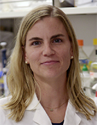 Laura Donlin合作论文数HSS Research Institute;Hospital for Special Surgery, Weill Cornell Medicine37
 Zhang Fan合作论文数Worcester Polytechnic Institute, University of Massachusetts35
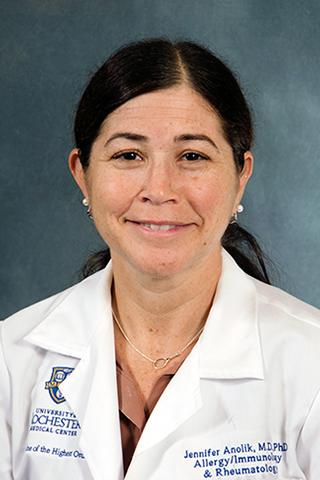 Jennifer Howitt Anolik合作论文数Anolik Lab, University of Rochester Medical Center32
 Ilya Korsunsky合作论文数Brigham & Womens Hosp, 75 Francis St, Boston, MA 02115 USA30
 Andrew Filer合作论文数Institute of Inflammation and Ageing, College of Medical and Dental Sciences, University of Birmingham30
 James A Lederer合作论文数Brigham and Women’s Hospital, Harvard Medical School27
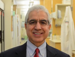 Hamid Band合作论文数Department of Pathology, Harvard Medical School26Ct Morita合作论文数25Sa Porcelli合作论文数24
 Susan M. Goodman合作论文数Department of Rheumatology, Hospital for Special Surgery;Weill Cornell Medical College24
 Robert L. Modlin合作论文数Department of Medicine, University of California, Los Angeles23Bykerk Vivian P合作论文数22
 Steven A. Porcelli合作论文数Department of Microbiology & Immunology, Albert Einstein College of Medicine;Department of Medicine, Albert Einstein College of Medicine21
 Gurdyal Besra合作论文数School of Biosciences, University of Birmingham21
 Samuel M Behar合作论文数Department Microbiology and Physiological Systems, University of Massachusetts Chan Medical School21Christopher C. Dascher合作论文数19M Sugita合作论文数18
 Larry Moreland合作论文数Division of Rheumatology, School of Medicine, University of Colorado18
 Lydia Lynch合作论文数17
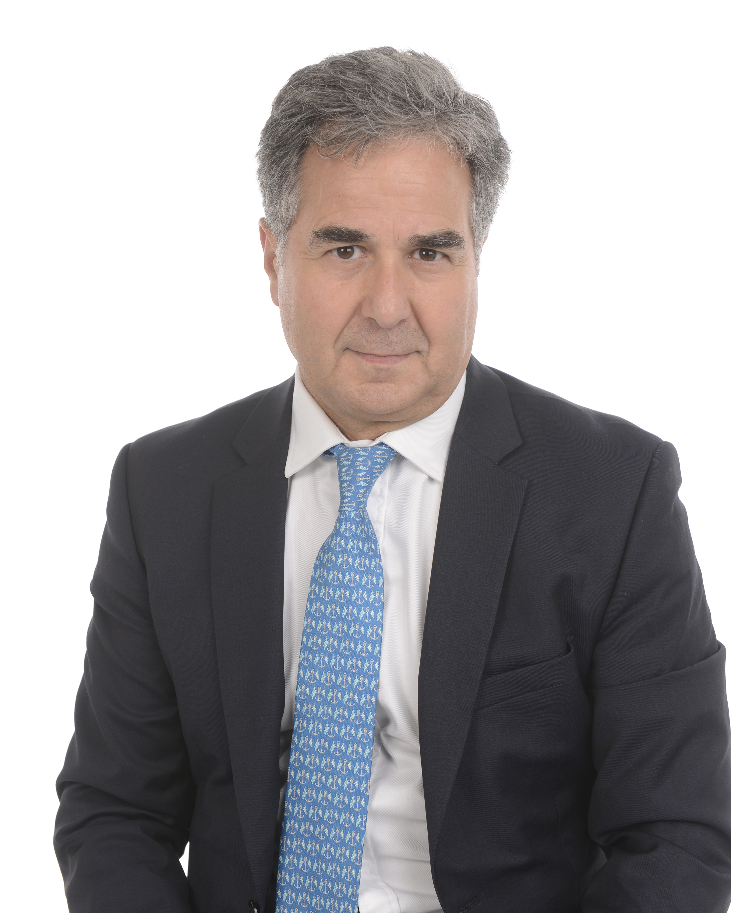 Costantino Pitzalis合作论文数William Harvey Research Institute, Barts and The London School of Medicine and Dentistry, Queen Mary University of London17
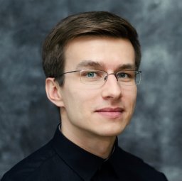 Slowikowski Kamil合作论文数Division of Rheumatology, Immunology, Allergy, Brigham and Women's Hospital and Harvard Medical School, Boston, MA, USA.17
 Christopher Buckley合作论文数Kennedy Institute of Rheumatology, University of Oxford;Department of Inflammation and Ageing, University of Birmingham;Institute of Inflammation and Ageing, College of Medical and Dental Sciences, University of Birmingham17
 Nir Hacohen合作论文数Center for Cancer Immunology, Massachusetts General Hospital;Mass General Research Institute;Broad Institute17
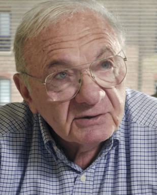 Jack Strominger合作论文数Department of Stem Cell and Regenerative Biology, Faculty of Arts and Sciences, Harvard University17
 Judith A. James合作论文数Oklahoma Medical Research Foundation;University of Oklahoma Health Sciences Center16
 D. Branch Moody合作论文数Faculty of Immunology, Harvard Medical School, Harvard University;Harvard Griffin Graduate School of Arts and Sciences, Harvard University;Department of Medicine, Brigham and Women's Hospital16
 V. Michael Holers合作论文数Department of Medicine-Rheumatology, School of Medicine, University of Colorado16
 Michael S. Krangel合作论文数Department of Immunology, School of Medicine, Duke University;Duke Cancer Institute15Shahnaz Shahinfar合作论文数15
 Ellen Gravallese合作论文数Division of Rheumatology, Brigham and Women's Hospital, Harvard Medical School15Jl Troy合作论文数14Parker C M合作论文数14
 Joel Guthridge合作论文数Oklahoma Medical Research Foundation14
 David A. Hodell合作论文数Godwin Laboratory for Palaeoclimate Research, Department of Earth Sciences, University of Cambridge14
 Stephen Anderson合作论文数Department of Molecular Biology and Biochemistry, School of Arts and Sciences, Rutgers–New Brunswick14Edward F. Dicarlo合作论文数Medical College, New York Hospital-Cornell University14Jenny E. Gumperz合作论文数14
 Barry R. Bloom合作论文数Department of Immunology and Infectious Diseases, Harvard T.H. Chan School of Public Health, Harvard University14
 Gary S. Firestein合作论文数Department of Medicine, School of Medicine, University of California13
 Jason H. Curtis合作论文数Department of Geology, University of Florida13Kevin D. Deane合作论文数Division of Rheumatology, University of Colorado Denver School of Medicine13William Apruzzese合作论文数Department of Orthopedics (WA, GSP),, Massachusetts General Hospital13BJ BALLERMANN合作论文数12
 Harris R Perlman合作论文数Rheumatology, Department of Medicine, Feinberg School of Medicine, Northwestern University12




