Supplementary Figure 7 contains dot plots showing KLK2 and STEAP1 IHC H-scores for AR+/NE-metastatic sites in the UW-RA cohort.
Supplementary Figure 12 contains additional RNA-seq analyses in AR+/NE-tumors from the UW-RA cohort and SU2C-IDT cohort.
Tier 1 druggable genome targets and their associated limma values from KLK2-high vs KLK2-low and STEAP1-high vs STEAP1-low differential expression analyses.
Differentially expressed genes from STEAP1-high versus STEAP1-low analysis in UW-TAN and ECDT cohorts.
Distribution of intra-tumoral and intra-patient hypergeometric heterogeneity indices for KLK2, STEAP1, and PSMA based on IHC H-scores across samples in the UW-RA cohort.
Supplementary Figure 9 contains donut plots showing the distribution of tumor expressing only one marker, both markers or neither marker for each KLK2-STEAP1, STEAP1-PSMA, and KLK2-PSMA pair across molecular phenotypes and across H-score thresholds.
Distribution of expression and co-expression between KLK2, STEAP1, and PSMA based on IHC H-scores across samples in the UW-RA cohort.
Supplementary Figure 10 shows UMAP projections of single-cell RNA-seq data from five additional AR+/NE-metastatic prostate cancer samples.
Gene set enrichment results from KLK2-high versus KLK2-low analysis in UW-TAN and ECDT.
Abstract Background: Metastatic castration-resistant prostate cancer (mCRPC) is a lethal disease with a major unmet clinical need to identify therapeutics that extend survival and biomarkers that predict treatment responses. The prostate-specific membrane antigen (PSMA) radioligand, 177Lu-PSMA-617 (177Lu-PSMA) is documented to increase overall survival (OS), but responses are highly variable. Analysis of cell-free DNA (cfDNA) from liquid biopsies offers a non-invasive window to profile tumor genomics and phenotypes, and identify markers predictive of 177Lu-PSMA outcomes. Methods: We interrogated a real-world prospective cohort of 140 patients with mCRPC meeting clinical guidelines for 177Lu-PSMA treatment. We analyzed pre-treatment and on-treatment circulating tumor DNA (ctDNA) by whole genome sequencing to quantitate ctDNA abundance and assess tumor-derived genomic features such as copy number alterations, somatic mutations, and structural rearrangements. We also employed a novel computational tool termed Proteus that uses ctDNA fragmentomic profiles to determine the expression of individual genes as well as expression programs that comprise a spectrum of relevant tumor phenotypes including mCRPC lineage subtypes, proliferation scores, and hypoxia signatures that may plausibly influence responses to 177Lu-PSMA. Tumor phenotype characteristics were integrated with PSMA-PET and SPECT imaging findings and evaluated for associations with clinical outcomes. Results: Pre-treatment ctDNA fraction was strongly associated of OS (multivariate HR=2.19, P=0.002), independent of prostate-specific antigen (PSA) response, and correlated positively with PSMA- and FDG-PET total tumor volume (R=0.59 and R=0.69, respectively). Poor responders were characterized by higher baseline ctDNA fraction, genomic instability, and overall tumor mutational burden. Deleterious alterations in the DNA repair gene ATM were associated with improved OS, whereas MYC amplifications were associated with poorer survival. Tumor phenotype features derived from ctDNA including cell cycle proliferation score, neuroendocrine differentiation, and complex copy number variation on chromosome 8 independently associated with clinical outcomes. Conclusions: Our findings demonstrate that baseline ctDNA analysis provides prognostic information for mCRPC patients undergoing 177Lu-PSMA therapy. ctDNA fraction and quantity, alongside specific genomic alterations and tumor phenotype features represent promising, non-invasively acquired biomarkers to guide patient selection, improve therapeutic monitoring, and further dissect genomic mechanisms of resistance to 177Lu-PSMA. Citation Format: Arthur P. McDeed, Robert Patton, Roman Gulati, Lukas Owens, Patricia Galipeau, Pooja Chandra, Aditya Pawar, Weston Hanson, Ruth Dumpit, Amir Iravini, Alireza Ghodsi, Delphine Chen, Michael T. Schweizer, Ruben Raychaudhuri, Michael Haffner, Gavin Ha, Peter S. Nelson. Prostate cancer phenotypes determined from circulating tumor DNA associate with outcomes to 177Lu-PSMA-617 therapy [abstract]. In: Proceedings of the American Association for Cancer Research Annual Meeting 2026; Part 2 (Late-Breaking, Clinical Trial, and Invited Abstracts); 2026 Apr 17-22; San Diego, CA. Philadelphia (PA): AACR; Cancer Res 2026;86(8_Suppl):Abstract nr LB006.
Metastatic prostate cancer (mPC) is characterized by molecular and phenotypic heterogeneity. With increasing guideline-driven use of metastatic biopsies, more mPC specimens are being evaluated in surgical pathology. However, unlike localized prostate cancer, no standardized framework currently exists to guide the diagnostic workup of metastatic biopsies or reliably determine phenotypic subtypes. While many mPCs retain conventional acinar features, a growing subset exhibits phenotypic plasticity - including loss of prostate epithelial identity and emergence of neuroendocrine or other divergent lineages. This phenotypic diversity often occurs in castration-resistant prostate cancer as a mechanism of resistance to chronic androgen receptor pathway inhibition and is characterized by genomic alterations and epigenetic reprogramming. This review outlines the histologic and molecular spectrum of mPC and proposes a practical, pathology-informed diagnostic approach integrating morphologic assessment and immunohistochemistry. Adoption of a standardized diagnostic framework and multidisciplinary integration will be useful for employing precision oncology in advanced mPC.
Distribution of KLK2 and STEAP1 single cell mRNA expression in cells from ARPC samples across MSK scRNA dataset.
Abstract Kallikrein 2 (KLK2) and six-transmembrane epithelial antigen of the prostate 1 (STEAP1) are two cell surface targets with relevance for prostate cancer therapy. The objective of this study was to characterize the expression landscape of KLK2 and STEAP1 in metastatic castration-resistant prostate cancer (mCRPC) and to define associated transcriptomic, genomic, and epigenomic features. We analyzed a total of 1,095 patient samples from three mCRPC cohorts, including in situ studies of rapid autopsy cases and patient-derived xenograft models. We found that KLK2 and STEAP1 expression is strongly enriched in androgen receptor (AR)–positive tumors and largely absent in neuroendocrine and double-negative phenotypes. Within AR+ tumors, pairwise comparisons revealed coexpression and high combined positivity rates for STEAP1, KLK2, and prostate-specific membrane antigen, suggesting that cotargeting any two of these antigens increases overall tumor coverage. Analysis of samples from a rapid autopsy cohort, which enabled assessment of intra- and intertumoral diversity, showed comparable degrees of expression heterogeneity for KLK2 and STEAP1. Antigen expression correlated positively with AR genomic alterations and serum prostate-specific antigen levels and negatively with RB1 and PTEN loss. Transcriptomic and epigenome analyses demonstrated distinct mechanisms governing antigen expression: KLK2 showed a strict AR dependence with coordinated AR/FOXA1/HOXB13 binding and enhancer activation, whereas STEAP1 was only partially AR-dependent and additionally regulated by locus-specific DNA methylation changes. Furthermore, KLK2 and STEAP1 expression states were associated with distinct transcriptional programs and immune microenvironmental features. Implications: These findings establish KLK2 and STEAP1 as key prostate adenocarcinoma-lineage antigens and provide critical insights to inform the rational design and clinical development of cell surface antigen–directed therapies in prostate cancer.
Results from predictive model integrating somatic alterations and serum PSA to estimate the probability that a sample is positive for the given antigen.
Supplementary Figure 11 shows additional KLK2 and STEAP1 IHC H-score boxplots stratified by alteration status.
PURPOSE:Treatment intensification with androgen receptor pathway inhibitors (ARPIs) has become the standard of care for patients with metastatic prostate cancer. However, there remains an unmet need to identify biomarkers for treatment resistance. Here, we identify SPEN inactivation as a driver of ARPI resistance. EXPERIMENTAL DESIGN:Pre-clinical studies were performed in LNCaP and VCaP cell lines. Data from a nationwide prostate cancer clinico-genomic database were extracted. Log-rank test and Cox proportional hazards models were used to compare time to next treatment (TTNT) on ARPI with/without SPEN mutations. SPEN immunohistochemistry was performed on a rapid autopsy metastatic tissue microarray. RESULTS:SPEN was identified as a top enzalutamide resistance hit in an unbiased genome-wide loss-of-function screen. SPEN inactivation results in upregulation of cell cycle proliferation and basal/stem cell activity as well as increased translation of pro-oncogenic genes. In a large patient cohort (N=6828), SPEN mutations are enriched following treatment with ARPIs (2.1% to 3.6%, p=0.001) and correlate with shorter TTNT on ARPI in patients with metastatic hormone-sensitive prostate cancer (6.4 vs 29.7 months, HR 2.67, p=0.02). In a metastatic rapid autopsy cohort (N=181), low SPEN H-score is associated with shorter time on abiraterone (5.0 vs 7.9 months, p=0.023) in metastatic castration-resistant prostate cancer. CONCLUSIONS:In real-world cohorts, loss of SPEN function across genomic, transcriptomic, and protein levels is associated with reduced benefit from ARPI therapy in metastatic prostate cancer. These findings identify SPEN inactivation as a clinically relevant biomarker of ARPI resistance that warrants prospective evaluation to guide treatment selection.
Supplementary Figure 2 shows representative IHC micrographs in benign and control samples.
 Evan Ya-Wen Yu合作论文数Clinical Research Division, Fred Hutchinson Cancer Center;Division of Hematology and Oncology, University of Washington School of Medicine;University of Washington Medical Center84
 Heather H. Cheng合作论文数Divisions of Human Biology,;Medicine, and;Divisions of Human Biology,;Medicine, and82
 Peter Nelson合作论文数Stuart and Molly Sloan Precision Oncology Institute, Fred Hutch Cancer Center;Human Biology Division, Fred Hutch Cancer Center;University of Washington School of Medicine62
 Petros Grivas合作论文数Department of Medicine, University of Washington59
 Todd Yezefski合作论文数48Montgomery Bruce合作论文数45
 Andrew Caleb合作论文数Department of Chemical and Biological Engineering, Princeton University43
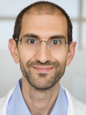 Emmanuel Antonarakis合作论文数Division of Hematology, Oncology, and Transplantation, Medical School, University of Minnesota;Masonic Cancer Center, University of Minnesota41
 Roman Gulati合作论文数Fred Hutchinson Cancer Research Center41
 Colin C. Pritchard合作论文数Department of Laboratory Medicine & Pathology, University of Washington39
 Michael C Haffner合作论文数Johns Hopkins Univ, Sidney Kimmel Comprehens Canc Ctr, Baltimore, MD USA37Montgomery Robert Bruce合作论文数36
 Lawrence D. True合作论文数Department of Laboratory Medicine & Pathology, School of Medicine, University of Washington;University of Washington Health Sciences Center34
 Schade George R合作论文数34
 Daniel W. Lin合作论文数Department of Urology, University of Washington33
 Jonathan Wright合作论文数Department of Urology, University of Washington31Funda Vakar-Lopez合作论文数30
 Ilsa Coleman合作论文数Fred Hutchinson Cancer Research Center29Ruben Raychaudhuri合作论文数28
 Jessica Hawley合作论文数Columbia University Irving Medical Center;Fred Hutchinson Cancer Center, University of Washington;Columbia University Irving Medical Center, University of Washington28
 Mario Alfredo Eisenberger合作论文数School of Medicine, Johns Hopkins University;Sibley Memorial Hospital27
 Eva Corey合作论文数Department of Urology, University of Washington27
 Colm Morrissey合作论文数Department of Urology, University of Washington26
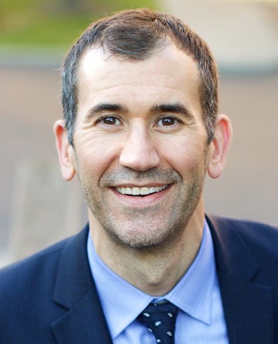 John Gore合作论文数Fred Hutchinson Cancer Center, University of Washington Medical Center;Department of Urology, American College of Surgeons, University of Washington;School of Medicine, University of Washington25
 John Lee合作论文数North Carolina State University24
 Sarah P Psutka合作论文数Harvard Medical School, Massachusetts General Hospital24
 Elahe Mostaghel合作论文数VA Puget Sound Health Care System;Medical Oncology Division, School of Medicine, University of Washington;Clinical Research Division, Fred Hutchinson Cancer Center22Peter S. Nelson合作论文数22Laura S Graham合作论文数21Sarah K. Holt合作论文数Fred Hutchinson Cancer Research Center21
 Navonil De Sarkar合作论文数Human Genetics Unit, Indian Statistical Institute19
 Neeraj Agarwal合作论文数Fred Hutchinson Cancer Research Center, University of Washington;Harborview Medical Center, University of Washington;Yoshio Inoue, Multicare Regional Cancer Center;Huntsman Cancer Institute, University of Utah;Sumanta K. Pal, City of Hope;Norris Comprehensive Cancer Center, University of Southern California;Ajjai Alva, University of Michigan;Karmanos Cancer Center, Wayne State University;Elaine T. Lam, University of Colorado;Morisani College of Medicine, University of South Florida;Hollings Cancer Center, Medical University of South Carolina;British Columbia Cancer Agency, University of British Columbia;Nicholas J. Vogelzang, Comprehensive Cancer Centers of Nevada;University of Texas Health Science Center at San Antonio;University of Washington, University of Texas Health Science Center at San Antonio18
 Delphine L Chen合作论文数Mallinckrodt Institute of Radiology, Washington University School of Medicine18
 Gavin Ha合作论文数18Ruth F Dumpit合作论文数17
 Amir Iravani合作论文数From the Mallinckrodt Institute of Radiology, Washington University School of Medicine;and Department of Molecular Imaging and Therapeutic Nuclear Medicine, Peter MacCallum Cancer Centre;From the Mallinckrodt Institute of Radiology, Washington University School of Medicine17
 Mehmet Asim Bilen合作论文数Winship Cancer Institute, Emory University;Department of Hematology and Medical Oncology, School of Medicine, Emory University17
 Maria Tretiakova合作论文数Department of Pathology, University of Chicago17
 Rana McKay合作论文数16
 Andrew John Armstrong合作论文数Department of Medicine, School of Medicine, Duke University16
 Leonidas N Diamantopoulos合作论文数Department of Medicine, University of Pittsburgh15
 Hung-Ming Lam合作论文数Univ Washington, Dept Urol, Seattle, WA 98195 USA15
 Ali Raza Khaki合作论文数School of Medicine, Stanford University15
 Ajjai Alva合作论文数Univ Michigan15
 Samuel Denmeade合作论文数School of Medicine, Johns Hopkins University;Genitourinary Oncology Division, Johns Hopkins Kimmel Cancer Center15Bruce Montgomery合作论文数14
 Tanya Barauskas Dorff合作论文数Department of Medical Oncology & Therapeutics Research, City of Hope14
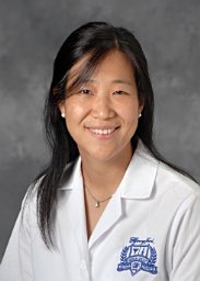 Hwang Clara合作论文数14Kilari Deepak合作论文数14




