As the brain transitions from wakefulness to sleep, processing of external information diminishes while restorative processes, such as glymphatic removal of waste products, are activated. Yet, it is not known what drives brain clearance during sleep. We here employed an array of technologies and identified tightly synchronized oscillations in norepinephrine, cerebral blood volume, and cerebrospinal fluid (CSF) as the strongest predictors of glymphatic clearance during NREM sleep. Optogenetic stimulation of the locus coeruleus induced anti-correlated changes in vasomotion and CSF signal. Furthermore, stimulation of arterial oscillations enhanced CSF inflow, demonstrating that vasomotion acts as a pump driving CSF into the brain. On the contrary, the sleep aid zolpidem suppressed norepinephrine oscillations and glymphatic flow, highlighting the critical role of norepinephrine-driven vascular dynamics in brain clearance. Thus, the micro-architectural organization of NREM sleep, driven by norepinephrine fluctuations and vascular dynamics, is a key determinant for glymphatic clearance.
Neuromodulators act on multiple timescales to affect neuronal activity and behavior. They function as synaptic fine-tuners and master coordinators of neuronal activity across distant brain regions and body organs. While much research on neuromodulation has focused on roles in promoting features of wakefulness and transitions between sleep and wake states, the precise dynamics and functions of neuromodulatory signaling during sleep have received less attention. This review discusses research presented at our minisymposium at the 2024 Society for Neuroscience meeting, highlighting how norepinephrine, dopamine, and acetylcholine orchestrate brain oscillatory activity, control sleep architecture and microarchitecture, regulate responsiveness to sensory stimuli, and facilitate memory consolidation. The potential of each neuromodulator to influence neuronal activity is shaped by the state of the synaptic milieu, which in turn is influenced by the organismal or systemic state. Investigating the effects of neuromodulator release across different sleep substates and synaptic environments offers unique opportunities to deepen our understanding of neuromodulation and explore the distinct computational opportunities that arise during sleep. Moreover, since alterations in neuromodulatory signaling and sleep are implicated in various neuropsychiatric disorders and because existing pharmacological treatments affect neuromodulatory signaling, gaining a deeper understanding of the less-studied aspects of neuromodulators during sleep is of high importance.
Impaired sleep is a common aspect of aging and often precedes the onset of Alzheimer’s disease. Here, we compare the effects of sleep deprivation in young wild-type mice and their APP/PS1 littermates, a murine model of Alzheimer’s disease. After 7 h of sleep deprivation, both genotypes exhibit an increase in EEG slow-wave activity. However, only the wild-type mice demonstrate an increase in the power of infraslow norepinephrine oscillations, which are characteristic of healthy non-rapid eye movement sleep. Notably, the APP/PS1 mice fail to enhance norepinephrine oscillations 24 h after sleep deprivation, coinciding with an accumulation of cerebral amyloid-β protein. Proteome analysis of cerebrospinal fluid and extracellular fluid further supports these findings by showing altered protein clearance in APP/PS1 mice. We propose that the suppression of infraslow norepinephrine oscillations following sleep deprivation contributes to increased vulnerability to sleep loss and heightens the risk of developing amyloid pathology in early stages of Alzheimer’s disease.
Extracellular potassium concentration ([K+](e)) is known to increase as a function of arousal. [K+](e) is also a potent modulator of transmitter release. Yet, it is not known whether [K+](e) is involved in the neuromodulator release associated with behavioral transitions. We here show that manipulating [K+](e) controls the local release of monoaminergic neuromodulators, including norepinephrine (NE), serotonin, and dopamine. Imposing a [K+](e) increase is adequate to boost local NE levels, and conversely, lowering [K+](e) can attenuate local NE. Electroencephalography analysis and behavioral assays revealed that manipulation of cortical [K+](e) was sufficient to alter the sleep-wake cycle and behavior of mice. These observations point to the concept that NE levels in the cortex are not solely determined by subcortical release, but that local [K+](e) dynamics have a strong impact on cortical NE. Thus, cortical [K+](e) is an underappreciated regulator of behavioral transitions.
Sleep is not homogenous but contains a highly diverse microstructural composition influenced by neuromodulators. Prior methods used to measure neuromodulator levels in vivo have been limited by low time resolution or technical difficulties in achieving recordings in a freely moving setting, which is essential for natural sleep. In this protocol, we demonstrate the combination of electroencephalographic (EEG)/electromyographic (EMG) recordings with fiber photometric measurements of fluorescent biosensors for neuromodulators in freely moving mice. This allows for real-time assessment of extracellular neuromodulator levels during distinct phases of sleep with a high temporal resolution.
Information transfer within neuronal circuits depends on the balance and recurrent activity of excitatory and inhibitory neurotransmission. Chloride (Cl − ) is the major central nervous system (CNS) anion mediating inhibitory neurotransmission. Astrocytes are key homoeostatic glial cells populating the CNS, although the role of these cells in regulating excitatory-inhibitory balance remains unexplored. Here we show that astrocytes act as a dynamic Cl − reservoir regulating Cl − homoeostasis in the CNS. We found that intracellular chloride concentration ([Cl − ] i ) in astrocytes is high and stable during sleep. In awake mice astrocytic [Cl − ] i is lower and exhibits large fluctuation in response to both sensory input and motor activity. Optogenetic manipulation of astrocytic [Cl − ] i directly modulates neuronal activity during locomotion or whisker stimulation. Astrocytes thus serve as a dynamic source of extracellular Cl − available for GABAergic transmission in awake mice, which represents a mechanism for modulation of the inhibitory tone during sustained neuronal activity.
Sleep has a complex micro-architecture, encompassing micro-arousals, sleep spindles and transitions between sleep stages. Fragmented sleep impairs memory consolidation, whereas spindle-rich and delta-rich non-rapid eye movement (NREM) sleep and rapid eye movement (REM) sleep promote it. However, the relationship between micro-arousals and memory-promoting aspects of sleep remains unclear. In this study, we used fiber photometry in mice to examine how release of the arousal mediator norepinephrine (NE) shapes sleep micro-architecture. Here we show that micro-arousals are generated in a periodic pattern during NREM sleep, riding on the peak of locus-coeruleus-generated infraslow oscillations of extracellular NE, whereas descending phases of NE oscillations drive spindles. The amplitude of NE oscillations is crucial for shaping sleep micro-architecture related to memory performance: prolonged descent of NE promotes spindle-enriched intermediate state and REM sleep but also associates with awakenings, whereas shorter NE descents uphold NREM sleep and micro-arousals. Thus, the NE oscillatory amplitude may be a target for improving sleep in sleep disorders.
Abstract Information transfer within neuronal circuits depends on the balance and recurrent activity of excitatory and inhibitory neurotransmission. Chloride (Cl-) is the major CNS anion mediating inhibitory neurotransmission and thus essential for balanced brain function. Sustained activation of inhibitory synapses leads to a significant reduction in the amplitude of IPSPs over time, reflecting a collapse of the Cl- gradient, due to Cl- influx into neurons through GABAA receptors. Astrocytes are key homeostatic glial cells populating all parts of the central nervous system (CNS). Despite astrocytic contribution to brain state transitions, the role of these cells in regulating inhibition remains unexplored. Here we show, for the first time, that astrocytes act as a dynamic Cl- reservoir regulating Cl- homeostasis in awake mice. Intracellular chloride concentration ([Cl-]i) in astrocytes is high and stable during sleep. In awake mice astrocytic [Cl-]i is lower and fluctuates in response to both sensory input and motor activity. Stimulation of astrocytic GABAA receptors induces Cl- efflux, providing an additional source of Cl- for inhibitory transmission. Increasing astrocytic [Cl-]i by optogenetic manipulations alleviates neuronal activity induced by locomotion or whisker stimulation. Our analysis shows that astrocytes serve as a dynamic source of extracellular Cl- available for GABAergic transmission in awake mice, which represents a novel mechanism for modulation of the inhibitory tone.
Summary We normally regard sleep and wake as two distinct opposing brain states, where sleep requires silence of wake-promoting structures such as the locus coeruleus (LC)-norepinephrine (NE) system. We set out to investigate how cortical NE dynamics and NE-related astrocytic activity relates to LC population activity during sleep states. We show that LC displays regular phasic activity bouts during NREM sleep leading to a slow oscillatory pattern of prefrontal NE levels of which the majority of NE increases does not lead to awakening. NE troughs link to sleep spindles and continued NE decline transitions into REM sleep. Last, we show that prefrontal astrocytes have reduced sensitivity towards NE during sleep. Our results suggest that dynamic changes in the activity of wake-promoting systems during sleep create alternation between crucial sleep processes and broadening of sensitivity towards incoming sensory input. Highlights Extracellular levels of norepinephrine display dynamic changes during NREM and REM sleep Phasic activity of locus coeruleus neurons during NREM underlies slow norepinephrine oscillations Spindles occur at norepinephrine troughs and are abolished by norepinephrine increases Increased spindles prior to REM reflect the beginning of a long-lasting norepinephrine decline REM episodes are characterized by a sub-threshold continuous norepinephrine decline The responsiveness of astrocytic Ca 2+ to norepinephrine is reduced during sleep
We continuously need to adapt to changing conditions within our surrounding environment, and our brain needs to quickly shift between resting and working activity states in order to allow appropriate behaviors. These global state shifts are intimately linked to the brain-wide release of the neuromodulators, noradrenaline and acetylcholine. Astrocytes have emerged as a new player participating in the regulation of brain activity, and have recently been implicated in brain state shifts. Astrocytes display global Ca2+ signaling in response to activation of the noradrenergic system, but whether astrocytic Ca2+ signaling is causative or correlative for shifts in brain state and neural activity patterns is not known. Here we review the current available literature on astrocytic Ca2+ signaling in awake animals in order to explore the role of astrocytic signaling in brain state shifts. Furthermore, we look at the development and availability of innovative new methodological tools that are opening up for new ways of visualizing and perturbing astrocyte activity in awake behaving animals. With these new tools at hand, the field of astrocyte research will likely be able to elucidate the causal and mechanistic roles of astrocytes in complex behaviors within a very near future.
 Maiken Nedergaard合作论文数Department of Neurology, Center for Translational Neuromedicine, University of Rochester Medical Center;Department of Neurosurgery, University of Rochester Medical Center12
 Kjaerby Celia合作论文数Synaptic Transmission I&H. Lundbeck A/S;Synaptic Transmission I&II, H. Lundbeck A/S10
 Natalie L Hauglund合作论文数Center for Translational Neuromedicine, University of Copenhagen8
 Hajime Hirase合作论文数Division of Neuron-glia Circuitry, Center for Translational Neuromedicine, University of Copenhagen7
 Pia Weikop合作论文数Division of Glial Disease and Therape, Nedergaard Laboratory, Center for Translational Neuromedicine;Neuropsykiatrisk Laboratorium Rigshospitalet5
 Verena Untiet合作论文数5
 Tessa Radovanovic合作论文数4Anastasia Tsopanidou合作论文数4Laura Rose合作论文数3
 Peter Kusk合作论文数Faculty of Health and Medical Sciences, University of Copenhagen3Camilla Dall合作论文数3Björn Sigurdsson合作论文数Ctr Translat Neuromed, Univ Copenhagen3N Petersen合作论文数Dept Neurosci, Univ Copenhagen3
 Fengfei Ding (丁凤菲)合作论文数Department of Pharmacology, School of Basic Medical Sciences, Fudan University3Saiyue Deng合作论文数2
 Birgitte Rahbek Kornum合作论文数Department of Neuroscience, Faculty of Health and Medical Sciences, University of Copenhagen2A. N. Zahid合作论文数2
 Sebastien Arthaud合作论文数2
 Alexei Verkhratsky合作论文数Division of Neuroscience, School of Biological Sciences, Faculty of Biology, Medicine and Health, The University of Manchester2
 Viviana Lo Martire合作论文数PRISM Lab, Univ Bologna2Beinlich Felix合作论文数2Ning Kang合作论文数2
 Jiesi Feng (冯杰思)合作论文数School of Life Sciences, Peking University2
 Yulong Li (李毓龙)合作论文数School of Life Sciences, Peking University;IDG/McGovern Institute For Brain Research, Peking University;PKU-THU Center for Life Sciences;Shenzhen Bay Laboratory2
 Chiara Berteotti合作论文数Department of Human and General Physiology, University of Bologna2
 Alessandro Silvani合作论文数Dipartimento di Fisiologia Umana e Generale, Università di Bologna2
 Christelle Peyron合作论文数2
 Morten Mørup合作论文数Department of Applied Mathematics and Computer Science, DTU2
 Antoine Adamantidis合作论文数Zentrum Fur Experimentelle Neurologie, Department of Neurology, Inselspital University Hospital Bern, University of Bern2Giovanna Zoccoli合作论文数Institute of Human Physiology, University of Bologna2H Birgens合作论文数1
 Christine Delle合作论文数1Noa Matosevich合作论文数Sackler School of Medicine & Sagol School of Neuroscience, Tel Aviv University1Andrea Grostøl Dietz合作论文数Center for Translational Neuromedicine, University of Copenhagen1Sisi Peng合作论文数1Georgios Foustoukos合作论文数Laboratory of Sensory Processing, Brain Mind Institute, Faculty of Life Sciences, Ecole Polytechnique Fédérale de Lausanne (EPFL), Lausanne, Switzerland1
 Yiyao Zhang (张艺瑶)合作论文数NYU, Neurosci Inst1Natalie Beschorner合作论文数1Qian Sun合作论文数1
 Antonio Ladrón-de-Guevara合作论文数Department of Biomedical Engineering, University of Rochester1
 Ryan Tisdale合作论文数Biosciences Division, SRI International1
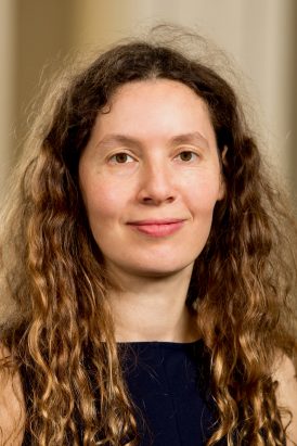 Ada Eban-Rothschild合作论文数Department of Psychology, College of Literature, Science, and the Arts, University of Michigan;Michigan Neuroscience Institute, University of Michigan;Michigan Institute for Computational Discovery and Engineering, University of Michigan1
 Wei Song合作论文数Department of Biological and Enviromental Engineering, Cornell University1Brown Peter合作论文数1
 Niels Henning Skotte合作论文数Child and Family Research Institute, University of British Columbia1
 Henrik Frederiksen合作论文数1
 Filip Mundt合作论文数1Lambert Marius合作论文数1
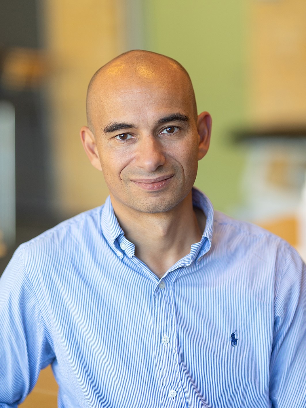 Tarec Christoffer El-Galaly合作论文数Department of Clinical Medicine, Faculty of Medicine, Aalborg Universitet;Department of Haematology, Aalborg Universitet Hospital;Klinisk Institut, Sundhedsvidenskabelige Fakultet, Aalborg Universitet1




