AIM We present our experience with micropercutaneous cystolithotripsy (mPCCL) using transillumination-guided access in children with bladder stones and evaluate the safety and efficacy of this procedure. METHODS Twelve children who underwent mPCCL for bladder stone removal in our department between January 2011 and September 2013 were included in this study. Percutaneous access was performed using the transillumination technique. The chief complaint, age and sex of the patients, stone size, operative time, complications, stone-free rate, and stone composition as determined by radiography diffraction analysis were recorded. RESULTS The mean age of the patients was 2.6 (1-7) years. One out of 12 patients was a girl. The mean stone size was 14 mm (7-32 mm). The mean mPCCL procedure time was 38.7 (15-65) minutes. The mean hospital stay was 1.4 (0.5-4) days. The stone-free rate after one mPCCL intervention was 91.6% and increased to 100% after two sessions of mPCCL. The notable complications were transient macroscopic hematuria in one patient, acute urinary retention in one patient, and the need for a second session of mPCCL in another patient. The stones were composed of calcium oxalate (3), ammonium acid urate (1), calcium phosphate (1), cystine (1), struvite (1), and unknown (5). CONCLUSION Transillumination-guided mPCCL is a safe and effective technique, does not require the use of ionizing radiation, is technically easy, and is a minimally invasive alternative for the management of bladder stones. It has many advantages, especially in pediatric patients, such as decreased urethral injury, no need for postoperative catheter insertion in most cases, and shorter operative time.
ABSTRACT Introduction: Double-J stent insertion during laparoscopic pyeloplasty is a difficult and time-consuming process and several techniques were defined to perform a double-J stent with an antegrade approach. In this study we present the technique (PICA) of antegrade double-J placement during laparoscopic pyeloplasty by using 14 gauge intravenous cannula. Surgıcal technıque: After we complete the suturing of the posterior wall of the anastomosis during laparoscopic pyeloplasty, we first puncture the abdominal wall with a 14-gauge “intravenous cannula” from a location that provides most suitable angle for inserting the double-J stent into the ureter. We remove the metal needle of the cannula, and the sheath which has an inner diameter of 5.2F remains over the abdominal wall. The double J stent is then advanced from inside the cannula sheath to the intraperitoneal area; under laparoscopic imaging the stent is gently grasped at its distal end using an atraumatic laparoscopic forceps to insert it into the ureter. The stent is then pulled down to its proximal end, and after the guidewire is removed, the proximal end of the double-J stent is placed inside the renal pelvis with an atraumatic forceps. With this technique we can apply the double-J stent in just one step. Additionaly we can use a 14-gauge IV cannula sheath as a trocar when needed during laparoscopic pyeloplasty to retract an organ or reveal an anastomosis line. Comments: Our new technique of antegrade double-J placement during laparoscopic pyeloplasty by 14 gauge intravenous cannula sheath, is very easy and quick to perform.
Objectives:To suggest a novel disposable percutaneous nephrolithotomy (PCNL) set that we named the ‘Economical One-shot PCNL Set’ (Ecoset), which consists of a single 30-F dilator, 30-F sheath, and 8-F polyurethane dilator, as use of a ‘one-shot’ dilatation technique during PCNL is becoming widespread.Patient and methods:The medical records of 42 patients with kidney stones who had undergone ‘one-shot’ PCNL from February 2014 to June 2016 were retrospectively reviewed and analysed. Demographic data, as well as the stone size, radiation exposure time, operation time, hospitalisation duration, rate of treatment success and complications, were recorded.Results:The mean (SD, range) age of the patients was 44.43 (16.54, 11–72) years. The mean (SD) stone size was 35.12 (17.53) mm. The mean (SD) operation time was 54.58 (22.24) min. The mean (SD) fluoroscopic screening time was limited to 154.72 (117.48) s. Treatment success was achieved in 32 (76%) patients. The mean (SD) hospital stay was 3.09 (0.75) days. None of the patients had any major complications. Bleeding requiring blood transfusion was required in three patients. The cost of a disposable dilatation set for a single PCNL operation with a balloon set, a standard Amplatz set, or an Ecoset is ∼$137, $120, or $27 (American dollars), respectively.Conclusions:The one-shot dilatation technique using the Ecoset for PCNL can be feasibly, safely, and effectively performed in almost every adult patient. The Amplatz dilator set and balloon dilator set have the disadvantage of relatively high cost, whereas the Ecoset is the cheapest ‘disposable set’ that can be used during PCNL surgery.
MicroRNAs (miRNAs) are small conserved non-coding RNA molecules that post-transcriptionally regulate gene expression. Although it is reported in many studies that there are associations between alterations of miRNA homeostasis and pathological conditions such as cancer, psychiatric and neurological diseases, cardiovascular disease and autoimmune disease, the effects of common genetic variants of these genes on male infertility are unclear. To better understand this effect, we performed a case-control study including a total of 108 infertile men with idiopathic azoospermia and 125 fertile control subjects. Real-time polymerase chain reaction was used to genotype six single-nucleotide polymorphisms (SNPs) of microRNA biogenesis pathway genes and the associations between individual and combined genotypes and idiopathic azoospermia were analysed. The results showed significant difference between the individual AA genotype frequency of the GEMIN3 (rs197388) gene in the patient and control groups, indicating that the AA genotype may be considered as indicative of a higher predisposition to idiopathic azoospermia. The combined genotype analysis, including six SNPs, revealed statistically significant differences between the patients and control subjects for some combinations. For example, the frequency of genotype distributions of the AA\CA-CC-TT-AT genotype combination for the XPO5-RAN-DICER1GEMIN3 combined loci was significantly different, and it may be considered a predisposition to idiopathic azoospermia. According to the obtained results, both individual and combined genotypes of SNPs from miRNA genes may be used to predict the risk of male infertility with idiopathic azoospermia.
This study aimed to investigate the effectiveness and reliability of micro-percutaneous nephrolithotomy (micro-PNL) for the management of kidney stones in pediatric patients. This retrospective study included 24 infants that underwent micro-PNL for renal stones between March 2012 and April 2015. The inclusion criteria included: age younger than 2 years, stone size <20 mm, absence of urinary tract malformations and active infection, and no coagulopathy. Stone-free rates were assessed one month postoperatively by ultrasonography (USG) and kidneys, ureters, bladder (KUB) radiography. Complications were classified according to the Clavien classification system. The mean age of the patients was 15.8 ± 7.8 months (range, 8–23) and the mean stone size was 13.5 ± 3.84 mm. Intra-renal access was achieved using USG in 11 patients and KUB fluoroscopy in 13 patients. The operation time and fluoroscopic screening time were 53.7 ± 10.35 and 1.4 ± 0.9 min, respectively. The mean hospital stay was 2.5 ± 0.8 days, and the mean drop in the hemoglobin level was 0.51 ± 0.34 g/dL. Bleeding requiring blood transfusion was not observed. A ureteral J stent was implanted perioperatively in four patients because of stone burden. Four patients complained of postoperative renal colic (Clavien grade 1) and postoperative fever was observed in two patients. Definitive success rates were as follows: the stone-free rate was 83.3 % (n = 20) and residual fragments were observed in four patients.
OBJECTIVE:The aim of this study was to compare the results of urine cultures obtained either from urethral, and percutaneous nephrostomy (PCN) catheters. MATERIALS AND METHODS:This study included 328 consecutive patients that underwent PCN at our institution with complicated urinary tract infections (UTIs) between July 2010 and April 2015. Results of urine cultures obtained from the urethral and nephrostomy catheters were compared. RESULTS:This study included 152 male and 176 female patients. Mean age of the patients was 46.2±24.3 years. The main indications were obstructive uropathy due to urolithiasis complicated with pyonephrosis 145 (44%), malignant disease (n=87; 26%), pregnancy (n=26; 8%), and anatomical abnormality (n=23; 7%). One hundred and twenty three patients had diabetes mellitus. The most common causative organisms were Escherichia coli, Klebsiella pneumoniae, and Pseudomonas aeruginosa. Blood cultures showed the same results for the PCN and bladder urine cultures. The bladder urine culture was positive in 304 patients, while the PCN urine culture in 314 patients. CONCLUSION:PCN is an important treatment for the management of pyonephrosis. Cultures from the PCN yield valuable information that is not available from urethral urine cultures, and is a guiding tool for antibiotic therapy selection.
Pelvic kidney stones remain a unique challenge to the endourologists. Treatment options include open surgery, extracorporeal shockwave lithotripsy, percutaneous nephrolithotomy (PNL), retrograde intrarenal surgery, and laparoscopy assisted PNL (LA-PNL). As a minimal invasive option, LA-PNL can decrease the risk for bowel and major vessel injury. Here, we describe our experience using the LA-PNL procedures with different combinations, to treat kidney stones in multiple patients with a pelvic ectopic kidney (PEK). Eight patients, with PEK, kidney stones, and no other treatment choice, but open surgery, were included in the study. Two different laparoscopic techniques such as mesocolon dissection and transmesocolic, and four different percutaneous procedures such as standard-PNL, mini-PNL, micro-PNL, and a PNL through the renal pelvis were used for stone extraction in these patients. The mean age of patients was 25.6 ± 12.9 years and mean stone size was 524.1 ± 430.3 mm2. Mean operation time was 150.5 ± 40.0 (77–210) min which was composed of retrograde catheterization (14.8 ± 2.9 min), laparoscopic procedure (48.7 ± 20.6 min) and PNL procedure (86.8 ± 31.1 min). Residual stones were seen in two patients (no additional treatment was need), while a ‘stone-free’ procedure was achieved in six patients (75.0 %). On the post-operative first month visit, a stone was observed on radiological examinations in only one patient (87.5 % stone-free). Mean hospitalization time was 2.8 ± 0.9 days. No perioperative or post-operative complication was observed in all patients. LA-PNL surgery is a safe and effective option for treatment of PEK stones, and has several alternative approaches.
The objective of this study is to assess the utility of the Guy, S.T.O.N.E., and CROES nephrolithometry scoring systems (SS), and compare the capability of each system to predict percutaneous nephrolithotomy (PNL) outcome in patients with anatomical abnormalities. We retrospectively collected medical records of patients with anatomical abnormalities who underwent PNL for the treatment of renal calculi by experienced surgical teams in four referral centers. All of the patients were graded by a single observer from each department based on preoperative computed tomography images using each SS. Patient demographics and outcomes were compared according to the complexity of the procedure as graded by each scoring system. A total of 137 cases with anatomical abnormalities [horseshoe kidney (n = 46), malrotation (n = 33), kypho and/or scoliosis (n = 31) and ectopic kidney (n = 27)] were assessed retrospectively. The mean stone burden, number, and density were 708.5 mm2, 1.7, and 791.8 HU, respectively. The mean procedure, fluoroscopy, and hospitalization times were 75.2 ± 35.3 min, 133.4 ± 92.3 s, and 3.5 ± 2.1 days, respectively. Stone-free status was achieved in 106 cases (77.4 %). A total of 17 (13.6 %) complications occurred postoperatively. The mean scores were 2.7, 7.2, and 219.1, for the Guy, S.T.O.N.E., and CROES systems, respectively. CROES score was the independent predictor of PNL success in cases with anatomical abnormalities [p: 0.001, OR 1.01, (95 % CI 1005–1021)]. The CROES scoring system is well correlated with the success of PNL in cases with anatomical abnormalities; the S.T.O.N.E. and Guy scoring systems failed to predict the outcomes of PNL in this specific patient population.
This study aims to investigate the role of urinary biomarkers in the determination of the potential risks of renal parenchymal tubular damage in adult patients who underwent percutaneous nephrolithotomy (PNL) with the indication of renal stone. A randomized and prospective controlled study was performed between June and December 2013. We enrolled 29 consecutive patients with renal calculi>2cm and who underwent PNL, as well as 47 healthy control subjects. Urine samples, including 2 h before surgery, 2 and 24 h after surgery were collected from the patient group. Freshly voided urine samples were collected from the control group. Kidney injury molecule-1 (KIM-1), neutrophil gelatinase-associated lipocalin (NGAL), N-acetyl-glucosaminidase (NAG), and liver-type fatty acid binding protein (LFABP) levels were measured from these urine samples. The mean KIM-1/Cr value that measured 24 h after the operation was statistically significant, higher than its preoperative (preop) level (p= 0.045). A significant difference was detected between the mean preop and postoperative (postop) 24 h NAG/Cr values (p< 0.001). Also, postop 24 h NGAL/Cr levels were statistically significant, higher than its preop levels (p= 0.013). According to the comparison of preop and postop levels, an increase in LFABP/Cr values secondary to surgical intervention was observed without any statistically significant difference. Besides the LFABP/Cr levels do not change after percutaneous kidney surgery, KIM-1/Cr, NAG/Cr, and NGAL/Cr levels increase postop period, especially at 24 h. Further studies with a larger series and repeated measurements should be performed to clarify if they can be used to demonstrate renal damage after percutaneous surgery or not.
During infancy, the renal parenchyma and pelvicalyceal system are relatively fragile. Therefore, percutaneous nephrolithotomy for the management of renal stones in this age group is a challenging procedure for urologists. Herein, we present the uneventful management of bilateral renal stones using micropercutaneous nephrolithotomy (microperc) administered to a 7-month-old infant with recurrent urinary tract infections. In this paper, the advantages and disadvantages of the microperc procedure are discussed. As far as we know, our patient is the youngest case in whom the microperc procedure has been performed.
OBJECTIVE:To compare outcomes of micro-percutaneous nephrolithotomy (PNL; microperc) with mini-PNL (miniperc) in the treatment of pediatric renal stones of sizes 10-20 mm. MATERIALS AND METHODS:Patients aged <18 years who underwent PNL for renal stones of sizes 10-20 mm between August 2011 and March 2014 in 3 referral centers were reviewed retrospectively. Patients were evaluated in the following 2 groups: microperc (group 1) and miniperc (group 2). Demographics and perioperative parameters (fluoroscopy and operation time, hemoglobin drop, and stone-free and complication rates) were retrospectively analyzed. RESULTS:A total of 119 patients were evaluated, including group 1 (n = 56) for microperc and group 2 (n = 63) for miniperc. We found mean stone sizes as 13.4 ± 3.4 and 14.8 ± 3.7 mm in the groups, respectively (P = .046). Mean operation and fluoroscopy times were 57.1 ± 31.2 minutes and 132.4 ± 92.5 seconds in the microperc group and 68.9 ± 36.7 minutes and 226.2 ± 166.2 seconds in the miniperc group, respectively (P = .110 and P <.001). Stone-free rates were similar in both groups (82.1% vs 87.3%; P = .433 and 92.8% vs 93.6%; P = 0673) on postoperative day 1 and at first-month follow-up. The mean hemoglobin drop in group 2 differed from that in group 1 significantly (P <.001). The difference of average hospitalization times was statistically significant (43.0 ± 15.4 vs 68.5 ± 31.7 hours; P <.001). CONCLUSION:Our outcomes show that microperc may be preferred as an alternative to mini-PNL for the treatment of pediatric kidney stones of sizes 10-20 mm with comparable success and complication rates, as well as shorter hospitalization and fluoroscopy times.
OBJECTIVE:To evaluated the damage effects of retrograde intra-renal surgery (RIRS) on kidney tissue by measuring kidney injury molecule-1 (KIM-1), neutrophil gelatinase-associated lipocalin (NGAL), N-acetyl-β-D-glucosaminidase (NAG), liver-type fatty acid binding protein (LFABP) expression.MATERIAL AND METHODS:We enrolled thirty consecutive patients (Group 1) who underwent RIRS that presented with renal calculi size < 2 cm. Forty-seven control patients (Group 2) with no signs or symptoms of urogenital disease were also enrolled for this study. Patients urine KIM-1, NGAL, NAG, and LFABP and creatinine levels were determined before the surgery, 2 hours after the surgery, and 24 hours after the surgery.RESULTS:Demographic data were established and found to be similar between the two groups. Two hours after the surgery KIM-1/Cr and NGAL/Cr levels had increased significantly in urine compared to levels before the surgery (P:0.04, P:0.02 respectively) and decreased 24 hours after the surgery. The NAG/Cr and LFABP/Cr levels did not change significantly after the surgery.CONCLUSION:According to acute kidney injury (AKI) markers, this study is suggests that RIRS is a safe method , KIM-1/Cr and NGAL/Cr levels were increased first 2 hours but returned to initial levels within 24 hours after the surgery.
PURPOSE:The aim of this study was to compare the positive aspects and complications of monoplanar and biplanar access techniques used in percutaneous nephrolithotomy (PCNL) based on the modified Clavien complication grading system.PATIENTS AND METHODS:The data from patients who underwent PCNL using either monoplanar access (group 1) or biplanar access (group 2) techniques were analyzed retrospectively. For the biplanar technique, puncture is adjusted based on different fluoroscopic projections, including vertical and 30 degree positions. For the monoplanar technique, a C-arm fluoroscope was brought into vertical position, the collecting system was visualized with a contrast agent, and the most appropriate calix was selected to extract the targeted stone.RESULTS:The monoplanar technique was performed for renal access in 310 patients (group 1), and the biplanar technique was used for renal access in 351 patients (group 2). There were no statistically significant differences between the two groups with regard to demographic data. The mean puncture time was significantly lower in group 1 (monoplanar) when compared with that of group 2 (biplanar, P=0.04). The overall success rates of the monoplanar and biplanar groups were 88% and 89% (including clinically insignifican residual fragments in 9% and 7%), respectively (P>0.05), and the complication rates of both groups were similar.CONCLUSION:The monoplanar access technique, which is safe to use, decreases puncture time, minimizes the surgeon's direct exposure time to radiation, and has similar success rates as the biplanar access technique.
Percutaneous nephrolithotomy is accepted as the standard management approach for kidney stones that are either refractory to extracorporeal shock wave lithotripsy or are >2 cm in diameter. The recently developed micro-percutaneous nephrolithotomy (microperc) technique provides intrarenal access under full vision using an optic instrument with a smaller calibration. A lesser amount of bleeding has been reported with the use of this method. Here we present a case of a bleeding complication on postoperative day 15 after a microperc procedure used to treat a left kidney stone. The complication led to retention of bloody urine in the bladder and required transfusion of 5 units of whole blood.
We aimed to present the outcomes of PNL surgery performed in infantile patients with small renal stones who were younger than 1 year of age. A single-center prospective trial was initiated and during the period between Jan 2013 and Jan 2015, PNL was applied to 20 renal units of 16 infants (6 girls and 10 boys), including 4 patients with bilateral kidney stones. PNL was performed in patients with renal stones larger than 2 cm, as well as stones resistant to SWL or renal stones that were undetectable during SWL. The mean age of the patients was 9.55 (5-12) months. Of the 20 renal units, 1 had complete staghorn stones, 3 had partial staghorn stones, 13 had renal pelvic stones, and 3 had lower pole stones. The mean stone size was 18.5 mm (range 12-36 mm). Mean operative time for PNL was 88 (25-135 min). Mean fluoroscopy time was estimated as 3.4 min. Mean hemoglobin loss was 0.72 g/L (0.2-3). The mean hospital stay was 4.1 days (2-8 days). On postoperative day 1, a complete stone-free state was achieved in 70% of renal units (14 of 20). At the end of the first postoperative week, the remaining two patients had insignificant residual fragments of 3 mm and were followed conservatively without any specific intervention. Thus, the total SFR was 80% (16 of 20) at discharge. In infants aged less than 1 year, minimal access tract dilation during PNL, the use of smaller caliber pediatric instruments, and the realization of this procedure by surgeons with adequate experience in adults carry utmost importance. In addition, special care should be taken to avoid hypothermia and radiation exposure during PNL.
To investigate whether aging affects surgical outcomes by comparing the results of two patient groups undergoing PNL: those over 60 and those under 60. A retrospective screen was made for patients undergoing conventional PNL surgery for renal stones performed in two separate centers between 2010 and 2013. 520 patients included were classified into age groups: patients aged 18–59 comprised Group-1 and those aged over 60 comprised Group-2. Those between 60–69 years (sexagenarian) were assigned to Group-2a; 70–79 years (septuagenarian) to Group-2b; and 80–89 years (octogenarian) to Group-2c. Patients' demographic characteristics (accompanying comorbidities, ASA scores, body mass indices and stone size) and perioperative values (duration of surgery and hospital stay, success and complication rates) were compared between the groups. Mean stone size was similar in groups (30.1 ± 15.5 vs. 31.5 ± 15.4 mm, p = 0.379). The mean ASA value for the patients in Group-1 was 1.61; significantly lower than that in the other groups (p = 0.000). The level of accompanying comorbidities in Group-1 was significantly lower than that of the other groups (p = 0.000). The mean duration of surgery, postoperative hematocrit drop, complication and success rate were statistically similar in Groups 1 and 2 (p = 0.860, p = 0.430, p = 0.7, and p = 0.66, respectively). The duration of hospital stay was significantly shorter in the patients in Group-1 compared to those in Group-2 (p = 0.008). In experienced hands, PNL can be safely and reliably performed in the treatment of renal stones in elderly patients.
OBJECTIVES: We aimed to compare ureterolithotripsy and inserting only ureteral-j stent in terms of efficiency safety and patient comfort in treatment of ureteral calculi in pregnant patients.METHODS: Seventy patients who developed hydronephrosis due to ureteral calculi during pregnancy and on whom endoscopic intervention was performed were included in the study In a center, the stones were broken up by ureteroscopy, and then ureteral stents (JJ) were placed if needed. In the other center, nothing was performed on the stones, and only ureteral stents (JJ) was placed. For the statistical analysis, Pearson's chisquared test and the Mann-Whitney U tests were used and the significance level was determined as p < 0.05.RESULTS: The average age of the patients was 26.2 years (18-39) and the average gestational week was 23.4 weeks (8-36). While no significant difference was found between the two groups in terms of the frequency of complications (p=0.381) and post-operative pyelonephritis (p=0.2), the need for additional intervention in the group on whom ureteroscopy was performed was found to be less (9.7% vs. 31%; p=0.032). Moderate or severe LUTS or flank pain during the period between the procedure and the birth was found to be significantly less in the group in which ureterolithotripsy was performed (14% vs. 55%; p=0.036).CONCLUSIONS: Ureterolithotripsy is a safe and more comfortable procedure than only ureteral double-j insertion on pregnant patients with ureterolithiasis.
 Mehmet Nuri Bodakci合作论文数82
 Necmettin Penbegul合作论文数Department of Urology, Dicle University School of Medicine74
 Ahmet Ali Sancaktutar合作论文数Faculty of Medicine, Dicle University71
 Haluk Soylemez合作论文数61Yasar Bozkurt合作论文数Diyarbakýr59Murat Atar合作论文数55Dağgülli Mansur合作论文数27
 Yıldırım Kadir合作论文数15Utangaç Mazhar合作论文数14Dede Onur合作论文数13Cakmakci Suleyman合作论文数10
 Abdulkadir Tepeler合作论文数Faculty of Medicine, Bezmialem Vakif University10Mehmet Yucel合作论文数9musa atar合作论文数8
 Mesrur Selcuk Silay合作论文数Faculty of Medicine, Bezmialem Vakif University8Armağan Abdullah合作论文数7
 Soner yalcınkaya合作论文数7S. Cakmakci合作论文数6Buldu İbrahim合作论文数Faculty of Medicine, Mevlana University5Mustafa Okan Istanbulluoglu合作论文数Konya Medical and Research Center, Baskent University5Nebahat Hatipoglu合作论文数4
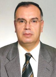 Erdal Mehmet Emin合作论文数School of Medicine, Mersin University4
 Firat Ugur合作论文数4
 Tuna Karatag合作论文数Department of Urology, Mevlana University4Yüksel Hatice合作论文数4
 Osman Evliyaoglu合作论文数Faculty of Medicine, Dicle University4
 Tevfik Ziypak合作论文数Departments of Urology School of Medicine, Ataturk University4
 Bulent Altunoluk合作论文数4Aytan Kar合作论文数4Tolga Akman合作论文数Department of Urology, Haseki Training and Research Hospital4Isa Ozbey合作论文数Departments of Urology School of Medicine, Ataturk University4
 Yahya Islamoglu合作论文数Department of Cardiology, Universty of Dicle Medical Center4Sahin Kabay合作论文数Faculty of Medicine Hospital, Dumlupinar University4Mahmut Balkan合作论文数4Galip Dedekarginoglu合作论文数3Ismail Yildiz合作论文数3Hamidi Cihad合作论文数3Dedeoglu Serhat合作论文数3Dağguli Mansur合作论文数3
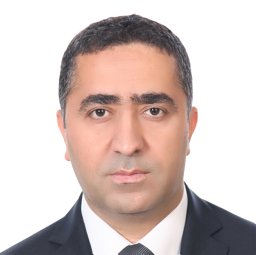 Evsen Mehmet Sıddık合作论文数Faculty of Medicine, Dicle University3
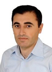 Halit Akbas合作论文数Department of Medical Biology, Harran University Faculty of Medicine3Turkcu Gul合作论文数3
 Alp Harun合作论文数Faculty of Medicine, Mustafa Kemal University Hatay3
 Aydin Rustemoglu合作论文数Faculty of Medicine, Gaziosmanpasa University3
 Senol Adanur合作论文数3Ugur Saracoglu合作论文数3Erol Aras合作论文数3
 Isen Kenan合作论文数Clinic of Urology Ofis cami sok, State Hospital of Diyarbakir3Gülay Dede合作论文数2




