PURPOSE:Freezing of gait (FOG) occurs in both Parkinson disease (PD) and cerebral small vessel disease (CSVD), but its mechanism in CSVD is unknown. We hypothesize that FOG arises from distinct neural substrates in these 2 disorders. MATERIALS AND METHODS:This cross-sectional study included 7 CSVD patients with FOG, 7 PD patients with FOG, and 20 healthy controls. All underwent clinical assessment and dual-tracer PET ( 18 F-FDG for metabolism; 11 C-CFT for dopamine transporter (DAT) binding). Group comparisons and clinical correlations were performed. RESULTS:CSVD-FOG patients showed relatively lower 18 F-FDG uptake in frontal-striatal and posterior cingulate regions, accompanied by relatively higher uptake in temporal-occipital-cerebellar areas after whole-brain normalization. By contrast, PD-FOG patients exhibited relatively higher striatal 18 F-FDG uptake and relatively lower uptake in parietal-temporal regions. Critically, DAT binding was severely reduced in PD, while relatively preserved DAT binding was observed in CSVD patients compared with PD. In the CSVD group, higher putaminal 18 F-FDG SUVr was associated with faster gait speed and lower FOG severity. CONCLUSIONS:FOG in PD and CSVD is associated with divergent dopaminergic and metabolic imaging patterns. PD-FOG is characterized by marked nigrostriatal DAT reduction, whereas CSVD-FOG shows relatively preserved DAT binding compared with PD and distinct whole-brain-normalized 18 F-FDG uptake patterns, suggesting involvement of neural networks beyond primary nigrostriatal dopaminergic pathways. These imaging differences may support etiology-informed diagnostic considerations and individualized management strategies.
This study aimed to investigate in vivo fibroblast activation in hypereosinophilic syndrome (HES) with cardiac involvement, and to explore the correlation between fibroblast activation protein inhibitor (FAPI) uptake and eosinophil-related biomarker-eosinophil extracellular traps (EETs). This study enrolled 20 patients diagnosed with HES and cardiac involvement. All patients underwent Al18F-FAPI-04 PET/CT imaging, transthoracic echocardiography, as well as measurement of absolute eosinophil count (AEC) and EETs levels. Among the 20 patients enrolled, 16 patients were diagnosed with reactive HES and 4 with idiopathic HES. Elevated FAPI uptake was observed across different stages of disease course. Myocardial Al18F-FAPI uptake was observed in all patients (20/20), with an extensive pattern in 15 and a patchy pattern in 5. Quantitative uptake was significantly higher in the extensive pattern group (maximum standardized uptake value (SUVmax): 6.58 [5.01–8.62] vs. 2.44 [1.91–2.76], p < 0.001). Segmental analysis revealed higher FAPI uptake in apical and mid-ventricular segments compared to basal segments (SUVmax: apical, 4.67 [1.94–6.89] vs. middle, 4.25 [2.33–5.61] vs. basal, 3.44 [2.46–5.23], p = 0.022). FAPI-PET/CT identified a higher proportion of involved myocardial segments (83.2
Background:This prospective study aimed to evaluate the prognostic value of 68Ga-DOTATATE PET/CT imaging in assessing myocardial macrophage infiltration in patients with autoimmune diseases and its relationship with clinical outcomes, specifically all-cause mortality and cardiovascular rehospitalization. Methods:A total of 36 patients with cardiac involvement due to autoimmune diseases were enrolled. All underwent 68Ga-DOTATATE PET/CT imaging to assess left ventricular mononuclear macrophage infiltration. Clinical data, including myocardial biopsy results and left ventricular SUV values (LVmax and LVmean), were recorded. Prognostic thresholds were identified using ROC curve analysis, while Kaplan-Meier survival were used to analyze the relationship between myocardial macrophage activity and clinical outcomes. Results:Of the 36 patients, 9 died and 9 were rehospitalized for cardiovascular reasons. ROC analysis demonstrated that LVmax ≥ 2.405 predicted all-cause mortality with an AUC of 0.96 (95% CI: 0.92-1.00, P < 0.0001), while LVmean ≥ 1.36 predicted cardiovascular rehospitalization with an AUC of 0.87 (95% CI: 0.79-0.96, P < 0.0001). High 68Ga-DOTATATE uptake thus significantly correlated with adverse clinical outcomes, surpassing traditional markers such as ejection fraction and inflammatory biomarkers. Moreover, 68Ga-DOTATATE imaging results were more consistent with the patients' clinical conditions compared to myocardial biopsy, highlighting its superior diagnostic utility in assessing diffuse myocardial inflammation. Conclusion:68Ga-DOTATATE PET/CT imaging offers a valuable, non-invasive, and quantitative method for assessing myocardial inflammation in autoimmune diseases. This technique shows potential for improving personalized treatment and prognosis by identifying patients at higher risk for adverse outcomes in autoimmune diseases.
This article presents a case study of a patient who visited the Geriatric Department of Peking Union Medical College Hospital due to "palpitations, shortness of breath for more than 2 years, limb weakness for 6 months, edema, and nocturnal dyspnea for 2 months". The patient exhibited decreased muscle strength in the limbs and involvement of swallowing and respiratory muscles, alongside complications of heart failure and various arrhythmias which were predominantly atrial. Laboratory tests revealed the presence of multiple autoantibodies and notably anti-mitochondrial antibodies. Following a comprehensive multidisciplinary evaluation, the patient was diagnosed with anti-mitochondrial antibody-associated inflammatory myopathy. Treatment involved a combination of glucocorticoids and immunosuppressants, along with resistance exercises for muscle strength and rehabilitation training for lung function, resulting in significant improvement of clinical symptoms. The case underscores the importance of collaborative multidisciplinary approaches in diagnosing and treating rare diseases in elderly patients, where careful consideration of clinical manifestations and subtle abnormal clinical data can lead to effective interventions.
Fibroblast activation protein (FAP)-targeted PET/CT can visualize activated fibroblasts, offering us the opportunity to identify myocardial fibrosis at an early stage. We aimed to establish a preliminary mapping of [68Ga]Ga-FAPI PET/CT in patients with non-ischemic cardiomyopathy (NICM), and to explore the correlation between myocardial FAPI uptake and cardiac function. Fifty patients with different subtypes of NICM were enrolled. All patients underwent [68Ga]Ga-FAPI-04 PET/CT. A positive PET scan was defined as increased left ventricular (LV) FAPI activity than blood pool. LV molecular volume (LVMV), SUVr and SUVmean were calculated using a semi-automatic quantification method. The correlations between FAPI-uptake values and ventricular function were analyzed. Among all patients, 64
Introduction The aim of this study was to describe the clinical features and treatment responses of Rosai-Dorfman disease (RDD) and Erdheim-Chester disease (ECD) with kidney involvement. Methods We retrospectively analyzed patients with RDD and ECD with kidney involvement from 2005 to 2023, evaluating kidney function changes, as well as computed tomography (CT), and metabolic responses. Results The study included 4 patients with RDD and 44 with ECD, with median ages of 58 and 51 years, respectively. Patients with RDD lacked kidney symptoms, whereas 27.3% of patients with ECD exhibited lower extremity edema. The median estimated glomerular filtration rate (eGFR) was 80.5 (63–125) in RDD and 100 (22–133) ml/min per 1.73 m2 in ECD. All patients with RDD had renal masses; 68.2% of ECD cases showed perirenal infiltration. Two patients with RDD received steroids (1 postnephrectomy), and 2 received lenalidomide and dexamethasone (RD) therapy. One patient with RDD (25%) showed eGFR improvement, and 3 (75%) had CT and positron emission tomography (PET)-CT responses. Of 34 patients with ECD, 26 were treated with interferon-alpha, 5 with BRAF inhibitor, and 3 with cytarabine. The rates of eGFR improvement, CT response, and PET-CT response were 14.7%, 5.9%, and 52.9%, respectively. Median follow-up was 27.0 months for RDD and 53.0 months for ECD. The 5-year overall survival was 66.7% for RDD and 81.8% for ECD. The median progression-free survival was 18.3 months for RDD and 59.4 months for ECD. Conclusion We described details of kidney involvement characteristics and treatment responses in RDD and ECD, highlighting the typical renal mass in RDD and perirenal soft tissue in ECD. Patients with ECD may experience irreversible kidney dysfunction, indicating the critical need for early diagnosis and timely treatment.
Objective To evaluate the heart response of Erdheim-Chester disease (ECD) through continuous follow-up within our large cohort, for which there is a lack of understanding. Methods We conducted a retrospective analysis of clinical data from patients with ECD with cardiac involvement diagnosed at our centre between January 2010 and August 2023. We assessed the heart response by integrating pericardial effusion and metabolic responses. Results A total of 40 patients were included, with a median age of 51.5 years (range: 29-66) and a BRAF(V600E) mutation rate of 56%. The most common imaging manifestations observed were pericardial effusion (73%), right atrium (70%) and right atrioventricular sulcus infiltration (58%). Among 21 evaluable patients, 18 (86%) achieved a heart response including 5 (24%) complete response (CR) and 13 (62%) partial response (PR). The CR rate of pericardial effusion response was 33%, while the PR rate was 56%. Regarding the cardiac mass response, 33% of patients showed PR. For cardiac metabolic response, 32% and 53% of patients achieved complete and partial metabolic response, respectively. There was a correlation between pericardial effusion response and cardiac metabolic response (r=0.73 (95% CI 0.12 to 0.83), p<0.001). The median follow-up was 50.2 months (range: 1.0-102.8 months). The estimated 5-year overall survival was 78.9%. The median progression-free survival was 59.4 months (95% CI 26.2 to 92.7 months). Patients who received BRAF inhibitors achieved better heart response (p=0.037) regardless of treatment lines. Conclusion We pioneered the evaluation of heart response of ECD considering both pericardial effusion and cardiac metabolic response within our cohort, revealing a correlation between these two indicators. BRAF inhibitors may improve heart response, regardless of the treatment lines.
A 47-year-old female with cardiac dysfunction and lymphadenopathy underwent 18FDG PET/CT and 68Ga-FAPI-04 imaging for tumor screening. Mild uptake in the left ventricular wall was detected on the oncology 18FDG PET/CT. True myocardiac-involvement could not be distinguished with physiological uptake. The following 68Ga-FAPI-04 showed intense heterogeneous uptake in the left ventricular wall, particularly in the septum and apex area, corresponding with the late gadolinium enhancement regions shown by cardiac MR. Intense uptake was also noted in the mediastinal and bilateral hilar lymph nodes. Endomyocardial biopsy demonstrated sarcoidosis.
A variety of external factors might seriously degrade PET image quality and lead to inconsistent results. The aim of this study is to explore a potential PET image quality assessment (QA) method with deep learning (DL). A total of 89 PET images were acquired from Peking Union Medical College Hospital (PUMCH) in China in this study. Ground-truth quality for images was assessed by two senior radiologists and classified into five grades (grade 1, grade 2, grade 3, grade 4, and grade 5). Grade 5 is the best image quality. After preprocessing, the Dense Convolutional Network (DenseNet) was trained to automatically recognize optimal- and poor-quality PET images. Accuracy (ACC), sensitivity, specificity, receiver operating characteristic curve (ROC), and area under the ROC Curve (AUC) were used to evaluate the diagnostic properties of all models. All indicators of models were assessed using fivefold cross-validation. An image quality QA tool was developed based on our deep learning model. A PET QA report can be automatically obtained after inputting PET images. Four tasks were generated. Task2 showed worst performance in AUC,ACC, specificity and sensitivity among 4 tasks, and task1 showed unstable performance between training and testing and task3 showed low specificity in both training and testing. Task 4 showed the best diagnostic properties and discriminative performance between poor image quality (grade 1, grade 2) and good quality (grade 3, grade 4, grade 5) images. The automated quality assessment of task 4 showed ACC = 0.77, specificity = 0.71, and sensitivity = 0.83, in the train set; ACC = 0.85, specificity = 0.79, and sensitivity = 0.91, in the test set, respectively. The ROC measuring performance of task 4 had an AUC of 0.86 in the train set and 0.91 in the test set. The image QA tool could output basic information of images, scan and reconstruction parameters, typical instances of PET images, and deep learning score. This study highlights the feasibility of the assessment of image quality in PET images using a deep learning model, which may assist with accelerating clinical research by reliably assessing image quality.
Objective Our study aimed to investigate the utility of 18F-FDG PET imaging in diagnosing and monitoring patients with anti–leucine-rich glioma-inactivated 1 antibody autoimmune encephalitis (anti-LGI1 AE). We also sought to understand the mechanisms of faciobrachial dystonic seizures (FBDSs). Patients and Methods We analyzed 18F-FDG PET scans from 50 patients with anti-LGI1 AE, using visual and semiquantitative methods, and compared these with 24 healthy controls. All patients tested positive for anti-LGI1 antibodies in serum or cerebrospinal fluid before PET imaging. The patients were divided into FBDS and non-FBDS groups to compare metabolic differences using voxel-based semiquantitative analysis. Finally, we separately analyzed PET images of patients with symptom recurrence. Results The sensitivity of 18F-FDG PET was superior to MRI (97.9% vs 63.8%, respectively; P < 0.001). Semiquantitative analysis revealed hypermetabolism in the basal ganglia, medial temporal lobe, and brainstem, and hypometabolism in most neocortical regions compared with healthy controls. The FBDS group exhibited hypometabolism in the frontal and temporal lobes compared with the non-FBDS group. Among 7 recurrent patients, 3 were confirmed as recurrence and 3 as sequelae by PET. One patient relapsed shortly after discontinuing corticosteroids when PET indicated active lesions. Conclusions 18F-FDG PET scans were more sensitive than MRI in detecting anti-LGI1 AE, which displayed a pattern of hypermetabolism in the basal ganglia and medial temporal lobe, as well as neocortex hypometabolism. Hypometabolism in the frontal and temporal lobes was associated with FBDS. Furthermore, 18F-FDG PET scans can differentiate recurrence from sequelae and guide the timing of immunotherapy cessation.
Objectives To compare neuroimaging characteristics of three types of histiocytoses, namely Langerhans cell histiocytosis (LCH), Erdheim-Chester disease (ECD), and Rosai-Dorfman disease (RDD), with central nervous system (CNS) involvement. Methods A total of 121 adult patients with histiocytoses (77 LCH, 37 ECD, and 7 RDD) and CNS involvement were retrospectively included. Histiocytoses were diagnosed based on histopathological findings combined with suggestive clinical and imaging features. Brain and dedicated pituitary MRIs were systematically analyzed for tumorous, vascular, degenerative lesions, sinus, and orbital involvement and for hypothalamic pituitary axis involvement. Results Endocrine disorders, including diabetes insipidus and central hypogonadism, were more common in LCH patients than in ECD and RDD patients ( p < 0.001). In LCH, tumorous lesions were mostly solitary (85.7%), located in the hypothalamic pituitary region (92.9%), and without peritumoral edema (92.9%), while in ECD and RDD, tumorous lesions were often multiple (ECD: 81.3%, RDD: 85.7%), their distribution was more widespread with meninges mostly involved (ECD: 75%, RDD: 71.4%), and they most likely presented with peritumoral edema (ECD: 50%, RDD: 57.1%; all p ≤ 0.020). Vascular involvement was an exclusive imaging characteristic of ECD (17.2%), which was not observed in LCH or RDD; this was also associated with a higher risk of death ( p = 0.013, hazard ratio = 11.09). Conclusion The typical characteristic of adult CNS-LCH was endocrine disorders with radiological findings limited to the hypothalamic pituitary axis. The pattern of multiple tumorous lesions with predominant involvement of meninges was the main manifestation of CNS-ECD and CNS-RDD, while vascular involvement was pathognomonic for ECD and associated with poor prognosis. Clinical relevance statement Involvement of the hypothalamic-pituitary axis is the typical imaging characteristic of Langerhans cell histiocytosis. Multiple tumorous lesions, predominantly involving but not limited to meninges, occur in most Erdheim-Chester disease and Rosai-Dorfman disease patients. Vascular involvement occurs only in Erdheim-Chester disease patients. Key Points • The different distribution patterns of brain tumorous lesions can help differentiate among LCH, ECD, and RDD. • Vascular involvement was an exclusive imaging finding of ECD and was associated with high mortality. • Some cases with atypical imaging manifestations were reported to further expand the knowledge on these diseases.
Background This pilot study is aimed to perform a pilot visualization study to investigate in vivo fibroblast activation in non-ischemic cardiomyopathies by 68 Ga-FAPI-04 PET/CT. Methods Twenty-nine consecutive patients with symptomatic non-ischemic cardiomyopathies who underwent 68 Ga-FAPI-04 PET/CT were prospectively recruited. Clinical characteristics and echocardiographic parameters were recorded. Cardiac uptake was quantified by standardized uptake values (SUV max , SUV mean , SUVR) and left ventricular metabolism volume. The relationship between 68 Ga-FAPI-04 uptake with clinical and echocardiography parameters was investigated. Results Heterogeneous 68 Ga-FAPI-04 uptake was observed in different subtypes of non-ischemic cardiomyopathies. Twenty-two (75.9%) patients showed elevated 68 Ga-FAPI-04 uptake in the left ventricle, and 10 (34.5%) patients also showed slightly diffuse elevated uptake in the right ventricle. Cardiac uptake values were significantly correlated with enlarged ventricular volume evaluated by echocardiography. Conclusion FAPI PET/CT presents a potential value for in vivo visualization and quantification of fibroblast activation on the molecular level. Further study is warranted for investigating the theranostic and prognostic value of elevated FAP signal.
Objective: Our study aimed to investigate the utility of F-18-FDG PET imaging in diagnosing and monitoring patients with anti-leucine-rich glioma-inactivated 1 antibody autoimmune encephalitis (anti-LGI1 AE). We also sought to understand the mechanisms of faciobrachial dystonic seizures (FBDSs).Patients and Methods: We analyzed F-18-FDG PET scans from 50 patients with anti-LGI1 AE, using visual and semiquantitative methods, and compared these with 24 healthy controls. All patients tested positive for anti-LGI1 antibodies in serum or cerebrospinal fluid before PET imaging. The patients were divided into FBDS and non-FBDS groups to compare metabolic differences using voxel-based semiquantitative analysis. Finally, we separately analyzed PET images of patients with symptom recurrence.Results: The sensitivity of F-18-FDG PET was superior to MRI (97.9% vs 63.8%, respectively; P < 0.001). Semiquantitative analysis revealed hypermetabolism in the basal ganglia, medial temporal lobe, and brainstem, and hypometabolism in most neocortical regions compared with healthy controls. The FBDS group exhibited hypometabolism in the frontal and temporal lobes compared with the non-FBDS group. Among 7 recurrent patients, 3 were confirmed as recurrence and 3 as sequelae by PET. One patient relapsed shortly after discontinuing corticosteroids when PET indicated active lesions.Conclusions: F-18-FDG PET scans were more sensitive than MRI in detecting anti-LGI1 AE, which displayed a pattern of hypermetabolism in the basal ganglia and medial temporal lobe, as well as neocortex hypometabolism. Hypometabolism in the frontal and temporal lobes was associated with FBDS. Furthermore, F-18-FDG PET scans can differentiate recurrence from sequelae and guide the timing of immunotherapy cessation.
Erdheim-Chester disease (ECD) involves multiple organs and tissues and has diverse manifestations, which makes it difficult to distinguish lesions caused by ECD from those caused by other diseases. Variable degrees of fibrosis are present in ECD. Therefore, we conducted a prospective cohort study to explore the ability of 68Ga fibroblast activation protein inhibitor (68Ga-FAPI) PET/CT to detect lesions in ECD patients. Methods: Fourteen patients diagnosed with ECD, as confirmed by histology, were included in this study. For every patient, 68Ga-FAPI PET/CT and 18F-FDG PET/CT were conducted within 1 wk. The positive rate and SUVmax of the lesions in the involved organs were compared between the examinations. Results: The most commonly involved organs were bone (100%), heart (57.1%), lung (57.1%), kidney (42.9%), and peritoneum or omentum (35.7%); other common manifestations were intracranial infiltration (50%) and cutaneous infiltration (35.7%). 68Ga-FAPI PET/CT detected 64 of 67 lesions in 14 patients, whereas 18F-FDG PET/CT detected 51 of 67 lesions (P = 0.004). The SUVmax for 68Ga-FAPI PET/CT was significantly higher than the SUVmax for 18F-FDG PET/CT of the heart (4.9 ± 2.4 vs. 2.8 ± 1.2, respectively; P = 0.050), lung or pleura (6.8 ± 4.9 vs. 3.1 ± 1.3, respectively; P = 0.025), peritoneum or omentum (5.7 ± 3.6 vs. 2.8 ± 1.7, respectively; P = 0.032), and kidney or perinephric infiltration (4.9 ± 1.2 vs. 2.9 ± 1.1, respectively; P = 0.009). Conclusion: The detectivity of 68Ga-FAPI PET/CT is superior to that of 18F-FDG PET/CT. Moreover, 68Ga-FAPI PET/CT has a better image contrast and higher SUVmax for lesions in multiple organs including the heart, lungs, peritoneum, and kidneys. 68Ga-FAPI PET/CT is a promising tool to assess pathologic features and disease extent in ECD patients.
BACKGROUND:Understanding the characteristics of intrinsic connectivity networks (ICNs) in terms of both glucose metabolism and functional connectivity (FC) is important for revealing cognitive aging and neurodegeneration, but the relationships between these two aspects during aging has not been well established in older adults.OBJECTIVE:This study is to assess the relationship between age-related glucose metabolism and FC in key ICNs, and their direct or indirect effects on cognitive deficits in older adults.METHODS:We estimated the individual-level standard uptake value ratio (SUVr) and FC of eleven ICNs in 59 cognitively unimpaired older adults, then analyzed the associations of SUVr and FC of each ICN and their relationships with cognitive performance.RESULTS:The results showed both the SUVr and FC in the posterior default mode network (pDMN) had a significant decline with age, and the association between them was also significant. Moreover, both decline of metabolism and FC in the pDMN were significantly correlated with executive function decline. Finally, mediation analysis revealed the glucose metabolism mediated the FC decline with age and FC mediated the executive function deficits.CONCLUSIONS:Our findings indicated that covariance between glucose metabolism and FC in the pDMN is one of the main routes that contributes to age-related executive function decline.
目的 对Erdheim-Chester病(Erdheim-Chester disease,ECD)的99m Tc-亚甲基二膦酸盐(methylene diphos-phonate,MDP)全身骨显像特点进行总结,以期为临床诊疗提供参考.方法 本研究为描述性分析,研究对象为 2015年1 月—2022 年12 月北京协和医院99m Tc-MDP全身骨显像示骨受累的ECD患者,总结病变累及部位、病变特征并分析骨骼病变评分与常见骨代谢分子标志物的相关性.结果 共入选 44 例出现骨受累的ECD患者.99m Tc-MDP全身骨显像示,全身骨骼均可受累,其中四肢骨(100%,44/44)、颅骨与颌面骨(84.1%,37/44)、肢带骨(65.9%,29/44)受累最为常见,肋骨与胸骨(38.6%,17/44)、脊柱(29.5%,13/44)受累相对少见.除累及部位具有特征性外,病灶呈双侧对称性分布是ECD患者99m Tc-MDP全身骨显像的另一重要特征.其中以四肢骨、颌面骨、锁骨病变呈双侧对称性分布的患者比例较高,尤其下肢骨受累,病变均为双侧对称(100%).四肢骨病变中,多为骨干和干骺端共同受累,81.8%(36/44)的患者下肢骨病变形成了以膝关节为中心的对称性干骺端和骨干高摄取表现;颌面骨病变主要表现为副鼻窦区域局灶性摄取增高,伴或不伴下颌骨弥漫性摄取增高,并可出现"林肯征"(61.1%,22/36).Spearman相关性分析显示,骨骼病变评分与血钙、血磷、碱性磷酸酶、甲状旁腺激素等骨代谢分子标志物均无线性相关(P均>0.05).结论 ECD可出现全身多处骨骼受累,其中以四肢骨尤其下肢骨受累最为常见,并常伴颅骨和颌面骨病变;膝关节周围干骺端和骨干对称性摄取增高是99m Tc-MDP骨显像最常见的特征,对ECD的诊断及鉴别诊断具有重要参考价值.
Objective:To evaluate the efficacy by using domestic recombinant human thyroid-stimulating hormone (rhTSH) in patients with differentiated thyroid cancer (DTC) before or after 131I therapy. Methods:From May 2019 to November 2020, a total of 24 patients with DTC (5 males, 19 females, median age 41 years) in Peking Union Medical College Hospital and Affiliated Tumor Hospital of Zhengzhou University were enrolled into the open-label, dose escalation phase Ⅰ study. All patients were divided into 4 domestic rhTSH dose groups: 0.9 mg×1 d (group A), 0.9 mg×2 d (group B), 1.8 mg×1 d (group C), 1.8 mg×2 d (group D) in succession, with 6 patients in each group. Each patient underwent rhTSH phase and thyroid hormone withdrawal (THW) phase. The end point included safety, tolerability, the quality of life (hypothyroidism symptom and sign score (Billewicz score), profile of mood states (POMS)), effectiveness (thyroid-stimulating hormone (TSH) and thyroglobulin (Tg) levels, diagnostic whole-body scan (Dx-WBS)) and pharmacokinetic characteristics (peak time, peak concentration) of rhTSH. Paired t test and Wilcoxon signed rank test were used for statistical analysis. Results:There were no dose-limiting toxicities, serious adverse events, or no grade ≥3 adverse events reported. The quality of life in rhTSH phase was significantly better than those in THW phase, including the lower Billewicz score (-53.00(-53.00, -53.00) vs -39.50(-47.00, -23.00); S=119.50, P<0.001) and the lower POMS score (91.92±12.06 vs 99.67±19.13; t=0.95, P=0.025). Serum TSH level was increased from 0.04(0.02, 0.11) mU/L (baseline) to 150.00(105.20, 173.31) mU/L 24 h after the last rhTSH administration, which was increased along with the elevation of rhTSH doses. In the THW phase, patients′ TSH levels were≥30 mU/L after 23 d (median) of THW, with the median of 73.51(57.22, 106.22) mU/L. Median Tg level of baseline was 0.10(0.10, 0.41) μg/L, which reached a peak of 0.85(0.12, 3.01) μg/L at 48 h after rhTSH administration. The peak Tg level in the THW phase was 0.88(0.15, 8.04) μg/L. The Dx-WBS consistency rate between rhTSH and THW phase was 95.8%(23/24). Conclusion:rhTSH is a safe and effective method to stimulate the serum Tg level and radioiodine uptake in patients undergoing post-operation or post- 131I assessment for DTC, as well as maintain a higher quality of life in comparison to THW phase.
 Fang Li (李方)合作论文数Peking Union Medical College Hospital27
 Xin-Xin Cao合作论文数18
 RuiXue Cui (崔瑞雪)合作论文数17
 Li Huo (霍力)合作论文数Peking Union Medical College Hospital13
 Daobin Zhou (周道斌)合作论文数Peking Union Medical College Hospital12Yanru Ma合作论文数10
 Jian Sun (孙健)合作论文数北京协和医学院北京协和医院7
 Jian Li (黎健)合作论文数Peking Union Medical College;School of Basic Medical Sciences, Peking University7
 Fengdan Wang (王凤丹)合作论文数Peking Union Medical College Hospital6
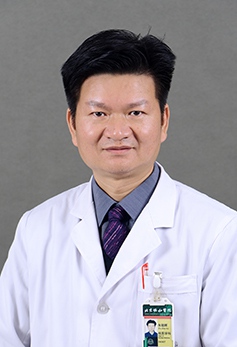 Zhaohui Zhu (朱朝晖)合作论文数Peking Union Medical College Hospital6
 Liying Cui (崔丽英)合作论文数Peking Union Medical College Hospital6
 Xiang Li (李响)合作论文数5
 Jing Yuan (袁晶)合作论文数Peking Union Medical College Hospital5
 Yining Wang (王怡宁)合作论文数Peking Union Medical College Hospital5Hongli Jing (景红丽)合作论文数5Huilei Miao合作论文数4Qiao Yang合作论文数4Ai-Lin Zhao合作论文数4MingQing Long (龙明清)合作论文数4LiBo Chen (陈黎波)合作论文数4JianTao Ba (巴建涛)合作论文数4Yimin Liu合作论文数4Hou Bo合作论文数4
 Zhiyong Liang (梁智勇)合作论文数Peking Union Medical College4
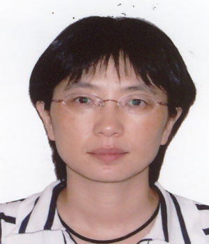 Yansong Lin (林岩松)合作论文数Department of Nuclear Medicine,Peking Union Medical College Hospital,Beijing ,P.R.China4
 Minghui Duan (段明辉)合作论文数Peking Union Medical College Hospital, Chinese Academy of Medical Sciences4Chenhao Jia合作论文数3Long Chang合作论文数3Ying ZHANG (张颖)合作论文数3Chenhui Mao合作论文数3Xue-Min Gao合作论文数3Yan Zhang合作论文数3Jing Gao合作论文数3
 Caiyan Liu合作论文数3
 Wei Chen (陈未)合作论文数Peking Union Medical College Hospital3Haiqun Xing (邢海群)合作论文数3
 Xue Lin (林雪)合作论文数Peking Union Medical College Hospital International Medical Services3
 Hongzhi Guan (关鸿志)合作论文数Peking Union Medical College Hospital3
 Feng Feng (冯逢)合作论文数Department of Radiology, Peking Union Medical College Hospital3
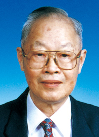 Shizhen Wang (王世真)合作论文数Institute of Radiation Medicine, Chinese Academy of Medical Sciences;Peking Union Medical College3
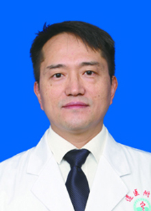 Jiong Cai (蔡炯)合作论文数Affiliated Hospital Of Zunyi Medical University3Stan Wang合作论文数2Jiangyu Ma合作论文数2Linwen Liu合作论文数2Menglin Liang合作论文数2Jingnan Wang合作论文数2Yue-Ying Mao合作论文数2




