Although recent work has made headway in understanding the neural temporospatial dynamics of conscious perception, much of that work has focused on visual paradigms. To determine whether there are shared mechanisms for perceptual consciousness across sensory modalities, here we test within the auditory domain. Participants completed an auditory threshold task while undergoing intracranial electroencephalography. Recordings from >2,800 grey matter electrodes were analyzed for broadband gamma power (a range which reflects local neural activity). For perceived trials, we find nearly simultaneous activity in early auditory regions, the right caudal middle frontal gyrus, and the non-auditory thalamus; followed by a wave of activity that sweeps through auditory association regions into parietal and frontal cortices. For not perceived trials, significant activity is restricted to early auditory regions. These findings show the cortical and subcortical networks involved in auditory perception are similar to those observed with vision, suggesting shared mechanisms for conscious perception.
Neural representations of perceptual decision formation that are abstracted from specific motor requirements have previously been identified in humans using non-invasive electrophysiology; however, it is currently unclear where these originate in the brain. Here we capitalized on the high spatiotemporal precision of intracranial EEG to localize such abstract decision signals. Participants undergoing invasive electrophysiological monitoring for epilepsy were asked to judge the direction of random-dot stimuli and respond either with a speeded button press (N = 24), or vocally, after a randomized delay (N = 12). We found a widely distributed motor-independent network of regions where high-frequency activity exhibited key characteristics consistent with evidence accumulation, including a gradual buildup that was modulated by the strength of the sensory evidence, and an amplitude that predicted participants' choice accuracy and response time. Our findings offer a new view on the brain networks governing human decision-making. Using intracranial EEG in human participants, the authors identify a functionally distinct set of brain regions which exhibited characteristic signatures of decision formation independently of the motor action associated with the choice.
Episodic memory function is predicated upon the precise coordination between the hippocampus and widespread cortical regions. However, our understanding of the neural mechanisms involved in this process is incomplete. In this study, human subjects undergoing intracranial electroencephalography (iEEG) monitoring performed a list learning task. We show sharp-wave ripple (SWR)-locked reactivation of specific semantic processing regions during free recall. This cortical activation consists of both broadband high frequency (non-oscillatory) and cortical ripple (oscillatory) activity. SWRs and cortical ripples in the anterior temporal lobe, a major semantic hub, co-occur and increase in rate prior to recall. Coincident hippocampal-ATL ripples are associated with a greater increase in cortical reactivation, show specificity in location based on recall content, and are preceded by cortical theta oscillations. These findings may represent a reactivation of hippocampus and cortical semantic regions orchestrated by an interplay between hippocampal SWRs, cortical ripples, and theta oscillations. ### Competing Interest Statement The authors have declared no competing interest.
The discrete events of our narrative experience are organized by the neural substrate that underlies episodic memory. This narrative process is segmented into discrete units by event boundaries. This permits a replay process that acts to consolidate each event into a narrative memory. High frequency oscillations (HFOs) are a potential mechanism for synchronizing neural activity during these processes. Here, we use intracranial recordings from participants viewing and freely recalling a naturalistic stimulus. We show that hippocampal HFOs increase following event boundaries and that coincident hippocampal-cortical HFOs (co-HFOs) occur in cortical regions previously shown to underlie event segmentation (inferior parietal, precuneus, lateral occipital, inferior frontal cortices). We also show that event-specific patterns of co-HFOs that occur during event viewing re-occur following the subsequent three event boundaries (in decaying fashion) and also during recall. This is consistent with models that support replay as a mechanism for memory consolidation. Hence, HFOs may coordinate activity across brain regions serving widespread event segmentation, encode naturalistic memory, and bind representations to assemble memory of a coherent, continuous experience.
Our continuous visual experience in daily life is dominated by change. Previous research has focused on visual change due to stimulus motion, eye movements or unfolding events, but not their combined impact across the brain, or their interactions with semantic novelty. We investigate the neural responses to these sources of novelty during film viewing. We analyzed intracranial recordings in humans across 6328 electrodes from 23 individuals. Responses associated with saccades and film cuts were dominant across the entire brain. Film cuts at semantic event boundaries were particularly effective in the temporal and medial temporal lobe. Saccades to visual targets with high visual novelty were also associated with strong neural responses. Specific locations in higher-order association areas showed selectivity to either high or low-novelty saccades. We conclude that neural activity associated with film cuts and eye movements is widespread across the brain and is modulated by semantic novelty.
AbstractElectrophysiological oscillations in the brain have been shown to occur as multicycle events, with onset and offset dependent on behavioral and cognitive state. To provide a baseline for state-related and task-related events, we quantified oscillation features in resting-state recordings. We developed an open-source wavelet-based tool to detect and characterize such oscillation events (OEvents) and exemplify the use of this tool in both simulations and two invasively-recorded electrophysiology datasets: one from human, and one from nonhuman primate (NHP) auditory system. After removing incidentally occurring event-related potentials (ERPs), we used OEvents to quantify oscillation features. We identified ∼2 million oscillation events, classified within traditional frequency bands: δ, θ, α, β, low γ, γ, and high γ. Oscillation events of 1–44 cycles could be identified in at least one frequency band 90% of the time in human and NHP recordings. Individual oscillation events were characterized by nonconstant frequency and amplitude. This result necessarily contrasts with prior studies which assumed frequency constancy, but is consistent with evidence from event-associated oscillations. We measured oscillation event duration, frequency span, and waveform shape. Oscillations tended to exhibit multiple cycles per event, verifiable by comparing filtered to unfiltered waveforms. In addition to the clear intraevent rhythmicity, there was also evidence of interevent rhythmicity within bands, demonstrated by finding that coefficient of variation of interval distributions and Fano factor (FF) measures differed significantly from a Poisson distribution assumption. Overall, our study provides an easy-to-use tool to study oscillation events at the single-trial level or in ongoing recordings, and demonstrates that rhythmic, multicycle oscillation events dominate auditory cortical dynamics.
The progress of therapeutic neuromodulation greatly depends on improving stimulation parameters to most efficiently induce neuroplasticity effects. Intermittent Theta Burst stimulation (iTBS), a form of electrical stimulation that mimics the natural brain activity patterns, has shown efficacy in inducing such effects in animal studies and rhythmic Transcranial Magnetic Stimulation (rTMS) studies in humans. However, little is known about the potential neuroplasticity effects of iTBS applied through intracranial electrodes in humans which could have implications for deep brain stimulation therapies. This study characterizes the physiological effects of cortical iTBS in the human cortex and compare them with single pulse alpha stimulation, another frequently used paradigm in rTMS research. We applied these stimulation paradigms to well-defined regions in the sensorimotor cortex which elicited contralateral hand or arm muscle contractions during electrical stimulation mapping in epilepsy patients implanted with intracranial electrodes. Treatment effects were evaluated using effective connectivity and beta oscillations coherence measures in areas connected to the treatment site as defined with cortico-cortical evoked potentials. Our results show that iTBS increases beta band synchronization within the sensorimotor network indicating a potential neuroplasticity effect. The effect is specific to the sensorimotor system, the beta frequency band and the stimulation pattern (no effect was found with single-pulse alpha stimulation). The effects outlasted the stimulation by three minutes. By characterizing the neurophysiological effects of iTBS within well-defined cortical networks, we hope to provide an electrophysiological framework that allows clinicians and researchers to optimize brain stimulation protocols which may have translational value.
Millions of people worldwide suffer motor or sensory impairment due to stroke, spinal cord injury, multiple sclerosis, traumatic brain injury, diabetes, and motor neuron diseases such as ALS (amyotrophic lateral sclerosis). A brain-computer interface (BCI), which links the brain directly to a computer, offers a new way to study the brain and potentially restore impairments in patients living with these debilitating conditions. One of the challenges currently facing BCI technology, however, is to minimize surgical risk while maintaining efficacy. Minimally invasive techniques, such as stereoelectroencephalography (SEEG) have become more widely used in clinical applications in epilepsy patients since they can lead to fewer complications. SEEG depth electrodes also give access to sulcal and white matter areas of the brain but have not been widely studied in brain-computer interfaces. Here we show the first demonstration of decoding sulcal and subcortical activity related to both movement and tactile sensation in the human hand. Furthermore, we have compared decoding performance in SEEG-based depth recordings versus those obtained with electrocorticography electrodes (ECoG) placed on gyri. Initial poor decoding performance and the observation that most neural modulation patterns varied in amplitude trial-to-trial and were transient (significantly shorter than the sustained finger movements studied), led to the development of a feature selection method based on a repeatability metric using temporal correlation. An algorithm based on temporal correlation was developed to isolate features that consistently repeated (required for accurate decoding) and possessed information content related to movement or touch-related stimuli. We subsequently used these features, along with deep learning methods, to automatically classify various motor and sensory events for individual fingers with high accuracy. Repeating features were found in sulcal, gyral, and white matter areas and were predominantly phasic or phasic-tonic across a wide frequency range for both HD (high density) ECoG and SEEG recordings. These findings motivated the use of long short-term memory (LSTM) recurrent neural networks (RNNs) which are well-suited to handling transient input features. Combining temporal correlation-based feature selection with LSTM yielded decoding accuracies of up to 92.04 ± 1.51% for hand movements, up to 91.69 ± 0.49% for individual finger movements, and up to 83.49 ± 0.72% for focal tactile stimuli to individual finger pads while using a relatively small number of SEEG electrodes. These findings may lead to a new class of minimally invasive brain-computer interface systems in the future, increasing its applicability to a wide variety of conditions.
Background: Paralysis and neuropathy, affecting millions of people worldwide, can be accompanied by significant loss of somatosensation. With tactile sensation being central to achieving dexterous movement, brain-computer interface (BCI) researchers have used intracortical and cortical surface electrical stimulation to restore somatotopically-relevant sensation to the hand. However, these approaches are restricted to stimulating the gyral areas of the brain. Since representation of distal regions of the hand extends into the sulcal regions of human primary somatosensory cortex (S1), it has been challenging to evoke sensory percepts localized to the fingertips. Objective/hypothesis: Targeted stimulation of sulcal regions of S1, using stereoelectroencephalography (SEEG) depth electrodes, can evoke focal sensory percepts in the fingertips. Methods: Two participants with intractable epilepsy received cortical stimulation both at the gyri via high-density electrocorticography (HD-ECoG) grids and in the sulci via SEEG depth electrode leads. We characterized the evoked sensory percepts localized to the hand. Results: We show that highly focal percepts can be evoked in the fingertips of the hand through sulcal stimulation. fMRI, myelin content, and cortical thickness maps from the Human Connectome Project elucidated specific cortical areas and sub-regions within S1 that evoked these focal percepts. Within-participant comparisons showed that percepts evoked by sulcal stimulation via SEEG electrodes were significantly more focal (80% less area; p = 0.02) and localized to the fingertips more often, than by gyral stimulation via HD-ECoG electrodes. Finally, sulcal locations with consistent modulation of high-frequency neural activity during mechanical tactile stimulation of the fingertips showed the same somatotopic correspondence as cortical stimulation. Conclusions: Our findings indicate minimally invasive sulcal stimulation via SEEG electrodes could be a clinically viable approach to restoring sensation.
Background and Objectives To identify the prevalence of EEG abnormalities in patients with coronavirus disease 2019 (COVID-19) with neurologic changes, their associated neuroimaging abnormalities, and rates of mortality. Methods A retrospective case series of 192 adult COVID-19-positive inpatients with EEG performed between March and June 2020 at 4 hospitals: 161 undergoing continuous, 24 routine, and 7 reduced montage EEG. Study indication, epilepsy history, intubation status, administration of sedatives or antiseizure medications (ASMs), metabolic abnormalities, neuroimaging pathology associated with epileptiform abnormalities, and in-hospital mortality were analyzed. Results EEG indications included encephalopathy (54.7%), seizure (18.2%), coma (17.2%), focal deficit (5.2%), and abnormal movements (4.6%). Epileptiform abnormalities occurred in 39.6% of patients: focal intermittent epileptiform discharges in 25.0%, lateralized periodic discharges in 6.3%, and generalized periodic discharges in 19.3%. Seizures were recorded in 8 patients, 3 with status epilepticus. ASM administration, epilepsy history, and older age were associated with epileptiform abnormalities. Only 26.3% of patients presented with any epileptiform abnormality, 37.5% with electrographic seizures, and 25.7% patients with clinical seizures had known epilepsy. Background findings included generalized slowing (88.5%), focal slowing (15.6%), burst suppression (3.6%), attenuation (3.1%), and normal EEG (3.1%). Neuroimaging pathology was identified in 67.1% of patients with epileptiform abnormalities, over two-thirds acute. In-hospital mortality was 39.5% for patients with epileptiform abnormalities and 36.2% for those without. Risk factors for mortality were coma and ventilator support at time of EEG. Discussion This article highlights the range of EEG abnormalities frequently associated with acute neuroimaging abnormalities in COVID-19. Mortality rates were high, particularly for patients in coma requiring mechanical ventilation. These findings may guide the prognosis and management of patients with COVID-19 and neurologic changes.
θ-Burst stimulation (TBS) protocols in transcranial magnetic stimulation studies have shown improved treatment efficacy in a variety of neuropsychiatric disorders. The optimal protocol to induce neuroplasticity in invasive direct electrical stimulation approaches is not known. We report that intracranial TBS applied in human sensorimotor cortex increases local coherence of preexistent β rhythms. The effect is specific to the stimulation frequency and the stimulated network and outlasts the stimulation period by ∼3 min.
Electrophysiological oscillations in the brain have been shown to occur as multi-cycle events, with onset and offset dependent on behavioral and cognitive state. To provide a baseline for state-related and task-related events, we quantified oscillation features in resting-state recordings. We used two invasively-recorded electrophysiology datasets: one from human, and one from non-human primate auditory system. After removing incidentally occuring event related potentials, we used a wavelet transform based method to quantify oscillation features. We identified about 2 million oscillation events, classified within traditional frequency bands: delta, theta, alpha, beta, low gamma, gamma, high gamma. Oscillation events of 1-44 cycles were present in at least one frequency band in 90% of the time in human and non-human primate recordings. Individual oscillation events were characterized by non-constant frequency and amplitude. This result naturally contrasts with prior studies which assumed such constancy, but is consistent with evidence from event-associated oscillations. We measured oscillation event duration, frequency span, and waveform shape. Oscillations tended to exhibit multiple cycles per event, verifiable by comparing filtered to unfiltered waveforms. In addition to the clear intra -event rhythmicity, there was also evidence of inter -event rhythmicity within bands, demonstrated by finding that coefficient of variation of interval distributions and Fano Factor measures differed significantly from a Poisson distribution assumption. Overall, our study demonstrates that rhythmic, multi-cycle oscillation events dominate auditory cortical dynamics.
 Stephan Bickel合作论文数11
 Ashesh D Mehta合作论文数Department of Neurological9
 Elizabeth Espinal合作论文数8
 Akash C Mishra合作论文数Int Ctr Theoret Sci, Tata Inst Fundamental Res4
 Charles E. Schroeder合作论文数Department of Neurosurgery, Columbia University College of Physicians and Surgeons;Translational Neuroscience Division, Nathan S. Kline Institute for Psychiatric Research3Samuel A. Neymotin合作论文数2
 Richard Ramdeo合作论文数2
 Joo-Won Kim合作论文数Baylor College of Medicine2Santosh Chandrasekaran合作论文数2
 Peter Lakatos合作论文数Department of Psychiatry, Grossman School of Medicine, New York University/The Nathan S. Kline Institute for Psychiatric Research2
 Griffith Erica Y合作论文数College of Physicians and Surgeons, Columbia University2
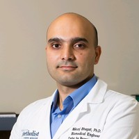 Nikunj Arunkumar Bhagat合作论文数Indian Institute of Science;The University of Texas Health Science Center at Houston2
 Gherman Sabina合作论文数Institute of Neuroscience and Psychology, University of Glasgow;Centre for Cognitive Neuroimaging, University of Glasgow;University of Glasgow, University of Glasgow2
 Barczak Annamaria合作论文数Nathan Kline Institute, New York University School of Medicine2
 Stephanie Jones合作论文数Department of Neuroscience, Brown University2
 Idan Tal合作论文数Gonda Multidisciplinary Brain Research Center, Bar-Ilan University2
 William Lytton合作论文数Wm. S. Middleton VA Hospital, University of Wisconsin2
 Chad E. Bouton合作论文数The Feinstein Institute for Medical Research2
 Haroon Anwar合作论文数Computational Neuroscience Unit, Okinawa Institute of Science and Technology2
 Junqian Xu合作论文数Baylor College of Medicine2
 Monica N O'Connell合作论文数Cognitive Neuroscience and Schizophrenia Program, Nathan Kline Institute for Psychiatric Research2
 Salvador Dura-Bernal合作论文数Centre for Robotics and Neural Systems, University of Plymouth2
 Alexander K. Smith合作论文数Department of Medicine, University of California2Tammy Mcginnis合作论文数2
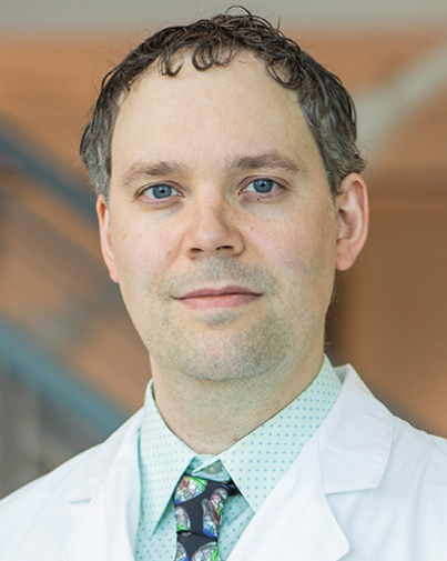 Matthew F. Glasser合作论文数Computational Imaging Research Center, Mallinckrodt Institute of Radiology, Washington University School of Medicine in St. Louis2Neeraj Singh合作论文数1Richard E Temes合作论文数1Tung Ming Leung合作论文数1Julia Z Ding合作论文数1Steven Pacia合作论文数1
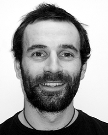 Simon P. Kelly合作论文数1Anup N Sonti合作论文数1
 Mark Aksen合作论文数1
 M. M. Gusso合作论文数1
 Lukas Hirsch合作论文数Department of Biomedical Engineering, The City College of New York1
 Christopher Micek合作论文数Worcester Polytechnic Institute1
 Ahmad A Ballout合作论文数Dept Neurol, Northwell Hlth1
 Maximilian Nentwich合作论文数1Ahmad Abdel-Aty合作论文数School of Medicine, Yale University1
 Serdar Akkol合作论文数Feinstein Institutes for Medical Research, Northwell Health1
 Aya Khalaf合作论文数University of Pittsburgh1
 Kun Wu合作论文数Department of Neurosurgery, Yale School of Medicine, Yale University1
 Dennis Spencer合作论文数Department of Neurosurgery, Yale School of Medicine, Yale University1
 Eyiyemisi Damisah合作论文数Department of Neurosurgery, Yale School of Medicine, Yale University1
 Jason Gerrard合作论文数Semmes Murphey Clinic & UTenn Health Sciences Center1
 Hal Blumenfeld合作论文数Neuroscience, Biological & Biomedical Sciences, Yale University1Najjar Souhel合作论文数1
 Redmond O'Connell合作论文数Inst Neurosci, Trinity Coll Dublin1Leszczynski Marcin合作论文数Department of Epileptology, University of Bonn Bonn1




