Complementary metal-oxide-semiconductor (CMOS)-based high-density microelectrode arrays (HD-MEAs) enable neuronal recordings with high spatiotemporal resolution. However, integrating polydimethylsiloxane (PDMS) microstructures onto HD-MEA surfaces to control network architecture is currently challenging and platform specific. Here, we present a protocol for PDMS fabrication, HD-MEA chip preparation, PDMS-HD-MEA microstructure alignment, and cell culture, including alternatives. Our results show reproducible formation of modular networks with characteristic activity patterns across different systems. This protocol supports engineering of defined neuronal architectures while maintaining compatibility with various HD-MEA systems. For complete details on the use and execution of this protocol, please refer to Sato et al.1.
There are a number of works on superhydrophobicity, or the Lotus Effect® as it is sometimes called. This chapter considers how wetting is affected by roughness, but in keeping with the nanotechnological outlook of the book focusses mostly on how the geometry or shape of the roughness changes the interaction with liquids. Simple considerations of superhydrophobicity would not reveal many of these effects as they only become apparent under conditions where the assumptions used in the simpler models do not apply. Some biological examples show complex geometries exploiting these effects.
Cardiovascular diseases (CVDs) are the leading causes of death worldwide, with approx. Twenty million deaths in 2021. Cardiovascular implants are among the most used biomaterials in the clinical world. However, poor endothelialisation and rapid thrombosis remains a challenge. Simple chemical surface modification techniques can be used to steer biological interactions without affecting the bioimplants' overall bulk characteristics such as radiopacity and flexibility. Although silanes are well studied for protein and cell interactions, the methodical investigation of cardiac endothelial cell (EC) alongside smooth muscle cell (SMC) to mimic natural arterial environments has been limited. In this study, these cells have been investigated on surfaces functionalized with methyl, amine, thiol, methacrylate, and fluorine organosilane groups. Cardiac EC and SMC growth was investigated with metabolic activity, time lapse imaging, and immunofluorescent staining techniques. The results demonstrated that the surfaces tested are able to selectively regulate the viability and growth of the cells. Aminosilane modified surfaces displayed 2-fold higher metabolic activity with HUVEC and 2-fold less metabolic activity with HCASMC cell lines, compared to tissue culture plastic controls. The amino-modification outperformed all other chemistries tested in terms of ability to promote the proliferation of ECs, while importantly reducing the activity of SMCs. This report demonstrates that aminosilane modified surfaces have the potential to be utilized in novel cardiovascular implants, which could improve biological integration in the short and possibly longer-term. The findings of this study suggest that specific chemical modifications of the surface can enhance endothelial cell activity while minimizing the proliferation of smooth muscle cells, which are often associated with thrombosis. This highlights the potential of carefully engineered surface chemistries to improve the clinical outcomes of cardiovascular implants.
The study of in vivo thrombus formation has principally been performed using intravital microscopy in mice and other species. These have allowed us to visualise the molecular and cellular processes that regulate thrombus formation inside the body. However current in vivo arterial thrombosis models are difficult to standardise between labs and frequently produce results that do not reliably translate successfully in human clinical trials. Here we provide a step-by-step description with accompanying video tutorials to demonstrate how to produce a 3D humanised thrombus-on-a-chip model, which uses perfusion of fluorescently-labelled human blood over a mechanically-injured human tissue engineered arterial construct (TEAC) within a 3D printed microfluidic flow chamber to replicate thrombus formation within a healthy artery. We also provide a written methodology on how to use 3D printing to produce a mechanical injury press that can reproducibly damage the TEAC as a stimulus for thrombus formation as part of a mechanical injury model. Perfusion of the uninjured TEAC with whole human blood containing DiOC6-labelled platelets without initiating notable thrombus formation. The mechanical injury press was shown to induce a reproducible puncture wound in the TEAC. Fluorescence microscopy was used to demonstrate that thrombus formation could be observed reproducibly around sites of injury. This humanised thrombosis-on-a-chip model can replace the use of animals in in vivo thrombosis models for preclinical assessment of anti-thrombotic therapies. This method also offers multiple scientific advantages: allowing new drugs to be directly tested on human blood from a diverse array of donors, facilitating use of a realistic and reproducible injury modality as well as removing the potential confounding effects of general anaesthetics in animal studies. The use of human thrombus-on-a-chip models combining TEACs offers a new methodology to reduce animal use whilst improving the predictive capabilities of preclinical trials of anti-thrombotic therapies.
The study of in vivo thrombus formation has principally been performed using intravital microscopy in mice and other species. These have allowed us to visualise the molecular and cellular processes that regulate thrombus formation inside the body. However current in vivo arterial thrombosis models are difficult to standardise between labs and frequently produce results that do not reliably translate successfully in human clinical trials. Here we provide a step-by-step description with accompanying video tutorials to demonstrate how to produce a 3D humanised thrombus-on-a-chip model, which uses perfusion of fluorescently-labelled human blood over a mechanically-injured human tissue engineered arterial construct (TEAC) within a 3D printed microfluidic flow chamber to replicate thrombus formation within a healthy artery. We also provide a written methodology on how to use 3D printing to produce a mechanical injury press that can reproducibly damage the TEAC as a stimulus for thrombus formation as part of a mechanical injury model. Perfusion of the uninjured TEAC with whole human blood containing DiOC6-labelled platelets without initiating notable thrombus formation. The mechanical injury press was shown to induce a reproducible puncture wound in the TEAC. Fluorescence microscopy was used to demonstrate that thrombus formation could be observed reproducibly around sites of injury. This humanised thrombosis-on-a-chip model can replace the use of animals in in vivo thrombosis models for preclinical assessment of anti-thrombotic therapies. This method also offers multiple scientific advantages: allowing new drugs to be directly tested on human blood from a diverse array of donors, facilitating use of a realistic and reproducible injury modality as well as removing the potential confounding effects of general anaesthetics in animal studies. The use of human thrombus-on-a-chip models combining TEACs offers a new methodology to reduce animal use whilst improving the predictive capabilities of preclinical trials of anti-thrombotic therapies.
Liquid biopsy is revolutionizing cancer management, with circulating tumor cells (CTCs), offering a transformative approach to screening, diagnosis, and treatment monitoring. However, existing CTC isolation methods relying on antigen expression or physical properties lack robustness, are operator-dependent, and suffer from automation challenges, leading to inconsistent and time-intensive analyses. A universal, unbiased methodology for CTC detection across tumor types is critically needed. Here, we present the first proof-of-concept study demonstrating the use of Fourier transform infrared (FT-IR) microspectroscopy to study cytospun blood samples coupled with a random forest (RF) classifier, for the detection of a single CTC in the blood of a lung cancer patient as confirmed via immunohistochemistry. Notably, our method utilizes glass coverslips as substrates, routinely employed in pathology departments, enabling seamless integration with histopathological analyses (e.g., staining, immunohistochemistry). Using FT-IR spectral data from in vitro growing lung cancer cells as a training model, we achieved precise CTC identification based on biochemical composition, specifically within the fingerprint region (1800 cm-1 to 1350 cm-1). This study introduces FT-IR microspectroscopy as a novel, label-free approach for CTCs detection in liquid biopsies, with the potential to redefine cancer diagnostics. By enhancing precision and accessibility in CTC identification, the clinical implementation of this methodology may represent a significant advancement in personalized oncology, offering a clinically viable tool for real-time cancer monitoring and improved patient stratification.
The clinical success of organoid‐based therapies depends critically on understanding and controlling how biomaterial surfaces influence organoid behavior and integration. Retinal organoids (ROs) are emerging as advanced in vitro models with potential for transplantation, yet the molecular underpinnings of their interactions with engineered biomaterials remain largely unexplored. Here, this study systematically examines the effects of surface chemical modifications: amine (NH2), hydroxyl (OH), phenyl (Ph), and methyl (CH3) groups presented via alkyl‐based self‐assembled monolayers, on RO attachment, migration, and differentiation at key developmental stages. This study's' findings reveal that surface chemistry is a major determinant of organoid response: Hydrophilic surfaces (OH and NH2) markedly enhance RO migration and retinal ganglion cell differentiation, while less wettable surfaces (Ph and CH3) restrict initial attachment. These results shed new light on the pivotal role of the biomaterial interface in directing 3D organoid development, offering actionable insights for the optimization of organoid delivery, integration, and function in regenerative medicine. A deeper understanding of these interactions will enable the rational design of biomaterial platforms and accelerate the translation of organoid models to clinical therapies.
The management of cancer patients has markedly improved with the advent of personalised medicine where treatments are given based on tumour antigen expression amongst other. Within this remit, liquid biopsies will no doubt improve this personalised cancer management. Identifying circulating tumour cells in blood allows a better assessment for tumour screening, staging, response to treatment and follow up. However, methods to identify/capture these circulating tumour cells using cancer cells' antigen expression or their physical properties are not robust enough. Thus, a methodology that can identify these circulating tumour cells in blood regardless of the type of tumour is highly needed. Fourier Transform Infrared (FTIR) microspectroscopy, which can separate cells based on their biochemical composition, could be such technique. In this feasibility study, we studied lung cancer cells (squamous cell carcinoma and adenocarcinoma) mixed with peripheral blood mononuclear cells (PBMC). The data obtained shows, for the first time, that FTIR microspectroscopy together with Random Forest classifier is able to identify a single lung cancer cell in blood. This separation was easier when the region of the IR spectra containing lipids and the amide A (2700 to 3500 cm-1) was used. Furthermore, this work was carried out using glass coverslips as substrates that are widely used in pathology departments. This allows further histopathological cell analysis (staining, immunohistochemistry, …) after FTIR spectra are obtained. Hence, although further work is needed using blood samples from patients with cancer, FTIR microspectroscopy could become another tool to be used in liquid biopsies for the identification of circulating tumour cells, and in the personalised management of cancer.
Collagen hydrogels are a rapidly expanding platform in bioengineering and soft materials engineering for novel applications focused on medical therapeutics, medical devices and biosensors. Observations linking microstructure to material properties and function enables rational design strategies to control this space. Visualisation of the microscale organisation of these soft hydrated materials presents unique technical challenges due to the relationship between hydration and the molecular organisation of a collagen gel. Scanning electron microscopy is a robust tool widely employed to visualise and explore materials on the microscale. However, investigation of collagen gel microstructure is difficult without imparting structural changes during preparation and/or observation. Electrons are poorly propagated within liquid‐phase materials, limiting the ability of electron microscopy to interrogate hydrated gels. Sample preparation techniques to remove water induce artefactual changes in material microstructure particularly in complex materials such as collagen, highlighting a critical need to develop robust material handling protocols for the imaging of collagen hydrogels. Here a collagen hydrogel is fabricated, and the gel state explored under high‐vacuum (10 −6 Pa) and low‐vacuum (80–120 Pa) conditions, and in an environmental SEM chamber. Visualisation of collagen fibres is found to be dependent on the degree of sample hydration, with higher imaging chamber pressures and humidity resulting in decreased feature fidelity. Reduction of imaging chamber pressure is used to induce evaporation of gel water content, revealing collagen fibres of significantly larger diameter than observed in samples dehydrated prior to imaging. Rapid freezing and cryogenic handling of the gel material is found to retain a porous 3D structure following sublimation of the gel water content. Comparative analysis of collagen hydrogel materials demonstrates the care needed when preparing hydrogel samples for electron microscopy.
Supplemental Figure S1. Comparison of clonogenic survival fractions of various cancer cell lines after treatment with SAR302503 and Ruxolitinib. Supplemental Figure S2. KRAS mutation by treatment sensitivities in NSCLC cell lines. Supplementary Figure S3. The response of NCIH1944 xenografts to ionizing radiation. Supplemental Figure S4. Top-ranked pathways in SAR sensitive NSCLC cell lines. Supplemental Figure S5. Cancer growth and proliferation gene network in SAR-sensitive NSCLC. Supplemental Figure S6. ROC analysis of SAR sensitivity by TSP-IRDS scores. Supplemental Figure S7. Distribution of TSP-IRDS scores in clinical NSCLCs. Supplemental Figure S8. TSP-IRDS predicts survival in clinical NSCLC. Supplemental Figure S9. TSP-IRDS scores predict the benefit of adjuvant cisplatin-based chemotherapy in clinical NSCLC. Supplementary Figure and Table Legends
Dried plant specimens stored in herbaria are an untapped treasure chest of information on environmental conditions, plant evolution and change over many hundreds of years. Owing to their delicate nature and irreplaceability, there is limited access for analysis to these sensitive samples, particularly where chemical data are obtained using destructive techniques. Fourier transform infrared (FTIR) spectroscopy is a chemical analysis technique which can be applied non-destructively to understand chemical bonding information and, therefore, functional groups within the sample. This provides the potential for understanding geographical, spatial and species-specific variation in plant biochemistry. Here, we demonstrate the use of mid-FTIR microspectroscopy for the chemical analysis of Drosera rotundifolia herbarium specimens, which were collected 100 years apart from different locations. Principal component and hierarchical clustering analysis enabled differentiation between three main regions on the plant (lamina, tentacle stalk and tentacle head), and between the different specimens. Lipids and protein spectral regions were particularly sensitive differentiators of plant tissues. Differences between the different sets of specimens were smaller. This study demonstrates that relevant information can be extracted from herbarium specimens using FTIR, with little impact on the specimens. FTIR, therefore, has the potential to be a powerful tool to unlock historic information within herbaria.
Thomas Hartung1,2*Lena Smirnova1Itzy E. Morales Pantoja1Akwasi Akwaboah3Dowlette-Mary Alam El Din1Cynthia A. Berlinicke4J. Lomax Boyd5Brian S. Caffo6Ben Cappiello7Tzahi Cohen-Karni8,9J. Lowry Curley7Ralph Etienne-Cummings3Raha Dastgheyb10David H. Gracias11,12,13,14,15,16Frederic Gilbert17Christa Whelan Habela10Fang Han18Timothy D. Harris19,20Kathrin Herrmann1Eric J. Hill21Qi Huang11Rabih E. Jabbour22Erik C. Johnson20Brett J. Kagan23Caroline Krall1Andre Levchenko24Paul Locke1Alexandra Maertens1Monica Metea25Alysson R. Muotri26,27Rheinallt Parri28Barton L. Paulhamus20Jesse D. Plotkin1Paul Roach29July Carolina Romero1Jens C. Schwamborn30Fenna Sillé1Alexander S. Szalay31,32,33Katya Tsaioun1Daniel Tornero34,35Joshua T. Vogelstein36Karl J. Wahlin37Donald J. Zack38,39,40,41
Developmental engineering (DE) aims to culture mammalian cells on corresponding modular scaffolds (scale: micron to millimeter), then assemble these into functional tissues imitating natural developmental biology processes. This research intended to investigate the influences of polymeric particles on modular tissue cultures. When poly(methyl methacrylate) (PMMA), poly(lactic acid) (PLA) and polystyrene (PS) particles (diameter: 5–100 µm) were fabricated and submerged in culture medium in tissue culture plastics (TCPs) for modular tissue cultures, the majority of adjacent PMMA, some PLA but no PS particles aggregated. Human dermal fibroblasts (HDFs) could be directly seeded onto large (diameter: 30–100 µm) PMMA particles, but not small (diameter: 5–20 µm) PMMA, nor all the PLA and PS particles. During tissue cultures, HDFs migrated from the TCPs surfaces onto all the particles, while the clustered PMMA or PLA particles were colonized by HDFs into modular tissues with varying sizes. Further comparisons revealed that HDFs utilized the same cell bridging and stacking strategies to colonize single or clustered polymeric particles, and the finely controlled open pores, corners and gaps on 3D-printed PLA discs. These observed cell–scaffold interactions, which were then used to evaluate the adaptation of microcarrier-based cell expansion technologies for modular tissue manufacturing in DE.
Tissue engineering approaches aim to overcome the limitations of organ transplants and facilitate tissue repair and regeneration, with demand now at a worldwide high for advanced therapies due to our global aging population. Neural tissue engineering is challenging with tissue dynamics and cellular complexity constraints necessary for tissue function. Here, a conductive, highly aligned, fibrous polycaprolactone/gelatin/polypyrrole/graphene scaffold is demonstrated for potential nerve tissue repair. A simple and efficient electrospinning technique with a rotating drum fabrication approach is utilized to create aligned fibrous structures with a diameter of 380 +/- 37 nm (no graphene) to 265 +/- 30 nm (up to 3 wt% graphene). The conductivity of the scaffold in wet conditions was found range from 0.76 +/- 0.1 S m(-1) with no graphene, to 3.96 +/- 0.2 S m(-1) with 3% wt graphene, with corresponding ultimate tensile strengths measuring 2.6 +/- 0.1-5.5 +/- 0.4 MPa, respectively. Samples were found to biodegrade during incubation in saline solution over 42 days by similar to 48.5%. Fibroblasts were used as a cell model to test for scaffold toxicity, with all samples presenting good cell adhesion and limited cytotoxicity. Overall, the results demonstrated an aligned fibrous platform with good mechanical and electrically conductive properties useful for tissue engineering applications, particularly for nerve tissue. Development of novel materials with a range of properties enabling optimization of cell adhesion through to tissue development will further support the development of regenerative medicine approaches.
Current in vitro thrombosis models utilise simplistic 2D surfaces coated with purified components of the subendothelial matrix. The lack of a realistic humanised model has led to greater study of thrombus formation in in vivo tests in animals. Here we aimed to develop 3D hydrogel-based replicas of the medial and adventitial layers of the human artery to produce a surface that can optimally support thrombus formation under physiological flow conditions. These tissue-engineered medial- (TEML) and adventitial-layer (TEAL) hydrogels were developed by culturing human coronary artery smooth muscle cells and human aortic adventitial fibroblasts within collagen hydrogels, both individually and in co-culture. Platelet aggregation upon these hydrogels was studied using a custom-made parallel flow chamber. When cultured in the presence of ascorbic acid, the medial-layer hydrogels were able to produce sufficient neo-collagen to support effective platelet aggregation under arterial flow conditions. Both TEML and TEAL hydrogels possessed measurable tissue factor activity and could trigger coagulation of platelet-poor plasma in a factor VII-dependent manner. Biomimetic hydrogel replicas of the subendothelial layers of the human artery are effective substrates for a humanised in vitro thrombosis model that could reduce animal experimentation by replacing current in vivo models.
Supplementary Table 1. Clonogenic survival of NSCLC cell lines after cytotoxic therapy.
Regeneration solutions for the osteochondral interface depth are limited, where multi-material implants have the potential to delaminate affecting the regeneration process and impacting the final integrity of tissue interface. Here we explore regionally mixed hydrogel networks, presenting distinct chemical features to determine their compatibility in supporting osteogenic or chondrogenic cell behaviour and differentiation. Poly(N-isopropylacrylamide) (pNIPAM) and poly(N-tert-butylacrylamide) (pNTBAM) hydrogels were assessed in terms of their chemical differences, mechanical strength, internal architecture, porosity and capacity to support cell viability, migration, and differentiation. pNTBAM polymerized with a Young’s modulus of up to 371 ± 31 kPa compared to the more flexible pNIPAM, 16.5 ± 0.6 kPa. Viability testing revealed biocompatibility of both hydrogels with significantly increased cell numbers observed in pNTBAM (500 ± 95 viable cells/mm2) than in pNIPAM (60 ± 3 viable cells/mm2) (P ≤ 0.05). Mineralization determined through alkaline phosphatase (ALP) activity, calcium ion and annexin A2 markers of mineralization) and osteogenic behaviour (collagen I expression) were supported in both hydrogels, but to a greater extent in pNTBAM. pNTBAM supported significantly elevated levels of chondrogenic markers as evidenced by collagen II and glycosaminoglycan expression in comparison to little or no evidence in pNIPAM (P ≤ 0.05). In conclusion, structurally similar, chemically distinct, acrylamide hydrogels display variable capacities in supporting osteochondral cell behaviours. These systems demonstrate spatial control of cell interaction through simple changes in monomer chemistry. Fine control over chemical presentation during the fabrication of biomaterial implants could lead to greater efficacy and targeted regeneration of semi-complex tissues.
In this review we provide a general review of current research themes where complex surface topography plays a role. Recent innovations and research possibilities have opened up using modern micro topography creation techniques. We are considering a large range of themes in limited depth in order to provide an introduction and to illustrate how broad the field is and to those already working in the area to help gain a broad perspective. Due to the breadth of scope this review will not go into much depth into any given area. This review aims to provide a comprehensive understanding and practical guidance on the modern use of micro and nano-topography to control water, adhesion, wettability, control of biological adhesion including implants, cell culture and biofouling and directional fluid transport. The broad range of functionality is highlighted in the many interfacial interactions, however, the manufacturing scale to allow these surfaces to be commonly used is still in development. In order to make it more accessible we have chosen to concentrate on open access articles to cite.
Mesenchymal stromal cells (MSCs) show immunosuppressive effects both via cell-to-cell contact (direct) with immune cells and by producing paracrine factors and extracellular vesicles (indirect). A key challenge in delivering this therapeutic effect in vivo is retaining the MSCs at the site of injection. One way to address this is by encapsulating the MSCs within suitable biomaterial scaffolds. Here, we assess the immunosuppressive effect of alginate-encapsulated murine MSCs on proliferating murine splenocytes. Our results show that MSCs are able to significantly suppress splenocyte proliferation by similar to 50% via the indirect mechanism and almost completely (similar to 98%) via the direct mechanism. We also show for the first time that MSCs as monolayers on tissue culture plastic or encapsulated within alginate, when physically isolated from the splenocytes via transwells, are able to sustain immunosuppressive activity with repeated exposure to fresh splenocytes, for as long as 9 days. These results indicate the need to identify design strategies to simultaneously deliver both modes of MSC immunosuppression. By designing cell-biomaterial constructs with tailored degradation profiles, we can achieve a more sustained (avoiding MSCs migration and apoptosis) and controlled release of both the paracrine signals and eventually the cells themselves enabling efficient MSC-based immunosuppressive therapies for wound healing.
 Glen McHale合作论文数School of Engineering, The University of Edinburgh16
 Neil J. Shirtcliffe合作论文数School of Biomedical and Natural Sciences, Nottingham Trent University16
 Michael Ian Newton合作论文数15
 Carole Perry合作论文数School of Science & Technology, Nottingham Trent University;Harvard University9
 Ralph R. Weichselbaum合作论文数Department of Radiation and Cellular Oncology, The University of Chicago7
 Rosemary A Fricker合作论文数7
 Nikolai N. Khodarev合作论文数7
 Morgan Alexander合作论文数School of Pharmacy, Faculty of Science, University of Nottingham6
 Mitchell C. Posner合作论文数Department of Radiation Oncology, The University of Chicago5
 Sean Pitroda合作论文数Division of the Biological Sciences, The University of Chicago;PersonaDx5
 Nicholas R. Forsyth合作论文数Department of Cell Biology, University of Texas Southwestern Medical Center;Fax: +1 214 648 8694, Tel: +1 214 648 3282;Department of Cell Biology, University of Texas Southwestern Medical Center5Xiaona Huang合作论文数4Suisui Song合作论文数4Melinda E. Stack合作论文数4Lucy Chen合作论文数4
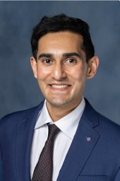 Akash D. Parekh合作论文数Department of Radiation and Cellular Oncology and Ludwig Center for Metastasis Research,4
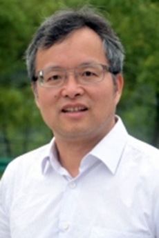 Hua Liang合作论文数Department of Statistics, Columbian College of Arts & Sciences, The George Washington University4
 Ying Yang合作论文数School of Life Sciences, Keele University4
 Shaun Atherton合作论文数4
 Nikolaj Gadegaard合作论文数James Watt School of Engineering, University of Glasgow4Nicola Doy合作论文数School of Science;Nottingham Trent University;Clifton Campus4Josep Sule-Suso合作论文数4
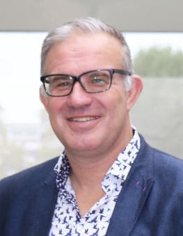 Mark Lewis合作论文数School of Sport, Exercise and Health Sciences, Loughborough University4Gene-Fu Liu合作论文数3daniel g van pittius合作论文数3Matthew J Köse-Dunn合作论文数3munyaradzi kamudzandu合作论文数3
 Kane Treadwell合作论文数3
 Rhys W. Lodge合作论文数Sch Chem, Univ Nottingham3
 Anna Piccinini合作论文数School of Pharmacy, Faculty of Science, The University of Nottingham3Rebecca Morgan合作论文数3
 Alasdair Kay合作论文数3
 Oksana Kehoe合作论文数3
 Andrew Capel合作论文数Department of Materials, Loughborough University3
 David Franklin Farrar合作论文数3Alan G.S. Harper合作论文数3
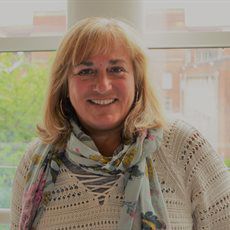 Alicia El Haj合作论文数School of Chemical Engineering, College of Engineering and Physical Sciences, University of Birmingham;Healthcare Technologies Institute, Institute of Translational Medicine, University of Birmingham3MI Newton合作论文数2Rupert Wright合作论文数2Srinivas Pillai合作论文数2
 Joshua C Price合作论文数2
 Lewis Dowling合作论文数2
 Mairead Hyland合作论文数2D. J. Merryweather合作论文数2Chari Divya M合作论文数2
 Stuart Iain Jenkins合作论文数2Orme Rowan P合作论文数2
 Ralf Warmuth合作论文数Department of Chemistry and Chemical Biology, School of Arts and Sciences, Rutgers, The State University of New Jersey2




